Video shot by Nick Andrussian | Mizzou Visual Productions Package produced by Evan Johnson | Bond LSC
As Mizzou seniors think about life after graduation, the research lab could serve as a proving ground for future plans.
That was the case for Bennett Flannagan, who graduated from Mizzou in 2024. He spent the last year as a research specialist I in the Paul de Figueiredo lab at Bond LSC, pushing himself and growing his expertise in preparation for graduate school.
His work paid off when he heard he was one of 20 applicants accepted into the Translational Biosciences PhD program at Mizzou’s School of Medicine for fall 2025.
How high-performance computing connects brain differences to apnea
Adobe Stock image
By Sophie Rentschler | Bond LSC
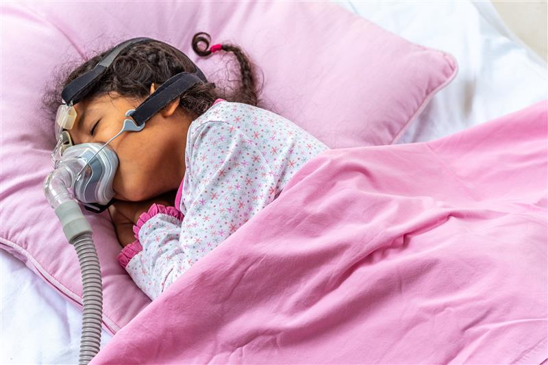
A good night’s rest lays the foundation for your daily performance but when a child’s body hampers airflow, that can lead to cognitive problems in their waking lives.
One recent Mizzou study looks at healthy neurological processes and how this differs for patients with obstructive sleep apnea. The study collected a wealth of patient information that can be utilized to help clinicians give a diagnosis quicker in the future.
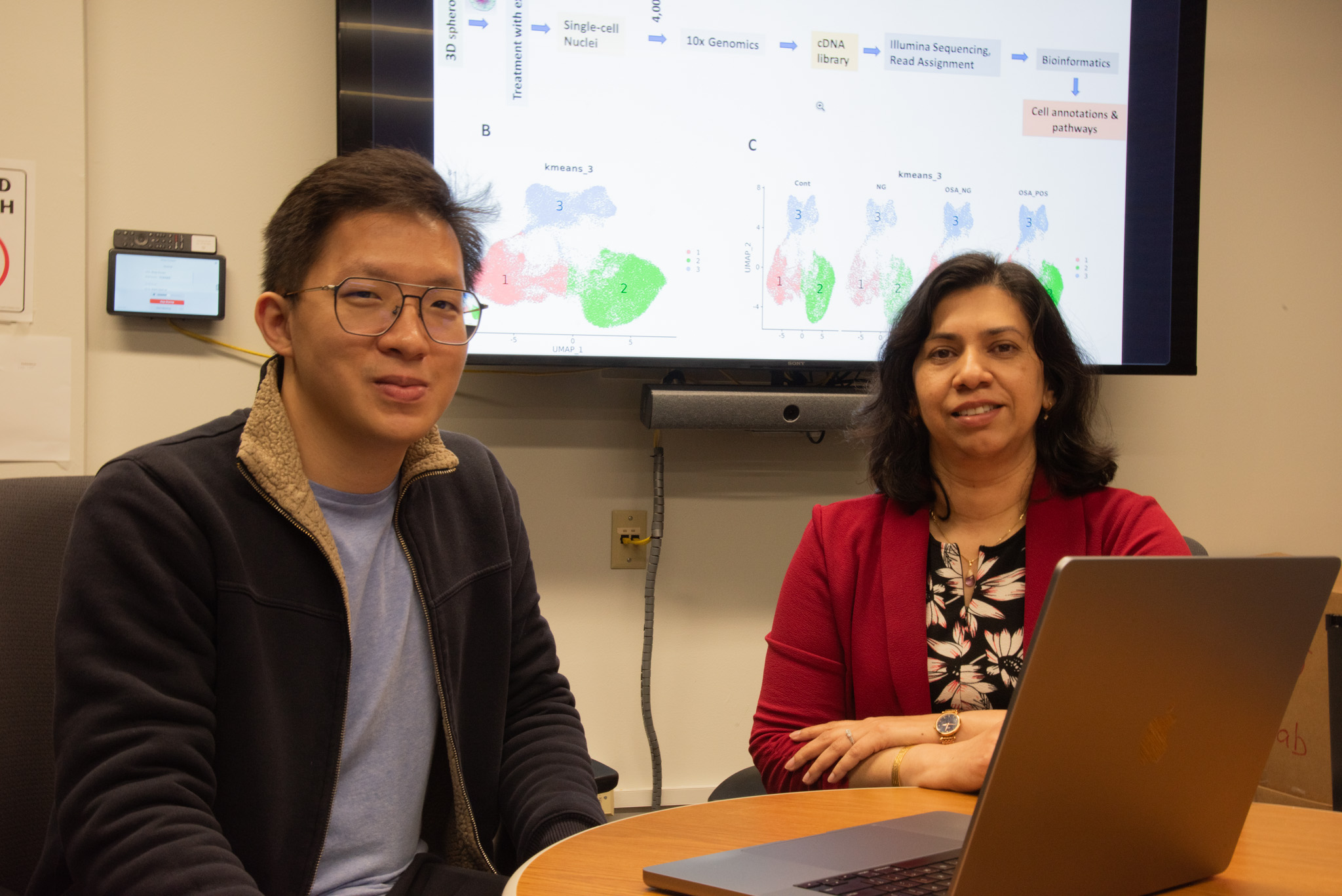
Yen On Chan, a graduate student in Trupti Joshi’s lab at Bond Life Sciences Center, uses computer models to understand children who have obstructive sleep apnea (OSA). Chan generates interactive visualizations for the lab with computing techniques thanks to his background in computer engineering. His 3D models illustrate the blood-brain barrier to compare patients without OSA, patients with OSA, and patients with both cognitive problems and OSA.
Yen On Chan, MU graduate student, and faculty lead for translational bioinformatics for NextGen Biomedical Informatics Trupi Joshi review visualizations from their study on April 2, 2025.
The Joshi lab worked alongside researchers from Marshall University, Abdelnaby Khalyfa and David Gozal, who see patients and collect information from them. The study focused on two structural differences induced by exosomes in children — those affecting the neurological components and the blood-brain barrier.
Exosomes are membrane-bound vesicles responsible for intercellular chatter, communicating signals to cells and carrying proteins between them. Exosomes travel through the blood-brain barrier (BBB). The BBB is a protective shell for the brain, separating the brain’s blood vessels and its cells. This neurological structure serves as a regulator for what molecules pass from blood to the brain, protecting it from harmful toxins and attacks from bacteria and viruses.
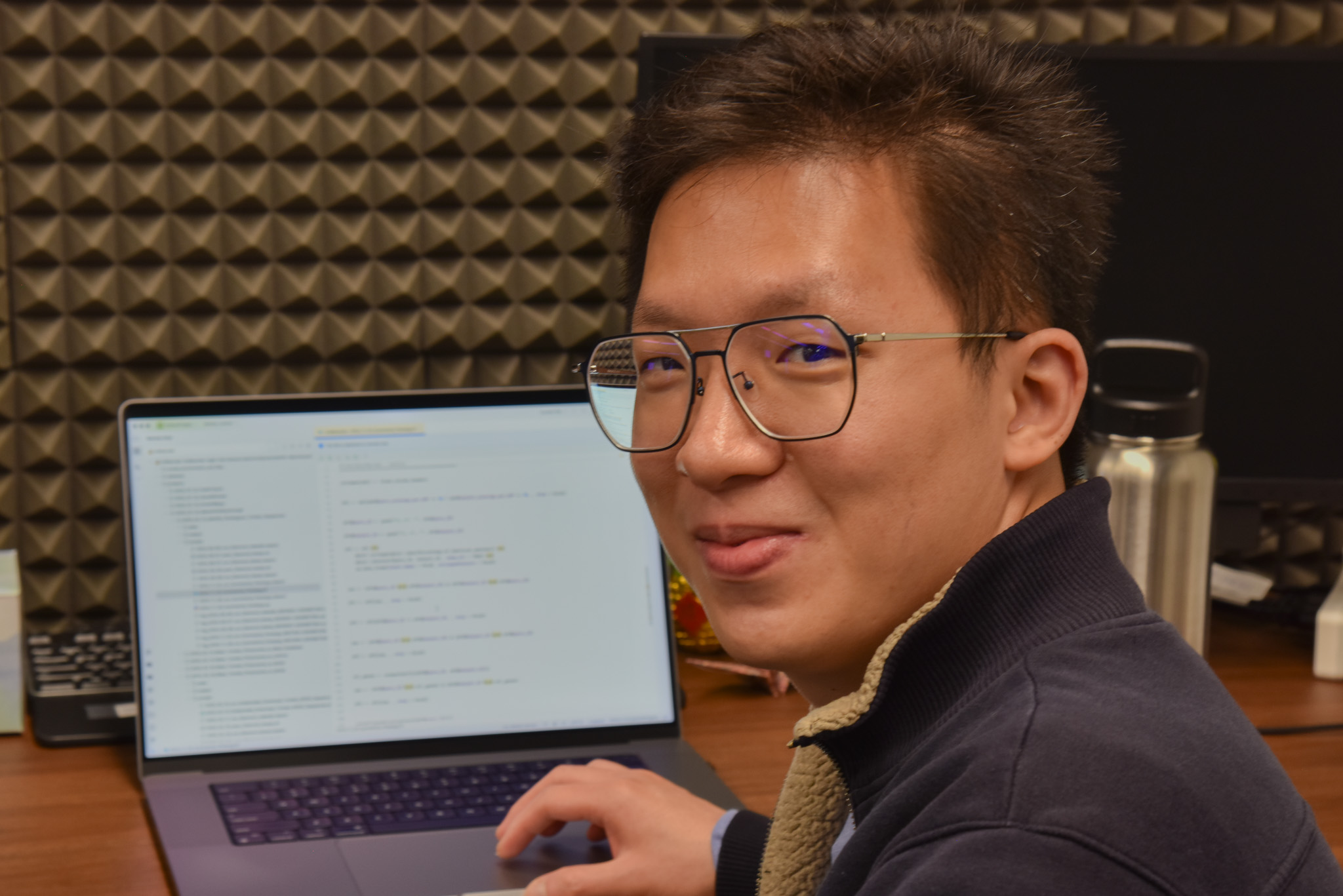
Chan entered the biology field with solely a computer engineering background. He was introduced to plant biology, later leading him to studying bioinformatics.
Yen On Chan, MU graduate student studying bioinformatics, works on his laptop on April 2, 2025.
“I enjoy programming and developing interactive visualizations (of the blood-brain barrier) which can help reveal deeper insights and enhance our understanding of its complex structure and functions,” Chan said.
Patient data from Joshi’s bioinformatics lab is so hefty that it must be run on a supercomputer.
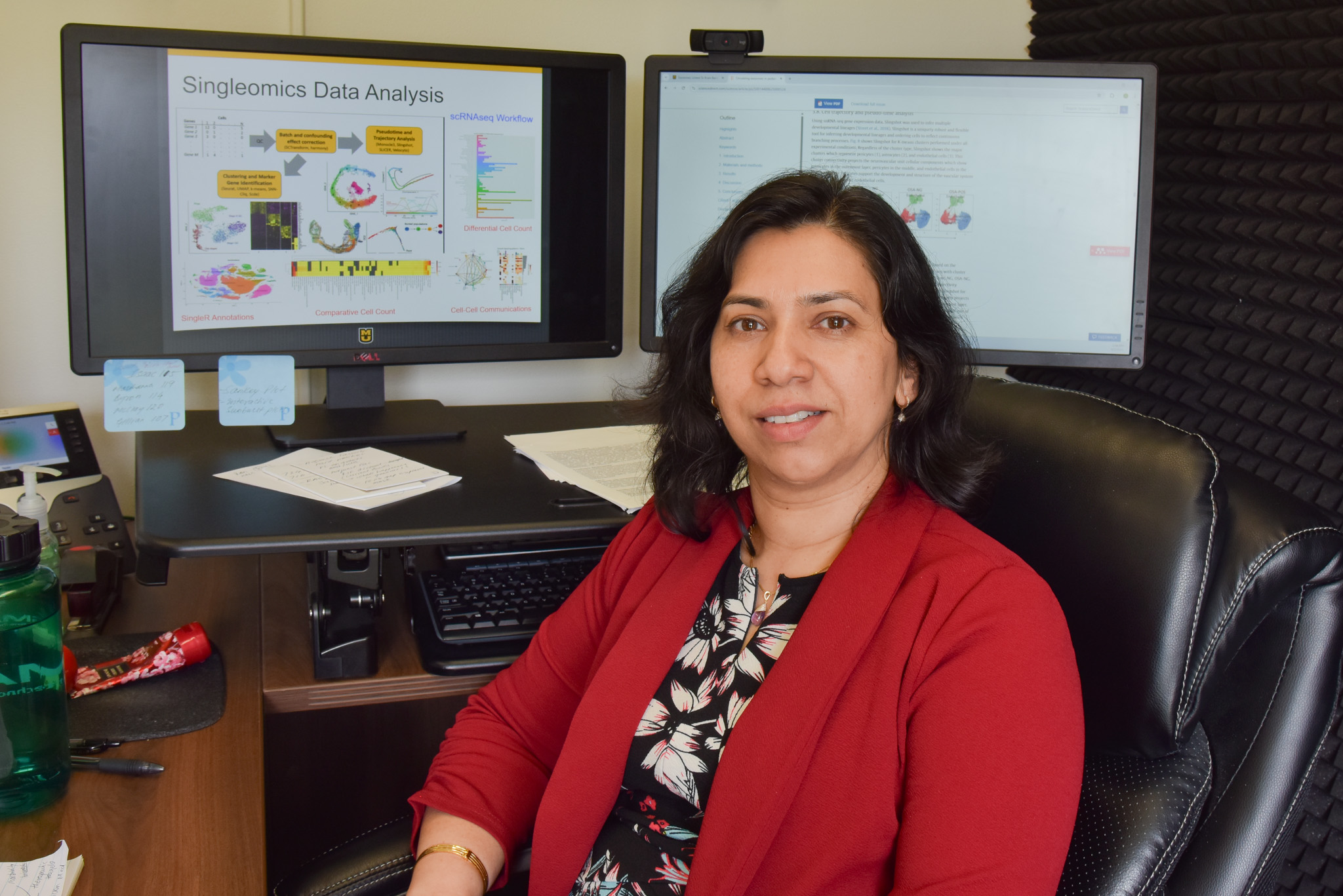
“You’re looking at terabytes of information,” said Joshi, faculty lead for translational bioinformatics for NextGen Biomedical Informatics.
Sleep apnea is a disorder where individuals stop breathing during sleep, which may lead to cognitive problems. It has been linked to anxiety, depression and decreased attention span; however, children more often see poor school performance and behavioral disturbances.
“OSA doesn’t just have an impact on sleep,” Joshi said. “It has an impact on systems beyond that.”
Joshi started her career as a clinician but said she entered the field of bioinformatics around the time the human genome was discovered in the early 2000s, and she has used data to explore a wide array of areas from soybeans to human diseases.
Trupi Joshi, faculty lead for translational bioinformatics for NextGen Biomedical Informatics, poses next to her lab’s visualizations for a childhood sleep apnea study on April 2, 2025.
That data led scientists in this study to conclude that exosomes from patients with OSA — whether it’s those without or with a cognitive deficit — carry out different functions than in those without OSA. Studying exosomes in children with OSA alongside the permeability of the blood-brain barrier can tell researchers how these neurological structures are being disrupted.
Joshi said studying that impact can allow for more personalized therapeutics. She added that because the blood-brain barrier and the neural system is so central to controlling so many activities, it’s integral to understanding this disorder.
“When the permeability (of the BBB) is increased, you have a lot more traffic of molecules going in and out,” Joshi said.
Wealth of data leads to personalized therapeutics
“When you have multiple conditions, you have to be able to overlap it, integrate it and see the differences,” Joshi said.
Chan makes that possible. Joshi said her collaborators are always pleased to find out that Chan is involved since he can write his own code, analyze data, and create his own data visualization tools.
“He can address the entire spectrum,” Joshi said about Chan’s contributions to the bioinformatics lab.
The future of this research lies within the wealth of data available to analyze with computing.
“The beauty of this large amount of data is with computer science and informatics, you can roll it into deep learning (artificial intelligence) models,” Joshi said.
Joshi said this modeling “is going to be helpful for clinicians because now they are better positioned to provide the best therapy that the kid needs, rather than having to wait 10 years later and then finding out.”
This study was published in March 2025 in the journal Experimental Neurology. Read more about it in Mizzou’s School of Medicine release.
This study was supported in part by grants through the Joan C. Edwards School of Medicine at Marshall University, the Missouri Department of Health and Senior Services, and the National Science Foundation.
Mizzou researchers genetically engineer plants to optimize microscopy
By Sophie Rentschler | Bond LSC
Gary Stacey’s lab is a breeding ground for model plants, curated to get the most precise image of the plant leaf tissues.
Those plants help scientists at Mizzou and the Environmental Molecular Sciences Laboratory (EMSL) bridge plant science and microscopy to capture high quality snapshots of a plant’s cellular structure. The collaborators recently co-published their latest contributions to lattice light-sheet microscopy imaging in The Biophysical Journal. It shows a way to better see plant plasma membrane-localized receptors that previously were obscured by the background glow of chlorophyll-like objects.
A plant that’s easier to see (under the microscope)
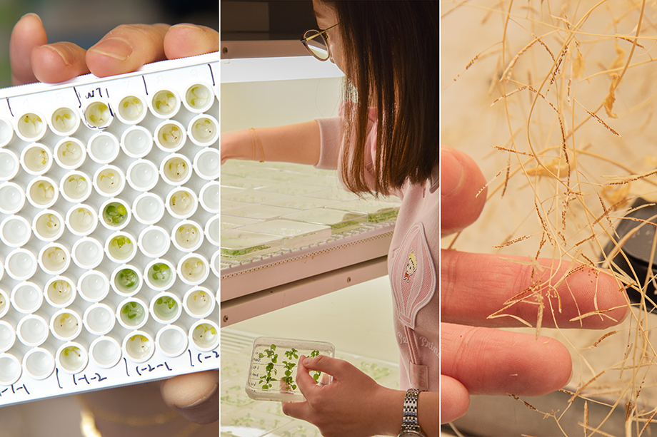
Mengran Yang’s life’s work is studying cellular proteins in plants, so she plays a big part in this partnership. Since she joined the Stacey lab in Bond Life Sciences at Mizzou as a postdoctoral fellow, Yang has immersed herself in the cellular components of models like Arabidopsis, a small mustard plant used ubiquitously in plant research. Whenever people know about Arabidopsis, Yang gleams with excitement.
“We want to use microscopic technologies to solve biology questions,” Yang said about the reason the collaboration came to be.
This collaboration with EMSL– a DOE Scientific User Facility located at the Pacific Northwest National Laboratory – will create precision imaging that can help future researchers understand subcellular location of proteins that contribute to a crop’s disease resistance or optimize farming to yield the best harvest, among other applications.
Yang has never visited Washington nor met the researchers from EMSL in person, but despite the distance she said she has vested trust in the team to share high resolution results.
Yang curates a handful of genetically modified or transgenic plants which produce thousands of seeds. She then sends the seeds off to a laboratory in Washington, where her collaborators grow their own plants optimized for light-sheet microscopy. Without this genetic modification, EMSL wouldn’t be able to analyze cellular components of plants with great detail let alone fulfill their quest to filter out chlorophyll.
Yang stashes the seeds of the genetically manufactured plants in Petri dishes where they emerge in their green leafy glory roughly 10 days later. She plants the newly sprouted seedlings that grow tall, decorated with white flowers. Afterwards, she tests the phenotype of the plants to ensure they are truly genetically modified. This process could take months.
She leaves the plants to shrivel, making it prime time to gather seeds. Yang is there for every stage of the plant’s life cycle, and she even sees them off as they are shipped across the country.
“Conducting this process with transgenic plants is very time consuming from phenotyping to harvesting,” Yang said. After seeing the images from EMSLs microscope, Yang added it makes her feel like her work is worth it.
One of the beauties of studying plants is because they “have more receptors localized on the plasma membrane compared to animals,” Yang said. This very factor lured her into studying Arabidopsis.
From modified plant to a clearer picture
The Stacey lab has spent more than a decade working with EMSL, because they need each other. This work wouldn’t be possible without the Stacey lab’s sharing its plant engineering know-how and EMSL contributing its national laboratory imaging prowess.
“Humans are social animals,” Stacey said about the natural urge to collaborate in the scientific research realm.
The researchers enhanced clarity in their pictures by getting around natural plant pigments that cloud their results. Chlorophyll — the compound that both gives the plant its color and is responsible for absorbing energy from the sun — is a nuisance for florescence microscopy imaging. It pollutes an image with background light and compounds the challenge the plant cell wall presents, which makes it difficult to tag internal cell parts with fluorescent markers needed to see plant receptors and other cell parts under a microscope.
Yang genetically engineers DNA that is inserted into the plant cell, making the receptors of the plant have the ideal functions for fluorescent viewing. The receptors are fused to a tag, thanks to this genetic process, which faces the outside of the cell making it easier for antibodies to bind to them. These antibodies are ready for fluorescent viewing. These tags glow when excited with a certain wavelength of light, making them stand out from other cell structures.
This method is the first of its kind, letting scientists see the location of a protein at a molecular resolution.
Because Yang’s research addresses fundamental questions in plant biology, she said it may appear “useless” to those not immersed in the field. But, seeing cell parts is a vital step to understanding how they contribute to things like plant defense. She joked that when she explains her research to her parents, she is met with looks of confusion.
“Progress (that impact humankind) is based on thousands or millions of basic research findings,” she said.
The study “Lattice light-sheet microscopy allows for super-resolution imaging of receptors in leaf tissue” published in the Biophysical Journal in February 2025. This research is funded by grants from the U.S. Department of Energy Biological and Environmental Research (DOE-BER), the National Science Foundation and the National Institute of General Medical Sciences of the National Institutes of Health.