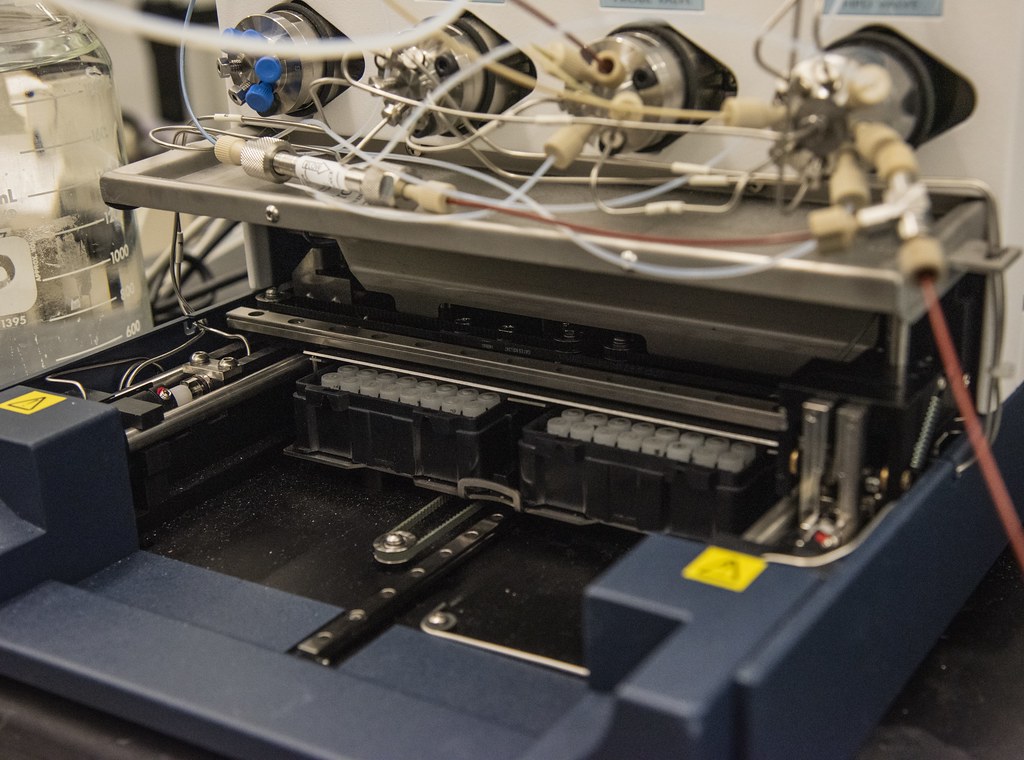
Michaela Beedy, Brian Thomas, and Margaret Beecher work on aptamers in the lab of Donald Burke. | Photo by Beni Adelstein, Bond LSC
Shrinking the Target: Developing Cancer Therapies
As cancer cells multiple and spread, doctors face finding treatments that destroy tumors while doing the least amount of damage.
This search for precision in cancer therapies is for good reason. It takes only a few minutes in a chemotherapy clinic to see the detriment of cancer drugs on the rest of the body.
“The issue with chemotherapeutic drugs is they have a lot of off-target effects,” said Brian Thomas, a MD-Ph.D. candidate working in the Donald Burke lab. “Our goal is to make them more targeted towards cancer cells using aptamers.”
Careful targeting of treatment is one important goal of cancer researchers, and aptamers are one way scientists at Bond Life Sciences Center work toward that goal.
Aptamers are short, single stranded DNA or RNA molecules that can bind to a target when it folds into a 3D structure. They can either serve as a vehicle for a cancer drug — delivering it to cancer cells while avoiding healthy cells — or bind itself to a cell’s receptors to interfere with its natural response.
In the Burke lab, the target of choice is epidermal growth factor receptors (EFGR), a protein that helps cells grow and spread. Mutations — mistakes in the DNA that makes this protein — can increase the number of EFGRs and cause cells to grow out of control, ultimately leading to cancer. The goal is to test how to best ensure aptamers successfully bind to cancer cells so they can be destroyed.
Margaret Beecher and Michaela Beedy worked in the Burke Lab over the summer.
Michaela Beedy, an undergraduate research assistant in the Burke lab works alongside Thomas. She is focusing on pairing aptamers with the chemotherapy drug doxorubicin to better target mutated cancer cells.
“Doxorubicin is really good at killing cells that are fast proliferating, which includes cancer cells and epithelial cells,” Thomas said. “It is also really good at killing cardiomyocytes, which is not a good thing.”
Margaret Beecher points out the tube containing the aptamer treated with doxorubicin.
The goal is to get doxorubicin to kill the cancer cells but to leave healthy cells like those heart muscle cells alone. Ideally, the cancer can be treated without posing too much risk to other vital organ functions.
Additionally, patients who undergo chemotherapy often develop resistance to a drug as newly mutated cancer cells become immune to the old therapy. That is why she is focusing on addressing specific aspects of what causes mutations.
Margaret Beecher, another Burke undergraduate, is testing a different hunch that could work in conjunction with Beedy’s work. Beecher investigates if dimeric aptamers might provide more of a targeted effect.
“Monomeric aptamers only have one spot that can bind to EFGRs while dimeric have two,” Beecher said. “Because there are two spots, this increases its avidity.”
Avidity is the combined strength of binding, and it is critical in getting a drug to interact with its appropriate target. This could be applied to Beedy’s doxorubicin approach.
“If we end up killing more EFGR-mutant positive cancer by using aptamer-doxorubicin treatment we could bring in the dimeric aspect as well to increase the effect of the therapy,” Beedy said.
The treatment in question is aptamer-doxorubicin conjugates, essentially a combination of the three.
Even if Beedy’s approach isn’t as effective at killing more mutant cells, Beecher’s research still comes in handy.
“If we saw less of a targeted effect because the aptamer wasn’t binding as well, maybe adding that dimeric aspect to it can help bring it back up to be on par with other treatments.,” Beecher explained.
New cancer therapies couldn’t come at a better time. Right now, the U.S. is in the midst of one of the worst cancer drug shortages in history due to manufacturing issues, leaving Thomas optimistic about the work in his lab.
“Right now, we’re doing in vitro…doing things in a dish. The ultimate goal is to get these treatments into animal models and, subsequently into humans to then finally create new therapies for patients in need.”
Donald Burke is a principal investigator at the Bond Life Sciences Center. He is a professor of molecular microbiology and immunology and a joint professor of biochemistry. Photo by Lauren Hines | Bond LSC
What started as an email correspondence between two aptamer enthusiasts rapidly snowballed into a hat trick of authorships for Donald Burke.
“I was contacted by a student in India asking if I would be an external advisor for her Ph.D. committee,” said Burke, a principal investigator at MU’s Bond Life Sciences Center.
Burke’s extensive research with sticky molecules called aptamers — totaling about 60 publications over 30 years — makes him an expert in the field of aptamer technologies. When Ph.D. student Shringika Soni at Amity University in Noida, India, near New Delhi, began characterizing the use of these molecules for drug testing in a literature review, she turned to Burke for advice on her work. The connection soon turned into a mentorship through regular email correspondence.
“I told her that I was interested in the kinds of things that she does, and I would be happy to interact with her about her science,” Burke said.
That project led Burke to collaborate on a project that attempts to create a rapid test to detect various drugs. This test would provide quicker information than waiting for the results of a blood or urine sample to be returned from a laboratory.
Starting out as a literature review, it soon turned into experimental research at Amity University. Burke’s role from halfway around the world was to ask tons of questions.
“There were a lot of back-and-forth emails around the general idea of defining precisely what her message was, what did she want to say about these systems, and as a particular case study might be mentioned, how much detail should be talked about in the review article,” Burke said.
While the research is not ready to be used commercially, Burke suggests potential applications could be for police officers who suspect drug use or for medical personnel trying to respond quickly to an affected individual.
“Maybe you’re suspicious that [someone is] on a particular drug. Having them spit onto a stick is just a whole lot less invasive and quicker,” Burke said.
Detection of the drugs falls to sticky molecules called aptamers. Essentially a chain of nucleotides folded into a particular shape, aptamers are selective in what they stick to, and each aptamer sticks to a specific compound. The researchers engineered their own aptamers that can stick to particular compounds found in certain drugs.
“The two major layers to this technology are designing the sensor components so that it will bind to the [compounds] really well and specifically,” Burke said. “The second component is to somehow turn that binding event into a detectable signal.”
But, aptamers just sticking to a drug is not enough because the researchers also need a signal to know the aptamer detected it.
To sense the chemical interaction, the researchers focused on electrical currents. With no drugs present, the aptamers do not stick to anything, and the electrical current flows. In contrast, when there are drugs present the aptamers stick to the compounds and block the electrical current from flowing. This allows the researchers to measure the drugs in the solution.
Due to Burke’s help on the project, the research team at Amity University offered to list Burke as a co-corresponding author.
“I just didn’t think that was appropriate, they were the ones leading the charge,” Burke said. “My role was to ask them questions from time to time and to push them to make sure they rounded out their arguments.”
This project adds to Burke’s long list of aptamer research fueled by his fascination with the properties of molecules driving living things.
“Just about every aspect of biology is driven by this choreography of who’s dancing with whom, by which molecules dance with which other molecules,” Burke said. “But why is it that some of the players refuse to dance with certain other players and others are just immediately drawn to certain other players?”
Burke’s research tries to answer these questions and determine why some molecules interact with specific compounds and why others do not.
“When you look at molecules the way they interact, it’s just so fun to watch the dance that they actually do,” Burke said.
Cynthia Tang is an M.D.-Ph.D. student in the Wan lab. Photo by Cara Penquite | Bond LSC
Cynthia Tang’s academic career is marked by her propensity to multitask. From earning a major and three minors during her undergrad to making a documentary while getting lab and clinical experience, she makes the most of her time.
Recently Tang received the Excellence in Public Health Award from the United States Public Health Service, and a $181.734 National Institutes of Health grant to be used over four years . . . all while getting an M.D. and Ph.D. simultaneously.
The funding from the grant goes towards Tang’s research on SARS-CoV-2, the virus responsible for the COVID-19 pandemic, and its effect in rural areas. By comparing DNA sequences of the virus from patients in rural and urban areas, Tang looks for differences in variants. Her goal is to understand how the virus has been evolving and why patients in rural areas experience more severe symptoms.
“I’ve always found infectious diseases really interesting,” Tang said. “This pandemic came up and it was exciting to be in the position to help contribute to the knowledge base of it.”
Cynthia Tang creates protein structure models for the spike protein on the Sars-CoV-2 virus. Along with 3D models, Tang creates phylogenetic trees to compare the genetic makeup of variants of the virus. Photo by Cara Penquite | Bond LSC
In a family of immigrants, Tang saw the challenges in health literacy and cultural differences affecting patient care in the U.S first-hand, and she also caught a glimpse into a global view of healthcare.
“I had an opportunity to travel to Vietnam and actually see some of the health disparities of different health care structures in different countries,” Tang said. “We were visiting my grandfather who was in the hospital, and it was alarming to see the contrast between the quality of health care and availability of health care there compared to some of the hospitals that we have here.”
Tang recalls noticing lack of supplies, physicians and space for patients. She saw similar issues in the U.S., and shined light on public health disparities here by creating a documentary about the challenges immigrants face in the U.S. healthcare system. She also held competitions across Washington University’s campus to teach students more about public health issues while working as a clinical research coordinator.
“I wanted to help improve health equity,” Tang said.
A bachelor’s in chemistry and minors in philosophy, international development and pre-health professions at the College of Idaho may start to explain Tang’s public health focus and her path to bench and clinical research.
“My minor in international development was really useful for me because that gave me a lot more exposure to the different economies of different countries of the world, their health status and how politics, economics and health care all interrelate in different countries,” she said.
Her research at Washington University led Tang to an interest in its clinical applications.
“I really enjoyed the aspect of working with patients, actually getting to talk to patients and hearing their stories,” Tang said. “I felt like with clinical research, you can see … slightly quicker results from bench to bedside compared to bench research, and that was also when I decided I was interested in becoming a physician.”
Part of an eight-year joint M.D.-Ph.D. program at Mizzou, Tang started with two years of pre-clinical medical school before transitioning to Ph.D. research and then will return to finish the clinical years of medical training.
When she came to Mizzou, Tang continued fighting health disparities for immigrants.
“We organized a group of volunteers that were able to translate COVID-19 information to different non-English speaking communities in Boone County,” Tang said.
Always one to tackle several projects at once, Tang plans to pursue a career as a pediatric physician scientist interfacing with patients while continuing lab research.
“I like the idea of doing both. As a physician, you get that one-on-one contact and you get to make a very direct contribution to someone’s life,” Tang said. “And then with research, you don’t have as much contact with patients, but what you do can affect populations.”
Investigators at Bond LSC take steps to apply basic research
By Cara Penquite | Bond LSC
Photo by Lauren Hines | Bond LSC
Scribbling in a lab notebook and planning experiments tucked between shelves of equipment, it’s easy to fixate on day-to-day lab operations. But scientists also face the challenge of finding how research can improve the world around us.
“The direction, the vision of the lab, ultimately comes from the principal investigator that bridges the research into applied directions,” said Jay Thelen, biochemistry professor and Bond LSC principal investigator
Despite the focus on basic research within the Bond LSC, many principal investigators choose to take their research to the next level with commercial partnerships.
Thelen’s lab researches ways to increase oil production in seeds and has three patents licensed to Yield 10 Bioscience, a sustainable crop innovation company who applies Thelen’s research to commercial crops. While seed oils like canola and soybean oil are known for their use in cooking, Thelen explains that increased production of these oils could play a larger role in sustainable fuel sources such as biodiesel and sustainable aviation fuel.
“We have to make more oil to balance out our need to eat it [and] our need to wean ourselves off of fossil fuels,” Thelen said. “To do that, we need to either plant more acres of oil seeds, or we have to raise the oil in existing oil seeds.”
Thelen researches enzymes with that potential application in mind. One is acetyl-CoA carboxylase, the enzyme which initializes the production of fatty acid chains found in plant oils.
“We’ve known this is an important enzyme, and we know that any tinkering you do with it has an impact on the oil production,” Thelen said. “In this case we’ve made new discoveries that permitted us to rationally engineer this enzyme to make it more active.”
Thelen suggests thinking of the enzyme as a “gatekeeper” to oil production which initializes the production of fatty acids and increases oil production. Thelen’s lab identified two different gene families that influence the activity of the enzyme in Arabidopsis and camelina plants. Yield 10 then applies these discoveries in other commercial plants.
While Thelen works closely with his commercial partner — having served on their scientific advisory board for three years and now stays in contact with Yield 10’s CEO to develop research projects — some labs stick with short-term arrangements.
Kamlendra Singh — assistant director of the Molecular Interactions Core at Bond LSC and Veterinary Pathobiology research assistant professor — studies HIV treatments. His lab identified a compound licensed by a commercial partner that targets the shell containing the virus’ genetic information.
Singh’s work in HIV started in 1994 with basic research investigating the enzyme that makes the viral DNA.
“I wasn’t into [studying] the drugs when I started working on HIV, I was mostly trying to understand how HIV enzymes works,” Singh said. “Once you know how the enzyme works, then you can target these enzymes for discovering the drugs.”
After years of studying how the enzyme works, Singh switched to HIV treatment. The first step to develop a treatment is to look for structures in the virus that the drug could potentially target to stop the viral replication. Singh targeted the shell around the virus’ genetic information known as the HIV capsid.
Building on previous research, Singh’s lab developed a compound able to bind the HIV capsid and prevent it from releasing the contained genetic information. Even with the licensing of his compound, Singh plans to continue researching ways to improve it.
“There are two reasons to keep working on it. One, well it’s my brainchild,” Singh said. “The second reason is as the company grows, we grow. We get more recognition and more funding. You can use it to [study] different viruses or use the same funding to improve upon it.”
While Singh plans to remain looking towards the applied side of his HIV research, he does not forget his roots in basic research.
“You have to put in time … [to] understand the system first, which is basic science, before you go to applied science,” Singh said.
Michael Roberts, a Chancellor’s Professor Emeritus of animal sciences and biochemistry who has had several patented projects, focuses on improving basic science projects and applies for patents if warranted.
“I don’t deliberately go into anything for commercial purposes,” Roberts said. “If I see something that I think does have commercial application, I’m happy to do it, but that is usually after you do [basic sciences].”
Whether starting a project with applications in mind or focusing on basic research, knowledge gained through research can be building blocks for the future.
“Science is simple. Even the most applied research project has its genesis in basic biology and basic research,” Thelen said.
One step into the Advanced Light Microscopy Core (ALMC) sounds an automated bell prompting Alexander Jurkevich, the core’s assistant director, to step out of his corner office into the open square room. With a friendly smile, Jurkevich coordinates biologists across MU’s campus to reveal the wonders of the microscopic world.
“Our mission is to provide researchers campus-wide with advanced microscopy instrumentation,” Jurkevich said. “We not only provide access to instrumentation, but we also train, advise users and support them during their early research at the core.”
The core hosts an annual image contest celebrates MU researchers’ microscopic imaging throughout the year. After being canceled for the past two years, this year contestants submitted their best images for consideration.
Christie Herd, a postdoctoral fellow in the Alexander Franz Veterinary Pathobiology lab won the Best of Show Award for her image of the La Crosse Virus in mosquito ovaries.
Image by Christie HerdDavid Porciani, assistant research professor under the supervision of Bond LSC’s Donald Burke, won Director’s Award for Best Technically Challenging Image with his image showing epidermal growth factor receptors in two types of resolution.
Image by David Porciani.
Janlo Robil, a graduate student in the Bond LSC’s Paula McSteen lab won Experts’ Choice Award with his image of hormones in a corn leaf.
Image by Janlo RobilA wonderful surprise
Herd was “blown away” when she got her colorful image that won the Best of Show Award. Not only had she tried imaging other viruses with less success, she also worked on different La Crosse samples with no luck.
“That image in particular, I was not expecting to see because the day before I was there for three hours and had to quit,” Herd said. “So, when I saw that image, I was surprised because I did not think I would see that much detail.”
Christie Herd is a postdoctoral fellow in Alexander Franz Veterinary Pathobiology lab. Herd won the Best of Show award for the 2022 ALMC Imaging Contest. Photo by Cara Penquite | Bond LSC
Herd’s image shows the La Crosse virus in developing eggs within mosquito ovaries. Herd uses La Crosse, a type of bunyavirus, as a model to study virus transmission from a female mosquito to her larvae to determine how viruses can remain transmitted within generations of mosquito populations. While La Crosse infects a small amount of humans a year, it transmits quickly, making it the perfect model to learn more about other bunyaviruses like Zika and Chikungunya.
“With bunyaviruses, they’re so multifaceted, and I like being able to research different aspects of them,” Herd said. “I just feel like they’re medically important.”
Herd dissects mosquitoes before imaging. She studies the transmission of bunyaviruses like the LaCrosse and Zika viruses from female mosquitoes to their progeny. Photo by Cara Penquite | Bond LSC
Herd’s image includes different colors to label different parts of the ovaries as well as the virus so she can see where in the ovaries the virus is traveling. Using a technique that allows her to see different focal planes, Herd can see where the virus is in every dimension, but it can be tricky to get high quality images.
“It does require a few hours of playing around, looking at the microscope,” Herd said with a chuckle. “It requires an investment of time, and sometimes there are days where it’s just not what you wanted and it doesn’t work … and you wasted days.”
Even with the challenges, the ability to see multiple dimensions of a sample at once is valuable for research projects like Herd’s.
“You just have to persevere and try again with a new set of samples,” Herd said.
From side project to passion
Although a deviation from his primary research, David Porciani’s project to image cell surface receptors slowly took over his focus.
“The biggest surprise was that I really had fun,” Porciani said. “This was not my primary project, but it became my primary project for a while.”
David Porciani is an assistant research professor under the supervision of Donald Burke. Porciani studies cell surface receptor interactions linked to lung cancer and his image won Director’s Award for Best Technically Challenging Image. Photo by Cara Penquite | Bond LSC
Porciani, an assistant research professor under the supervision of Donald Burke, studies molecules on the surface of cancer cells which are receptors for growth factors. These receptors act as a lock, with growth factors as a key. When the growth factors and receptors come together, the cells divide and create more cells. In lung cancer, there are more of these receptors, which can lead to uncontrolled tumor growth.
“This receptor, EGFR, has been widely studied,” Porciani said. “But for me there is definitely an interest because it’s one of the markers in lung cancer.”
Porciani tags those receptors with small molecules called fluorophores that glow under the light of the microscope, so he can see where the receptors are and how they move. However, the fluorophores cannot attach to the receptors alone, so he used aptamers — synthetic keys created by researchers that can bind receptor locks with specificity similar to the natural growth factors. Ultimately, the aptamers clip the fluorophores to the receptors.
“If you can follow the motion of the receptors, these receptors are kind of dancing,” Porciani said.
In his image, each dot is a different receptor made visible by the attached fluorophore.
However, fluorophores can become bleached, rendering them invisible after being exposed to the laser from the microscope for a while. If the synthetic keys, or aptamers, are still bound to those receptors, they cannot be imaged any longer. To address this, Porciani developed an aptamer which attaches to the receptor for a shorter time and then detaches so that even if the fluorophore bleaches, another aptamer can replace it and so receptors can be imaged for longer time.
“We engineered an aptamer with lower affinity that could work with this approach,” Porciani said. “By having lower affinity aptamers we can still determine localization of a high number of receptors and their motion.”
Porciani shows the process of imaging cells using super-resolution techniques. When a laser hits the cells with the fluorophore specific wavelength, the fluorophores glow while the rest of the cell remains dark, and Porciani can see where the fluorophores, and consequently the tagged receptors are in the cells. Photos by Cara Penquite | Bond LSC
For his winning image, Porciani split the image to show the difference between single molecule resolution on the bottom right and a lower resolution image on the top left.
“With [lower] resolution, you don’t have a single molecule solution. If there are two molecules close together you will see them as just one single dot,” Porciani said. “But with the image, after software analysis with the image on the bottom right, then you have single molecule resolution.”
With this technique — made possible with microscopes at ALMC in Bond LSC — Porciani saw his efforts come together.
“At the beginning, I was just focused on the aptamer engineering from a high affinity aptamer to low affinity aptamer, and making them was the fun part to play with the structure,” Porciani said. “But when we started doing the imaging experiments at the Bond Life Sciences Center I realized that it was not just fun, but it was actually meaningful and this approach could have lots of biomedical applications.”
The artist’s touch
Although passionate about biology and microscopy, Janlo Robil decided to submit his image based on aesthetics.
“I chose this one because I am also a graphic artist, and I appreciate the color and composition,” Robil said.
Janlo Robil is a Ph.D. Candidate in the Paula McSteen lab. Robil studies the hormones involved in corn leaf development. Photo by Cara Penquite | Bond LSC
Robil’s image shows a developing corn leaf with different colors labeling different plant hormone response proteins involved in stimulating growth. His unique image of an entire leaf, which is just under a millimeter in length, required piecing together images of sections of the leaf.
“About 0.75 millimeter, that’s still big in the microscope,” Robil said. “This is kind of difficult to make because it means that you need to tile several images [together] and sometimes it takes up to an hour just to [get] an image.”
The experiment requires planning ahead since the plants containing the fluorescent proteins must be crossed with plants with genetic mutations to determine the roles of the hormones in the leaf development.
To tag the plant proteins with fluorescent proteins requires planning ahead since the plants must be grown with genetic mutations.
“The beautiful image is actually a result of the expression of fluorescent proteins that are tagging the hormone response protein and also the hormone transport protein,” Robil said.
Robil looks at images from a confocal microscope. Using a laser, confocal microscopy brings clarity to Robil’s images. Photo by Cara Penquite | Bond LSC
Initially from the Philippines where the agricultural staple is rice, Robil came to Mizzou interested in genetic mechanisms to make rice plants more productive. One way to enhance the rice plants is to make the rice leaves more similar to corn leaves, so Robil found interest in the McSteen lab’s project understanding the role of hormones in corn leaves.
“This project is perfect for me because I am studying the leaf and also integrating genetics,” Robil said. “And I love microscopy so much. I had quite a good amount of training starting in 2021 on confocal microscopy, and that’s why I was able to image this.”
Hari Krishnan holds a handful of A. pavonina seeds. Known for their bright color, the seeds are known among many Asian and African communities as coming from the red bead tree. Photo by Cara Penquite | Bond LSC
By Cara Penquite | Bond LSC
An energetic and fulfilling day starts with a spread of healthy meals, and many people rely on nutrition labels to meet their daily quota of vitamins and nutrients. But how did scientists measure the Vitamin C in an orange or the protein content in peanuts for the label?
Finding out what is in food we eat starts with scientists like Hari Krishnan, a USDA-ARS molecular biologist and MU adjunct professor of plant science and technology. Krishnan calculated the type and degree of nutrients packed into seeds from red bead trees with the help of the Advanced Light Microscopy Core in Bond Life Sciences Center.
“We are highly dependent on the food that we eat,” Krishnan said. “Anything we can do in order to either improve nutrition or quality or finding alternative sources of food is very useful.”
Commonly known for its pods of seeds as bright as red M&M’s, the red bead tree is known as Adenanthera pavonina in research communities.Krishnan took an interest in A. pavonina seeds — turning away from his usual soybean research — to see if they could be a potential alternative protein source in developing countries. He quickly found a different, unusual characteristic.
“I’ve never seen a seed have such a high content of trypsin inhibitor,” Krishnan said.
Our bodies make trypsin to break down protein in our intestines, but trypsin inhibitors block the enzyme from its job of protein digestion. That makes it hard for us to benefit from nutritious proteins in the seeds. Although many Asian and African communities already incorporate A. Pavonina seeds in their diet, its unusual amount of trypsin inhibitors limits nutritional benefits.
“Soybeans have probably less than five percent of the entire seed protein made of trypsin,” Krishnan said. “This particular legume is 20 to 25 percent, which is fairly very high.”
Krishnan turned to Alexander Jurkevich, associate director of MU’s Advanced Light Microscopy Core, to find out where in the plant’s cells the trypsin inhibitors were located. Jurkevich and Krishnan used glowing molecules to mark the trypsin inhibitors and see where they are stored in the cells.
Hari Krishnan sits in his lab at MU Curtis Hall. A USDA-ARS molecular biologist and MU Adjunct Professor of plant science and technology, Krishnan’s research looks at the nutritional value of legumes. Photo by Cara Penquite | Bond LSC
Tagging protein here starts with an antibody. The first antibody acts like one side of a strip of velcro that can attach to a second antibody that also carries a molecule of a fluorescent dye. Through the double antibody system, the glowing, fluorescent flag attaches to show scientists which parts of the cell have trypsin inhibitors.
While Krishnan developed the antibody, Jurkevich used his expertise in light microscopy to take images of the glowing cells.
“This cooperation is very important, because modern research technologies are very complex and a single person cannot learn and excel at all techniques available in life sciences,” Jurkevich said.
Understanding the amount and locations of trypsin inhibitors, Krishnan looked to reduce inhibitors so the seeds would be more nutritious for humans.
Roasting or boiling the seeds breaks down the trypsin inhibitors, but Krishnan warns that it may break down other essential amino acids as well. In his work with soybeans, Krishnan looks for a way to grow plants without trypsin inhibitors at all.
“We were able to find some [soybean] mutants, which have lower levels of this trypsin inhibitor,” Krishnan said.
Natural mutants with lower levels of trypsin inhibitors could be cultivated on a larger scale to produce seeds that are easier for humans to digest.
Since trypsin inhibitors are not ideal for consumption, Krishnan decided to think outside of the box. He turned to USDA-ARS research scientist and entomologist Adriano Pereira to find another use for the seeds.
Pereira tested the seed proteins as an insecticide against corn rootworm larvae and found that it stunted larval growth but did not necessarily cause mortality. While it is still early in this research, there may be some insecticidal properties from the seed.
“It’s something that has to be investigated to make sure […], but we assume that since it is a trypsin inhibitor it could be inhibiting on some level the trypsin during the growth of the larvae,” Pereira said.
As Krishnan’s first look into A. pavonina seeds, the research opened many new questions yet to be answered, but he plans to continue investigating the role of trypsin inhibitors in soybeans and other plant protein sources.
“[Research] is a never-ending investigation, and one thing leads to another. It’s always interesting,” Krishnan said. “I come to the lab everyday thinking of what I’m going to end up doing today.”
The Advanced Light Microscopy Core provides technical imaging expertise to researchers who need state-of-the-art imaging for their experiments. That includes confocal, super-resolution, digital light-sheet and widefield microscopes, image analysis and processing and sample preparation.
The Baker lab poses for a group photograph. The lab has been working with specialized pro-resolving lipid mediators in efforts to help patients with Sjögren’s Disease. Photo by Karly Balslew, Bond LSC
By Karly Balslew | Bond LSC
Saliva is probably not the first thing that comes to mind when we think about eating our favorite foods. The clear liquid washes away food debris and bacteria, and it plays a vital role in maintaining our dental hygiene and oral health.
You may take it for granted, but for patients with Sjögren’s disease, life without saliva is challenging.
“We’ve seen firsthand how patients suffer from Sjögren’s disease and what the consequences are in the oral cavity,” said Olga Baker, a Bond Life Sciences Center principal investigator studying the syndrome.
The autoimmune disease causes our body to attack the glands that produce tears and saliva. This destruction at the hands of our immune system causes dry mouth and dry eyes as it eventually kills the cells that perform this valuable service to our bodies. Without saliva, tooth decay and painful diseases like gingivitis are almost inevitable.
Sjögren’s disease affects approximately 4 million people in the United States. The severity of the disease varies in patients, but it destroys all exocrine glands and there is currently no cure. While the cause of the disease is still unknown, researchers are turning efforts to alleviate patient discomfort.
The Baker lab focuses on a class of drugs called specialized pro-resolving lipid mediators. These medications are derived from essential fatty acids and play an important role in decreasing inflammation and recovering salivary function.
Harim Tavares Dos Santos — a post-doctoral fellow in the Baker lab and lead author on a recent study of this drug —has seen success with restoring salivary functions with the specialized pro-resolving lipid mediator drug resolvin D1 (RvD1). Resolvins act as anti-inflammatory mediators and restores saliva flow rates. The lab will test a variety of drugs from the specialized pro-resolving lipid mediators’ class to see which one would yield the best results, but some receptors they affect within the salivary glands are unknown.
“The first step is to see if we know all the receptors for other drugs,” Tavares Dos Santos said. “Then, we test the different drugs to see which ones activate the receptor to possibly decrease inflammation and reach the goal of getting the saliva back.”
RvD1 activates the receptor ALX/FPR2 Tavares Dos Santos explains. This then triggers a signaling mechanism that promotes the survival of salivary gland cells and protects the cells tight junctions while increasing saliva secretion. Researchers used six human minor salivary from female subjects in this experiment to determine the expression of pro-resolving lipid mediators in patients with and without Sjögren’s syndrome.
Previously, the lab tested these drugs on mice models with Sjögren’s-like features and saw they can recover salivary function. However, researchers need more data to find other receptors that can maximize the positive response from the drug.
But mice aren’t the same as humans, and even when researchers test on humans it can be difficult to find patients with Sjögren’s disease willing to participate in the research.
“We don’t have a lot of literature in salivary glands to look for. Basically, what is known about resolvins and salivary glands is generated here in the lab,” Tavares Dos Santos said.
“Another challenge is that no one knows what causes the disease, so the research is mainly trial and error,” Baker said.
Baker explains that studies show people with Sjögren’s disease could have a genetic predisposition for it, but there are also environmental factors like viruses that could trigger the genetic components that lead to Sjögren’s disease.
“In the end, we want to create a drug that has all those properties to cure the patient so hopefully in the future, we can get a product on the market,” Baker said.
The Baker lab will present their progress with Sjögren’s syndrome at the 2023 Gordon Research Conference in Ventura, California. Both Baker and Tavares Dos Santos will co-chair the international conference that attracts researchers studying salivary glands and exocrine biology.
“This is a very important conference that will elevate Missouri,” Baker said. “The whole thing is really exciting.”
Olga Baker is a professor of Otolaryngology-Head and Neck Surgery and Biochemistry as well as a Bond Life Sciences Center principal investigator. Harim Tavares Dos Santos is a Bond Life Sciences Center post-doctoral fellow.
Data connects all: ‘Champion collaborator’ Xu bridges research disciplines with bioinformatics
By Cara Penquite | Bond LSC
Dong Xu extracts wonder from numbers with a keyboard and eager teams of scientists at his fingertips.
With his salt-and-pepper hair visible above the cubicle walls and his voice softly but steadily articulated, the beauty of bioinformatics takes shape in his mind although it might not be inherently evident in the rows of computers tucked into a small first floor lab.
Xu weaves a multifaceted masterpiece of research methodologies and makes sense of a sea of data from cell biologists, plant scientists, engineers and many more with hundreds of publications to show for it.
“For research, collaborating is key because the nature of research requires many views, skills [and] knowledge,” Xu said.
A Bond Life Sciences Center researcher and a newly endowed Curators professor of bioinformatics in the School of Engineering, Xu harnesses data to better understand biological systems such as revealing a better picture of protein dynamics or analyzing genetic codes for individual cells. With advanced data interpretation and machine learning, the Xu lab opens opportunities for more in-depth research across Mizzou and the world.
With so much existing on a microscopic level in living creatures and new technologies to collect data continually evolving, massive amounts of information are extracted in biological research. Xu’s expertise is invaluable to interpret massive amounts of information, but he takes it a step further predicting how pieces of biological systems interact.
His recent efforts include using deep learning — a subset of artificial intelligence and machine learning — to understand how the shape of protein binding sites change when molecules bind to other areas of the protein. This research, recently published in Nature Communications, and represents one of many collaborations the Xu lab is known for — in this case with Jilin University in Changchun, China.
The Xu lab’s help is crucial to researchers whose specialties lie in biological systems rather than computer science. Bond LSC Director Walter Gassmann, recognizes Xu’s niche.
“Biology has become so complex that you can’t be expert in everything,” Gassmann said. “The way to move forward is to collaborate with people, and that’s what Bond LSC is all about — bringing people with different disciplines under one roof, letting them rub shoulders and figuring out how best to solve a problem.”
With computational analysis being key to many research studies, the Xu lab collaborates with various labs at the research center.
“Dong is the champion collaborator [with] so many connections in the center,” Gassmann said.
Xu makes note of the particular importance of working together when mentoring young researchers. Students in the Xu lab work in teams and alongside biologists, allowing the researchers to learn from one another.
“There is actually an African saying, ‘it takes a village to raise a child,’” Xu said. “The same is true for mentoring students. It really takes many people to mentor a student. That includes not only community members and collaborators, but also peers.”
Xu fosters that shared work ethic by giving credit where it is due when it comes to research. While other institutions sometimes only give the lead investigator full credit for a project, Mizzou recognizes the work put in by each collaborator for any given project.
“MU is a very collaborative environment,” Xu said. “It really supports interdisciplinary research, [and] I don’t take it for granted. Interdisciplinary research usually relies on administrative support.”
Bioinformatics was more of an afterthought to Xu despite its prominence in his life now. His studies started with bachelor’s and master’s degrees in physics, and it was not until he started Ph.D. studies that his focus shifted.
“I was working on the biophysics of saturated proteins, and that’s a computational analysis,” Xu said. “I became very interested in computational work, so since then I’ve been working on this computational biology, or bioinformatics, for about 30 years.”
Xu’s drive to know all things data is revealed in his excitement to talk science.
With a small chuckle he noted that research does not usually bring money and fame. While he could use his computer skills to gain such rewards working for tech companies, he chooses to pursue science instead.
“To be a scientist requires passion,” Xu said. “You really need passion and to believe in the impact, the value of research. I do feel a reward in that regard.”
The Xu lab develops machine learning, a subset of artificial intelligence that involves teaching computers to mimic human intelligence. That starts with developing technology to collect data and facilitate its analysis.
This stretches into deep learning, more advanced machine learning that involves layers of neural networks. The input information is organized through these networks as the computer makes sense of the information.
People encounter deep learning in their day-to-day lives. The algorithm that sorts photos by faces in your phone’s photo app — that’s deep learning.
“It can start from these pixels and then can retrieve information like eyes [and] nose, and then can [find] a match,” Xu said. “It’s not only deep layers, but also the complex architecture of the network, so that’s why it’s called deep learning.”
A similar process is used in the Xu lab’s work. Rather than sort pixels to recognize faces, Xu’s work sorts data — like amino acid sequences — to predict protein interactions.
By finding trends in large sets of data, deep learning can speed up existing processes.
Mark Hannink, a Bond LSC principle investigator, recently saw this in action. Xu developed techniques to read scientific articles and generate diagrams of protein pathways described in the database. While people can read and summarize these articles, the process takes time and leaves the latest of the diagrammed pathways outdated.
With deep learning, each time a new article is published, the diagram could automatically update.
Working closely with Xu, Hannink saw his mentoring approach in action.
“What I’ve been impressed with is how good of a mentor he is to the students working on this project,” Hannink said.
While high-impact research is one goal, Xu also takes care to develop scientific minds.
“Not only [do] we produce papers [and] software, we also really need to produce high-quality, next-generation researchers,” Xu said. “So, our goal is to mentor people to be successful, and many of our lab members are really on the right track to be successful.”
Pigs may have a reputation for being lazy and dirty but to immunologist John Driver, they are the key to understanding influenza in humans.
“Pigs are a great animal to study influenza in because they are susceptible to getting the flu,” Driver said. “They are like a mixing vessel for influenza viruses.”
Swine have their own strains of influenza virus but can also contract strains from other species like birds and humans. When that happens, these different flu viruses can recombine and create new variants in the animal that can occasionally give rise to viruses that cause human pandemics.
Driver arrived at Bond LSC in mid-January as part of an initiative to create an influenza center in Columbia with Bond principal investigator Henry Wan. While Wan works on the molecular aspects of how new influenza viruses emerge, Driver’s work focuses on the host immune system.
“We’re really interested in understanding how to prevent the next pandemic and learn how to control influenza in livestock,” Driver said.
The researchers will study how influenza viruses move between animal species and recombine into new viruses inside different hosts.
Originally from South Africa, Driver received his undergraduate degree in Animal Science from the University of Pretoria. In 2000 he traveled to Lexington, Kentucky to work at Alltech Inc, an animal feed additive company, as an intern. Before coming to Mizzou, Driver was a faculty member in the Animal Science Department at the University of Florida for ten years.
Driver hasn’t always worked with pigs. Earlier in his career, he preferred a smaller test model, the mouse.
Once he left Kentucky, he moved north to work at the Jackson Laboratory in Bar Harbor, Maine. The laboratory’s Mouse Genome Informatics database supplies information on mouse genetics and biology internationally. Here he discovered his love for immunology.
“I was totally immersed in mouse genetics and Type 1 autoimmune diabetes. It was a wonderful place to be for learning about all sorts of genetic models for various diseases,” Driver said.
Driver decided he wanted to dig deeper into his research and go beyond what a mouse model could offer him after six and a half years at the Jackson Laboratory. This landed him at the University of Florida studying pig models.
While studying influenza in mice and other small animals like ferrets is possible, Driver notes there are a lot of drawbacks compared to using pig models.
“A mouse can’t transmit the flu to another mouse and if a mouse does get the flu, it dies very easily,” he said. “Pigs transmit the flu just like we do. They cough, sneeze and get infections like humans. When we study a pig, we get a pretty good idea of what is going to happen in humans.”
Understanding viruses in pigs not only aids the swine industry but sheds light on developing vaccines and how to mitigate flu in humans as well. Mizzou is home to the National Swine Resource Center where researchers can create genetically edited pig models to study many diseases. This center is one factor that attracted Driver to MU.
“I came here to be more productive at what I do,” Driver said. “We have more animals to study [here], and we can make new models to ask more questions.”
Driver is excited to dive into the research and work with his students in the lab. His lab will look at how different cells in the pig immune system react during an influenza infection and build up resistance over time.
“I want to make real progress in understanding the host-pathogen interactions involved in influenza immunity,” he said. “I’m interested in studying rare types of immune cells and how they regulate disease resistance of the host.”
Being hands-on in the lab with his students is something Driver prioritizes. Even though his students are just starting with basic training he has enjoyed working with them.
“I like to be with my students in the trenches and running experiments with them,” he said. “It’s very rewarding to see students grow, mature, and learn things.”
Whether at his previous lab in Florida or his new home at Bond, Driver is looking forward to leading the next generation of scientists and innovators.
“It’s nice to know that they’re going to have very bright futures in science and be future leaders and I had a small contribution to play in getting them there.” Driver said.
COVID-19 virus particles have spike proteins, represented in red, that attach to receptors on host cells. Antivirals block the receptors on host cells so the virus cannot infect more cells. | Creative Commons Photo
By Cara Penquite | Bond LSC
Vaccines were the light at the end of the tunnel throughout the COVID-19 pandemic, but virus mutations threaten to extinguish hope of a quick end to the pandemic. Kamlendra Singh turns towards antivirals as the next step.
“There will be a time we will find an antiviral which will be very difficult for the virus to mutate [and avoid],” Singh said, “That’s what we are after.”
The Singh lab studied COVID-19 antiviral compounds that prevent binding between the virus and host cells with help from Siddappa Byrareddy, professor and vice-chair of research in the Department of Pharmacology Experimental Neuroscience at the University of Nebraska Medical Center (UNMC). The first COVID-19 antiviral compound Singh’s team discovered during their in a preliminary study conducted in mouse models, has been filed for a patent while they continue to search for the antivirals that target different proteins of the virus. These compounds would prevent the virus from entering cells even after exposure to the virus.
The antiviral compound disrupts the interaction between the ACE2 receptor on the surface of host cells and the spike protein on the virus so the virus cannot infect cells. ACE2 acts as a doorway into the cell where COVID-19 binds, enters and takes over the cell. Once inside, the virus hijacks the cell and uses it to create more virus. Additionally, the virus releases its genome into the host cells, activating cell defense mechanisms which can be more dangerous than the infection itself.
While vaccines prepare the immune system to fight off COVID-19, Singh’s antivirals simply block this doorway.
“The very first thing was to find the compound that can inhibit viral entry because it is the first step of the infection,” Singh said. “If you can block the very first step then you can block everything else.”
Vaccines act as a practice round for the body where the immune response learns which antibodies are effective against the virus. But when the virus mutates, antibodies built up from vaccination or prior infection may be less effective against the mutated virus.
The antiviral compounds discovered by Singh and his team do not have this problem since they bind to the host cell’s receptors where few mutations occur. Additionally, the compounds can change their shape in response to those mutations that may occur.
“We call it ‘wiggling and jiggling,’” Singh said. “The compound has the capability to change it’s shape conformation.”
This ability to change shape is not unique to COVID-19 antivirals. Drawing on previous experience working on HIV antivirals, Singh explains that shape changing properties are common in molecules with single bonds between their component atoms.
“If you have a single bond somewhere, then they can change or they can reorient and bind someplace,” Singh said.
Singh believes antivirals may be the key to fighting COVID-19 even with emerging mutants. While vaccines mitigated COVID-19 rates and hospitalizations, they also might have played a role in the creation of new mutants.
“It’s probably too early to say, but it looks like these variants are probably evolving under the pressure of antibodies,” Singh said. “Those antibodies may have been induced by either direct infusions giving the antibodies [to patients], or they may have been induced by other vaccines or by previous infection.”
While viruses also mutate under the pressure of antivirals, Singh hopes to find an antiviral that is difficult for the virus to avoid through mutation.
“We have been working on developing a better compound using the compounds we discovered, and we have found one more compound that has at least 10 times better efficacy against [SARS-Cov-2],” Singh said.
Kamlendra Singh is a research assistant professor of molecular microbiology and immunology and assistant director of the Molecular Interactions Core at Bond LSC. Singh wished to express his sincere gratitude to Prof. Byrareddy (University of Nebraska Medical School). Without their collaboration, the discovery of the antiviral compounds would not be possible. Singh is also thankful to two students – Saathvik Kannan (a Hickman High School student) and Austin (a Mizzou undergrad). Without the help of these two talented young scientists, the research would not have been so successful. Finally, Singh mentioned that the support from the Bond Life Sciences Center has been extremely valuable in his research.