

Figuring out how a virus takes over cells could help with gene therapy

By Mariah Cox | Bond LSC
When we catch a cold or contract the flu, we usually attribute it to picking up a virus from a friend or someone we know. Our bodies’ built-up immune systems have a way of attacking viruses to help us stay healthy, but sometimes viruses can hide.
A study published in eLife by lead researcher Kinjal Majumder, a postdoctoral fellow in the Bond LSC lab of David Pintel, sought to understand where a virus goes when it hijacks a host cell. This basic science may one day help researchers use viruses to better treat human diseases.
In general, when cancer-causing viruses infect a host cell, they translocate their genetic material into the DNA of healthy cells, taking over and causing disease in the host organism.
Majumder and his collaborators developed an experimental system to identify the ‘zip codes’ within the nucleus where the virus localizes.
“When looking at the mouse DNA in infected cells we noticed that the virus seems to associate with particular sites,” Majumder said. “When we mapped where those sites were, we found that the virus localizes to regions of the genome that are prone to DNA breaks. If you unravel all the DNA, which comes out to be about six meters in length, there are very distinct sites where the virus seems to associate.”
Through this research, Majumder was able to figure out where the virus goes when it infects a host cell and why it is attracted to those certain locations. These DNA sequence sites are prone to having breaks which is indicated by the presence of DNA repair machinery, making them more vulnerable to viruses. His current research focuses on how viruses hijack these break sites and take over the repair machinery to replicate.
“Viruses are very intelligent,” Majumder said. “What we’re now trying to do is to understand how they move about in the nucleus. We found out the where, we think we know the why, now we want to know how they know to attach to these sites.”
In order to conduct his research, Majumder is using a simple ‘test-tube’ model virus called minute virus of mice (MVM), which is a rodent parvovirus, to look at host-virus interactions. This model is particularly easy to work with in a lab setting and results are easily transferable to other viruses in animals and humans.
Other cancer-causing viruses that function similarly to parvovirus, such as human papillomavirus (HPV) and hepatitis B, have also been found to integrate into breaks in DNA chains and drive cancer progression.
“There is a close relationship between a replicating virus and the cell’s DNA repair machinery which normally protects us from cancer. Now we are starting to see that MVM localizes to the sites in the nucleus which contain a lot of these repair proteins,” Majumder said. “It’s an interesting phenomenon and we are starting to study the mechanism of how this might be happening.”
Majumder adapted an experimental system called chromosome conformation capture assay by chemically cross-linking virus DNA and the cellular DNA that are close together and generating hybrid DNA fragments in a test tube. After sequencing these fragments, Majumder and his team could determine the exact sites that the viruses associated with.
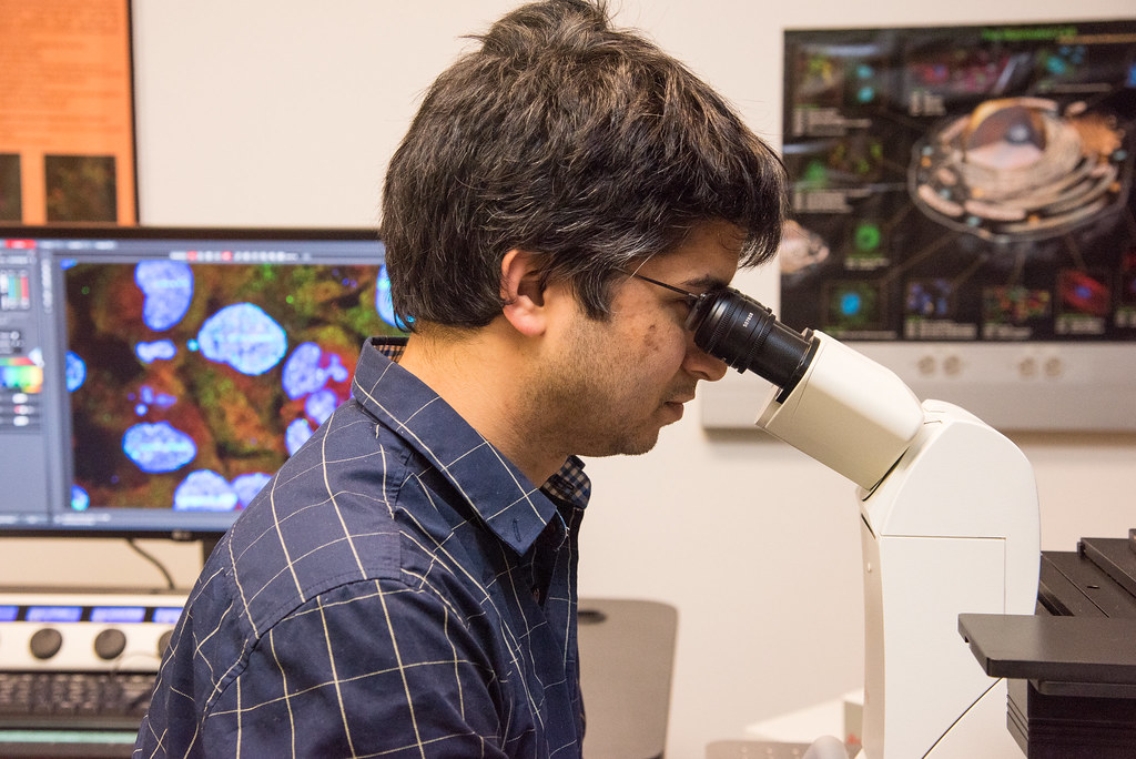
In order to confirm the location of these sites, they used powerful confocal microscopes in MU’s Advanced Light Microscopy Core. By using a laser to make a DNA break in a cell, Majumder could also show that virus localized near the induced breaks.
“If we can figure out how the virus is going to those regions, it opens up the possibility of finding whether other viruses, such as HPV or hepatitis B, utilize similar mechanisms to go to those sites,” Majumder said. “Knowing that may help study how oncogenic viruses cause cancer.”
Understanding how viruses hijack host cells and utilize cellular DNA breaks may allow future researchers to design improved cancer therapies.
Parvoviruses, in a broader sense, are used in oncolytic therapies to treat cancers using viruses. Cancer cells can arise due to dysregulation of DNA repair machinery in cells. They can also occur due to mutations in DNA breaks that can cause translocations. Parvoviruses have the ability to replicate in cancer cells, which is why they are being used as gene therapy vectors in clinical trials to target cancer.
“Knowing where gene therapy vectors stay long-term and how they interact with DNA repair proteins is important because they can express throughout their lifetime,” Majumder said. “For example, if you have cystic fibrosis, you have a mutation in one particular gene, so you don’t make specific proteins and as a result, you get a lot of health problems. Gene therapy adds a wild-type piece of DNA to express the correct version of the protein.”
For his Ph.D., Majumder looked at how the genome folds upon itself, gets packaged into the nucleus, and how this packaging regulates immune system development. His previous research on the genome is currently helping him advance his work with viruses.
“When I started working with viruses, I realized I could easily utilize those techniques to map how viral DNA interacts with the host’s DNA,” Majumder said.
Majumder hopes a better understanding of how viruses interact with DNA damage responses will move forward gene therapy.
“If you know how these viruses interact with the host’s proteins in the nucleus, you can design more effective gene therapy vectors.”
This research was published in the Journal “eLife” in July 2018 and was funded by the National Institute of Allergy and Infectious Disease of the National Institutes of Health.