Researchers from MU, the University of Maryland and the Pacific Northwest National Laboratory are building a microscope that doesn’t yet exist.

By Mariah Cox | Bond LSC
Tiny neon dots speckle a black backdrop – and no, this isn’t a Hasbro Lite Brite. Rather, these fluorescent dots indicate something about plants that scientists research and help them see the genes, traits and molecules they study amid thousands of possibilities.
To help in seeing that, a new imaging microscope will allow researchers to better pinpoint molecular interactions in plants they have a hard time highlighting to overcome the obstacle plant scientists face with wavelengths of light they can’t necessarily see.
“When you think about imaging, you think about what you can see with your eyes. But, there are a whole variety of other things you can image that aren’t visible to the human eye,” said Gary Stacey, a Bond Life Sciences Center principal investigator who is working to help develop a new microscope technology to view fluorescent quantum dot markers beyond the range of visible light, into the infrared spectrum.
Stacey, along with collaborators from the University of Maryland and the Pacific Northwest National Laboratory (PNNL), was awarded a combined $2.25 million grant from the Department of Energy (DOE) to develop a novel microscope for ‘multiplexed super-resolution fluorescence imaging in plants.’
The call for the development of this new imaging hardware was borne out of the need for a more precise measurement of enzyme function, tracking of metabolic pathways and monitoring the transport of materials and signaling processes within and among cells in plants. Right now, the emission spectrum of plant pigments limits the usefulness of and the number of fluorescent colors that can be detected in a single experiment.
Stacey and his collaborators were one of six groups to be chosen for a total $13.5 million investment from the DOE for new bioimaging approaches. For bioenergy, using quantum dots in combination with other novel technologies could enhance imaging techniques to allow scientists new ways to re-engineer plants and microbes for bioenergy conservation and production.
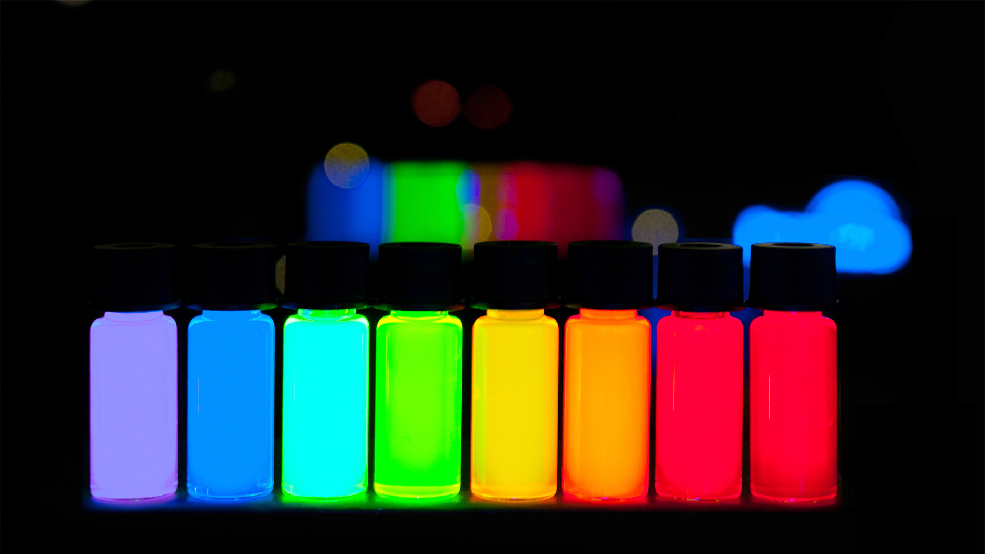
Quantum dots are small particles that are only a few nanometers in size, one nanometer equals one billionth of a meter, and are used as fluorescent biological labels in cells. These labels can be tagged to particular molecules, cell parts or genes of interest to a researcher.
“Think about [fluorescence] as a black light. If you have a room that’s completely dark with fluorescent paint on the wall and you turn on a black light, then you will be able to see where the paint is on the wall,” Stacey said. “It’s the same concept for quantum dots. One application is localizing where a virus is or label it and watch it move into a cell to try to understand the mechanism by which it moves.”
However, the mechanics behind quantum dots don’t make it simple. When exciting a single molecule, it will fluoresce and emit light but will do so in a diffuse pattern. This makes it difficult to see the molecule itself.
Additionally, plants absorb 490-700nm of light — essentially covers the entire visible range of light — allowing them to photosynthesize. As a result of absorbing these wavelengths of light, they also auto fluoresce, which is natural emission of light by structures inside plants cells such as chloroplasts.
The problem, then, is that viruses labeled with fluorescent probes in leaves are difficult to see because of the natural fluorescent glow coming from the plant.
For Stacey and his collaborators, the idea is to go beyond the visible light spectrum and use infrared light, which is above the visible light spectrum. Infrared light is most commonly known for its use in heat lamps.
“With infrared light, there would be no autofluorescence and so when you shine infrared light on a leaf, it would appear black,” Stacey said.” If you shine an infrared light on a fluorescent molecule, it would emit light and show up against a black background, making it very easy to see.”
The problem, though, is being able to distinguish one fluorescent molecule from another when they are close together. Because the researchers will be using infrared light, which has a longer wavelength, the imaging resolution decreases.
To get around that obstacle, the researchers will be using super-resolution microscopy to compensate for the resolution loss. The use of this technology will allow them to pinpoint the center of the fluorescence.
“It should be a big breakthrough. We would be able to look at single molecules interacting against a black background without any interference from autofluorescence,” Stacey said.
Stacey’s collaborators each contribute to the project in their unique way.
Zeev Rosenzweig from the University of Maryland, who is an expert in quantum dots, will be making the dots and labeling them with probes that absorb infrared light. Galya Orr from the Environmental Molecular Sciences Laboratory (EMSL), PNNL, in Richmond, Washington, has expertise in fluorescent microscopy and she, and her colleagues, will build the microscope.
An attractive part of submitting a proposal for the grant is the microscope’s prospect of being used as part of the EMSL user facility, which will ultimately allow researchers from all over the world to use the microscope when fully developed.
The researchers are excited to begin work on this project because they’re building a microscope that doesn’t yet exist. The microscope will expand the capabilities of researchers all over the world.
Stacey is appreciative that he gets to work with researchers from multiple disciplines. Namely, because he learns more about science from the expertise of others.
“That’s what makes it exciting because you’re constantly learning. The great thing about science is that you’re learning every day. It’s nice to get into these new areas especially where you don’t feel comfortable and learn new stuff,” Stacey said.
This work is funded by the Department of Energy for innovating new bioimaging approaches for bioenergy. The grant is split among the collaborators at the University of Maryland, University of Missouri and the Environmental Molecular Sciences Laboratory at the Pacific Northwest National Laboratory. Specifically, $1.5 mil. is to be used by researchers at the University of Maryland and the University of Missouri and $750,000 by researchers at EMSL.

