Video shot by Nick Andrussian | Mizzou Visual Productions Package produced by Evan Johnson | Bond LSC
As Mizzou seniors think about life after graduation, the research lab could serve as a proving ground for future plans.
That was the case for Bennett Flannagan, who graduated from Mizzou in 2024. He spent the last year as a research specialist I in the Paul de Figueiredo lab at Bond LSC, pushing himself and growing his expertise in preparation for graduate school.
His work paid off when he heard he was one of 20 applicants accepted into the Translational Biosciences PhD program at Mizzou’s School of Medicine for fall 2025.
ST. LOUIS, MO – The Academy of Science – St. Louis is proud to announce the recipients of the 27th Annual Outstanding St. Louis Scientists Awards, recognizing individuals and organizations that have made significant contributions to the advancement of science, engineering, and technology. The awards ceremony will be held on April 3, 2025, at the Missouri Botanical Garden.
Since its inception, the Outstanding St. Louis Scientists Awards has honored some of the brightest minds in the region, celebrating their exceptional achievements, groundbreaking discoveries, and lasting impact on science and society. This year’s honorees represent a diverse array of disciplines, from plant science and medicine to artificial intelligence and STEM education.
2025 Award Recipients
Fellows Award (Outstanding Achievement in Science)
● Dr. Steven Levine, Bayer – A global leader in ecotoxicology, Dr. Levine has made pioneering contributions to environmental safety assessments for crop protection.
● Dr. Ram Dixit, Washington University – A distinguished biologist whose research on the cytoskeleton is enhancing our understanding of cell shape and plant morphogenesis.
George Engelmann Interdisciplinary Award (Collaborative Science Achievement)
● Dr. Allison Miller, Donald Danforth Plant Science Center & St. Louis University – An innovative leader in plant science, engaged in interdisciplinary approaches to explore biology, evolution and root-shoot interactions to support sustainable agriculture systems.
Innovation Award (Exceptional Potential in Science, Engineering, or Technology)
● Dr. Peng Bai, Washington University in St. Louis – A trailblazer in battery research, developing new methods to improve energy storage and efficiency.
● Dr. Phani Chavali, Bayer – A machine learning and AI expert revolutionizing plant breeding to accelerate crop development and genetic gain.James B. Eads Award (Excellence in Engineering or Technology)
● Dr. Bing Yang, University of Missouri & Donald Danforth Plant Science Center – A world-renowned researcher in plant genome engineering, advancing CRISPR/Cas technology for disease-resistant crops.
Peter H. Raven Lifetime Achievement Award (Career of Service in Science, Engineering, or Technology)
● Mary E. Burke, MA, AAAS Fellow, Retired CEO, The Academy of Science – St. Louis – A transformative leader in STEM education and outreach, expanding science accessibility across the region.
Science Educator Award (Excellence in Science Education)
● Dr. Farzana Hoque, St. Louis University – A dedicated medical educator and mentor, advancing diversity, equity, and health awareness in the medical sciences.
● Peggy James Nacke, Retired Director of Special Projects and Events, The Academy of Science – St. Louis
– A champion of citizen science, spearheading groundbreaking STEM initiatives like the region’s first e-Science Fair and BioBlitz programs.
Science Leadership Award (Leadership in Advancing Science and Scientists)
● Dr. Thomas Eickhoff, Bayer – A mentor and industry leader driving innovation in agriculture and digital farming.
● MilliporeSigma, the Life Science business of Merck KGaA, Darmstadt, Germany – A St. Louis-based global leader in life sciences and pharmaceutical development, committed to STEM education and workforce development.
Deborah Patterson Award (Broadening Participation in STEM – Inaugural Award)
● Deborah Patterson, Former President of the Monsanto Fund – A tireless advocate for inclusive STEM education, instrumental in establishing STEMpact and STEMSTL.
A Celebration of Science and Innovation
The Outstanding St. Louis Scientists Awards Ceremony will bring together the region’s scientific, academic, and business communities to celebrate the honorees’ remarkable achievements. The event will also highlight the Academy’s ongoing commitment to fostering science literacy and inspiring the next generation of innovators.
For more information about the event, sponsorship opportunities, or to purchase tickets, please visit
www.academyofsciencestl.org or contact Executive Director Kate Polokonis (kpolokonis@academyofsciencestl.org).
About The Academy of Science – St. Louis Founded in 1856, The Academy of Science – St. Louis is a non-profit organization dedicated to promoting science literacy, education, and collaboration throughout the region. Through public seminars, student programs, and community engagement, the Academy continues to inspire scientific curiosity and discovery.
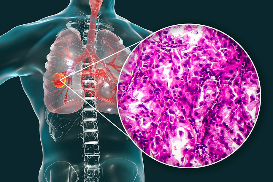
Non-small cell lung adenocarcinoma occurs in the glandular tissue of the lung and is illustrated here with a histopathology light micrograph. | Adobe Stock
By Roger Meissen | Bond LSC
Treating lung cancer is tricky business.
Not only is it more deadly than other cancers due to late diagnosis, but resistance also grows quickly against its few existing treatments and therapeutics, so new approaches are vital to higher survival.
That’s especially true for one subset of lung adenocarcinoma (LUAD), and University of Missouri scientists have shown promising progress toward understanding what drives this cancer growth and developing a way to treat it.
Using aptamers — short strands of DNA or RNA — a team from the Donald Burke lab at MU’s Bond Life Sciences Center decreased tumor growth and viability in mice by binding it with mutated receptors on the surface of cancer cells in this oncogene positive of non-small cell lung cancer. Their work published in Nature’s Precision Oncology.
“The aptamer folds up into a 3D structure that actually targets these mutated surface receptors that are always on, always signaling, in these cancer cells,” said Brian Thomas, lead author and Mizzou MD-PhD candidate in the Burke lab. “That binding event prevents growth and slashes proliferation to prevent the survival of these cancer cell lines.”
Intended to contribute to the federal Cancer Moonshot that aims to reduce cancer mortality 50 percent or more by 2050, the lab’s developments show promise for a novel therapeutic for a difficult to treat type of cancer. Current first line treatments involve tyrosine kinase inhibitors (TKIs) that prevent mutated epidermal growth factor receptor (EGFR) in LUAD from causing uncontrolled cell growth. However, resistance to these drugs typically grows in 12-18 months, and second- and third-line treatments frequently don’t work.
“EGFR is present on cell surfaces, but mutant EGFR is only present on cancer. This receptor is always on because of these mutations, and they cause uncontrolled growth, progression and cancer survival,” Thomas said. “Essentially, we show that when our anti-EGFR reagent, our aptamer, binds with this receptor, EGFR, it competed with FDA-approved antibodies — specifically cetuximab —commonly used to treat types of cancer including colorectal cancer.”
Because this aptamer competed with a clinically relevant antibody, Thomas and colleagues thought that it might have anti-cancer properties in certain cancers.
Aptamers aren’t a new technology. The small molecules of DNA or RNA were first considered by scientists in 1990 due to their potential to selectively bind to very targeted areas on a cell, and they show promise as therapeutics or as vehicles to deliver cancer drugs and treatments for other diseases. Their promise lies in being relatively inexpensive, readily scalable and their relatively low level of toxicity since the body’s adaptive immune system doesn’t recognize them.
Only two aptamers have been approved for use by the Food and Drug Administration (FDA) in the past 35 years — one in 2004 and one in 2023— but both provide treatment for an eye condition called macular degeneration. This low number of FDA-approved treatments from aptamers boils down to a few challenging shortcomings to the molecules.
“Their two primary limitations are that since aptamers are DNA or RNA, they can get chopped up and disposed of by things in the body called nucleases, and that they are very small,” Thomas said. “So, instead of staying in the body, they will be filtered out by the kidneys and essentially peed right out, which is why we injected our (aptamer) reagent directly into subcutaneous mouse tumors.”
Future research can be targeted at overcoming these obstacles. Thomas said coupling the aptamer to make it larger and exploring different delivery methods could make a treatment like this viable. “We can potentially make the aptamer bigger by appending it to some larger molecule that that can keep it in the body and keep it from getting filtrated, he said. “For lung cancer, researchers could also explore how to aerosolize it. Getting a patient to inhale it through an intranasal drip or nebulizer treatment, we can get high doses of oligonucleotides into the lung that way.”
Read more about this work in Thomas’ Behind the Paper article on Nature’s website.
Collaborators on this study include Bond LSC principal investigator Donald Burke and former Burke lab member David Porciani as well as Bond LSC principal investigator Trupti Joshi, graduate student Sania Awan of Mizzou’s Institute for Data Science and Informatics and Mizzou NextGen Precision Health researcher Mark Daniels.
Plant scientists recommend concerted approach to global food security
Adobe Stock image
By Roger Meissen | Bond Life Sciences Center
Climate change presents increasing dangers to crops, and plant scientists across the world recognize rapid changes are needed to prepare for its threats.
That’s the message a coalition of plant and agriculture researchers detailed recently in Trends in Plant Science. Organized by Michigan State University’s Plant Resilience Institute (PRI), their paper spelled out how farmers, scientists and policymakers must work closely together to develop crops that can withstand increasingly harsh environmental conditions.
“As rising temperatures and extreme weather events threaten crop yields and nutritional quality, our ability to feed a growing population becomes more and more uncertain,” said Seung Y. “Sue” Rhee, MSU Research Foundation Professor and PRI Director. “The urgency is clear: without climate-adapted crops, we face risks of famine, mass migration and global conflict.”
Ron Mittler, a Bond Life Sciences Center researcher and plant biologist at the University of Missouri, joined 20 experts to make recommendations on how to best address these dangers. Mittler’s science brings together the effects of many types of environmental stresses — from salinity and flooding to heat and drought — on the overall health of plants.
Ron Mittler, Bond LSC principal investigator and Curators’ Distinguished Professor of Plant Science and Technology | photo by Roger Meissen, Bond LSC
“We’re running experiments subjecting plants to 5-6 different stresses in all possible combinations, and what we’re finding is you don’t really have to have a strong stress, but rather a combination of different low-level stresses to actually topple a plant,” Mittler said. “We call it the multifactorial stress principle where we see — even with low levels of stressors — the more complex the environmental stress combinations become, the more rapid the plant deterioration.”
While scientists have advanced understanding of how plants handle environmental stresses, turning that knowledge into solutions for farmers is difficult due to financial, logistical and technical hurdles, according to the authors. These challenges are even greater in developing countries, where limited resources hinder solutions tailored to local needs. Improving plant resilience isn’t just a scientific issue, the authors said — it’s also a societal one that requires public support, clear communication and favorable policies.
The researchers propose several practical recommendations to leverage plant resilience to fight climate change and secure food supplies globally. They call for closer collaboration between scientists and policymakers and to establish research partnerships between the U.S., Europe and developing countries. The group recommends adopting a “farm to lab to farm” approach, where real farming challenges inform research, and discoveries are quickly applied back in the field. They also stress the importance of engaging the public about new technologies and being open about their benefits and risks to build trust. Finally, they advocate for science-based, efficient regulations to expedite the adoption of innovations.
“It can take a long, long time — often more than 10 years — to get a plant that shows more resistance to climate change to market, and within those 10 years things could change so that this newly developed transgenic plant may not be good enough anymore,” Mittler said. “Climate change doesn’t care about rules, and things will deteriorate faster than we can respond to under the regulations that we have now, so we think things need to be rethought to make them much friendlier to development and distribution of solutions.”
The group of 21 co-authors from nine countries formed as an outcome of the First International Summit on Plant Resilience, spearheaded by the PRI earlier this year. The summit promoted global cooperation in plant resilience research efforts, bringing together preeminent plant scientists from diverse disciplines. Together, they created a roadmap to position plant resilience research as a cornerstone of global climate change solutions.
Rhee remains optimistic about the future of plant resilience.
“By prioritizing innovation and working as a global community, we can create agricultural systems that not only withstand climate change but also ensure a sustainable, healthy future for generations to come,” she said.
Bond LSC lab reveals how a missing iron protein can cause muscle weakness
By Roger Meissen | Bond LSC Aging brings muscle weakness seen in the lack of strength of a handshake or the sureness of movement.
That atrophy is no accident, and it traces back to how cells, particularly their energy-producing components, decline in function as we climb in years.
One University of Missouri researcher’s latest discovery, published this week in Proceedings of the National Academy of Sciences, shows a distinct cellular reason why this weakness occurs.
His lab revealed how a muscle cell’s mitochondria fail to generate enough energy for skeletal muscles due to one iron–sulfur protein. This understanding could one day help lead to treatments for diseases like Duchenne muscular dystrophy — the most common type of muscular dystrophy in children — and muscle deterioration associated with aging.
“We followed the phenotype, the muscle weakness in our mice, to this protein,” said Ron Mittler, a principal investigator at Mizzou’s Bond Life Sciences Center and a plant biologist. “What we found is that CISD3 proteins — also found in our bodies — are important for regulating the levels of iron in the mitochondria, and previously nobody knew what they were doing.”
The importance of a single protein
CISD3 — (conserved iron-sulfur domain-containing protein 3) — exists solely inside a cell’s mitochondria, the organelle responsible for creating the energy for the cell. To figure out its function, the Mittler lab started by disabling the genetic code for it in test mice. Lab manager, Linda Rowland, noticed a particular lack of strength in these knockout mice compared to normal mice. Lab members confirmed the knockout mice were weaker through physical observation and strength measurements then used a series of tests to prove why. By analyzing the proteins in the model mouse and studying the structure of them, they found a markedly lower concentration of the CISD3 protein.
Ron Mittler, Bond LSC principal investigator and Curators’ Distinguished Professor of Plant Science and Technology | photo by Roger Meissen, Bond LSC
“Mitochondria in muscles are highly energetically active, but we found they were in very bad shape in these knockout mice,” Mittler said. “After that, we completed proteomic and structural studies, because we wanted to see what this CISD3 protein actually does.”
In this process, they noted that the reduced CISD3 binds closely with several proteins in respiratory chain complex I and II and transfers one of its iron clusters for its metabolic process. Without this chemical binding and transfer, some important cell respiration processes within the mitochondria don’t happen, reducing how much food is converted into an energy form that cells can use.
“Basically, complex I is almost shut down completely in the knockout mice, and that’s why they have weak muscle and die earlier,” Mittler said.
To confirm this interaction, Mittler’s lab partnered with scientists at Rice University and University of Texas to compare CISD3 protein with proteins from the respiratory complex one. The computational biology approach analyzes how proteins may have evolved together. When two proteins interact, scientists see fewer mutations over time. Using AI models, they predicted and ranked the likelihood of protein-to-protein interaction. They saw almost no mutations between these proteins, confirming that CISD3 binds with the NDUFV2 protein of the respiratory complex I.
Finally, the researchers utilized equipment at the Roy Blunt Precision Medicine building to further examine the metabolic function in skeletal muscle cells. Using Seahorse analyzers and muscle fibers they generated, they measured the rate of respiration, glycolysis and many other processes. With the knockout mouse muscle cells missing CISD3, they saw very little respiration and elevated glycolysis — where sugars are broken down into energy without needing oxygen like in respiration.
From plants and cancer to muscles
It’s been a long interaction between Mittler and iron-sulfur proteins.
He first happened upon this protein family in 2007 after he received a National Science Foundation grant to study proteins of unknown function. As a plant biologist, he detailed how CISD1 made model plants more resistant to oxidative stress when overexpressed. Oxidative stress is detrimental to cells because of increased levels of reactive oxygen species that make cells deteriorate faster.
“The plants actually looked bigger, they were happier,” Mittler said. “We initially didn’t know anything about this plant protein, and when we found the same protein in mammals, we then asked where that protein was important in animals.”
That question led him to study cancer where his team found lots of these iron-sulfur proteins, and when Mittler moved to the University of Texas in 2010, he shifted to also understand the protein in animals.
“Cancers like breast cancer are known to have what scientists consider an ‘iron addiction’ and thrive on having a lot of it, therefore they need a lot of all three of these proteins,” he said. “If you want a lot of cell proliferation, like in cancers, you need a lot of these iron clusters and reactive oxygen.”
This current study is his first foray into normal development instead of disease and destruction.
Moving research from lab to startup
Regardless of species, all this knowledge of sulfur-iron proteins has given Mittler the information and experience to pursue applications.
“For this study, there are a lot of applications from the standpoint to developing drug therapies for disorders such as Duchenne’s Muscular Dystrophy, or for CISD3, it’s possible to develop a genetic screen for embryos that doesn’t have respiratory complex I working,” he said.
He recently founded a startup company called MitoMed to develop drugs based on these sulfur-iron proteins.
“I’m trying to develop drugs to fight cancer because I think, for us, this is what will make a big difference,” he said. “We already have one type of drug with almost no side effects; we’ve done all the mouse work and the idea now is to get this to clinical trials.”
The paper “CISD3/MiNT is required for complex I function, mitochondrial integrity, and skeletal muscle maintenance” published in Proceedings of the National Academy of Sciences on May 23, 2024. Collaborators from the University of Missouri, Rice University and the University of Texas worked on this paper. This work was partially supported by grants from the National Institutes of Health, the National Science Foundation and the U.S.-Israel Binational Science Foundation.
Kim Jasmer, assistant research professor of biochemistry, in the lab of Gary Weisman at Bond LSC | photo by Roger Meissen, Bond LSC
By Sarah Gassel | Bond LSC
In the spring of 2009, Kim Jasmer, a swimmer from the University of Washington, arrived at the University of Missouri for the Missouri Grand Prix, one of a seven-meet series featuring elite swimmers from all over the world. Between swimming her own races and cheering on teammates, Jasmer had another important task on her agenda.
The athlete had scheduled a meeting with the now-retired Mizzou Professor of Biological Sciences, Steve Alexander, to discuss his work investigating genetic mechanisms underlying resistance to chemotherapeutics, topics quite far from her sport. The meeting began a long and sometimes winding road into research at Mizzou while allowing her to continue her swimming career, which ended after the 2012 Olympic Trials.
Jasmer, now an assistant research professor working with Bond LSC’s Gary Weisman, just made another important step down that path. She recently received her first National Institutes of Health (NIH) grant as the principal investigator. The two-year R03 award, which started in March, aims to uncover more information on an important receptor — the P2Y10 receptor — that could play a role in Sjögren’s disease.
“Grants are particularly fun because you get to dream up how you would answer a question,” said Jasmer. “I do like piecing everything together and the creativity of it.”
Sjögren’s disease is a common autoimmune disease that affects millions of Americans. The chronic condition — where immune cells attack and destroy the salivary and lacrimal glands that produce saliva and tears — mainly affects the eyes and mouth, though those with Sjögren’s have a wide range of symptoms.
Swimming in her own lane
Jasmer, who grew up in North Bend, Oregon, was following two of her passions — swimming and molecular and cellular biology — at the University of Washington when she saw Dr. Alexander’s work. She was interested in finding out how grad school at Mizzou could allow her to continue her passions, not just her studies.
“When I was looking at graduate schools, I wanted to keep swimming, so I looked at places where I could do both,” Jasmer said.
She started a biological sciences Ph.D. in 2009, and between hours doing laps and hours in the lab, Jasmer gained endurance in both. Her doctorate focused on cancer research, specifically looking at oxidative stress responses in melanoma, and after receiving the degree in 2015, she joined the lab of Michael Petris as a postdoc studying the influence of copper-dependent enzymes in the formation of tumors.
However, she was also interested in studying the immune system. In 2016, Gary Weisman’s lab — right across the hall from Michael Petris — received a grant for research on Sjögren’s disease and was looking for a postdoc to perform the studies. Knowing her interest in the subject, they approached her about the position. She accepted and began working half-time in each lab, eventually transitioning full-time to studying salivary glands.
“I didn’t start out intending to study salivary glands, but I landed in that community of researchers,” said Jasmer. “It’s a very small and supportive niche of scientists.”
The time since then has seen the Weisman lab learn more and more about the cellular workings of the disease, eventually leading to Jasmer submitting an R03.
The grant marked the first time Jasmer wrote and received an NIH grant independently. Although had previously written NIH grants collaboratively Weisman, this one was composed of her individual ideas, making the award all the more validating. Globally, NIH is the largest public funder of biomedical research, and the support an NIH grant provides can get a research project off the ground. But it is also highly competitive — on average, only about 20 percent of NIH grant applications are funded.
“I was kind of in shock because it feels like my career path has been so long, and there’s a lot of rejection that comes with it,” said Jasmer. “When one comes through and works out, it’s very exciting.”
The process of writing a grant is no easy feat, either. There are many questions that must be asked and answered when composing an application. It is not only necessary to propose what the research will potentially reveal but also the process needed to get this information, down to the techniques and analyses that will be used. Essentially, grant writing is akin to a much more complex and scientific version of the board game Clue.
Getting a spot on the podium
Recently, she’s seen her research make an impact in journals with her work being published in the Journal of Oral Biology and Craniofacial Research.
Currently, there are no curative therapies for Sjögren’s. Jasmer wants to eventually be able to apply her research to develop solutions. However, she says many questions must first be answered, and additional research must be done before reaching this goal.
“The downstream is that, if everything pans out, the P2Y10 receptor could be a great target, and we can work with the medicinal chemists to identify compounds that could target it and develop a therapy for Sjögren’s,” said Jasmer.
While this goal seems like a far way off, she has also come a long way through her leg of the relay so far.
Research illuminates how one of the most prevalent zoonotic diseases infects cells
MU undergraduate Raymond Preston shows an inoculated agar plate he uses in the Paul de Figureiredo lab to study bacterial and mechanisms. | photo by Sarah Gassel, Bond LSC
By Sarah Gassel | Bond LSC
When bacteria invade a host, they employ unique strategies to weaken the host’s cells for optimal infection. For the bacterial pathogen Brucella, this means manipulating internal cell machinery to subvert host function and favor infection.
New research at the University of Missouri and Texas A&M University reveals a specific mechanism not previously observed that this major public health concern uses to achieve this takeover. Their study published in the journal Cell Host & Microbe March 25, 2024.
“Brucella is a very smart pathogen, and a lot of how it manipulates a host’s function is largely unknown,” said Qingming Qin, a study author in the lab of Paul de Figueiredo, a principal investigator at MU’s Bond Life Sciences Center.
Brucella causes brucellosis, one of the most common zoonotic diseases worldwide. More prevalent in resource-limited countries, it primarily infects cattle, pigs, goats, sheep and dogs, but humans can contract the disease through eating or drinking unpasteurized milk or cheese from the animals. Brucellosis has symptoms similar to malaria and infects an estimated 2.1 million people globally each year, according to the . The frequency of illness relates to the pathogen’s complex mechanisms for infection. In addition to the public health concerns raised by brucellosis, its economic impacts are also a major driver of poverty in developing countries.
Generally, pathogens are able to evade recognition by the immune system in a host cell by disrupting a cell’s processes. They do this by targeting specific cell parts with necessary roles, such as proteins and sugars. Sugar molecules, called glycans, are building blocks of cell components. In addition to providing the cell with energy, glycans allow these components to function properly.
Because of their widespread role throughout the cell, sugars are an ideal target for manipulation by bacteria. De Figueiredo joined with researchers at Texas A&M, Texas Tech, CIRAD (French Agricultural Research Centre for International Development) and other colleagues to reveal a new mechanism bacteria use to do this.
“Our results demonstrate the potential of systems biology to enhance our understanding of bacterial ultimate adaptation to their hosts and to imagine new therapeutic tools,” said Damien Meyer, a study author and principal investigator at CIRAD UMR ASTRE.
Many bacterial pathogens have secretion systems, which operate like syringes that can inject material into a desired host cell. In the case of Brucella, it injects proteins into the cell to manipulate the cell’s machinery.
Known as an effector protein, it influences the cell’s systems to weaken and allow for successful bacterial infection and reproduction. Different effector proteins affect different parts of cell systems, which gives researchers insight into how to hinder a bacterial invasion.
“Studying the mechanisms underlying Brucella-host interaction can provide us a lot of new insight on how Brucella can infect the host cells and find a strategy of how to prevent infection and — even after infection — how we can stop it,” said Qin. “That’s our long-term goal.”
Using software to predict effector proteins, researchers made progress in this goal when they discovered Rhg1, an effector protein not previously identified.
To reveal the function of Rhg1, scientists performed tests that showed Rhg1 interacting with proteins associated with the oligosaccharide transferase (OST) complex. The OST complex is cell machinery that helps produce proteins by adding N-glycans — sugar molecules that specifically modify proteins. N-glycans attach to specific sites on the amino acid chain that makes a protein to determine how it folds. Proteins folded differently carry out different tasks, making N-glycans essential for the overall functioning of the cell. The amino acids are not processed with N-glycans pre-attached; rather, this is completed by the OST complex which catalyzes the transfer and attachment of N-glycans to the amino acids. According to the study, the Rhg1 effector protein targets the OST complex, manipulating these specific N-glycan sugars.
This novel discovery gives additional insight into Brucella’s complex strategies.
“It opens a lot of doors to look at different connections,” said Qin.
This finding also allowed researchers to gain a better understanding of the protein malfunctions that occur in the host cell during infection.
Researchers observed that Rhg1’s modification of the OST complex causes an overall reduction of N-glycan sugars. This deficiency within the amino acid chains results in misfolded proteins that cannot be utilized. The accumulation of unfolded or misfolded proteins in the lumen of the endoplasmic reticulum (ER) compartment causes a cell response known as the unfolded protein response (UPR). During this process, defective proteins are chopped into small blocks that are recycled under normal conditions.
The lower illustration show how Rhg1 from the bacteria interacts with the OST complex of the cell and leads to incorrectly folded proteins that benefit Brucella bacteria.
But, Brucella bacteria use this response to their advantage. They use this recycled material for replication, suggesting the bacteria purposefully seek to activate the response. Because of the misfolding, Brucella benefits even more when cells reduce specific proteins that recognize pathogens, which may also help the bacteria avoid activating immune responses from host cells.
Observing Rhg1 revealed to scientists how the pathogen may be smarter than previously thought, and expanding understanding of this infection process could pave the way to prevent human brucellosis by improving current treatments.
“We definitely would like to make good use of this discovery one day in the future,” Qin said.
Cell Microbe and Host journal published “Brucella-driven host N-glycome remodeling controls infection” March 25, 2024. Co-authors include by Ana-Lucia Cabello, Kelsey M. Wells, Wenjing Peng, Hui-Qiang Feng, Junyao Wang, Damien F. Meyer, Christophe Noroy, En-Shuang Zhao, Hao Zhang, Xueqing Li, Haowu Chang, Gabriel Gomez, Yuxin Mao, Kristin L. Patrick, Robert O. Watson, William K. Russell, Aiyung Yu, Jieqiang Zhong, Fengguang Guo, Mingqian Li, Mingyuan Zhou, Xiaoning Qian, Koichi S. Kobayashi, Jianxun Song, Suresh Panthee, Yehia Mechref, Thomas A. Ficht, Qing-Ming Qin, and Paul de Figueiredo.
This work is supported by grants from Texas A&M Clinical Science Translational Research Institute, the Defense Advanced Research Projects Agency, the NIH, the National Science Foundation, the Bill and Melinda Gates Foundation, the University of Missouri NextGen Precision Health Endowment and the National Institute of Child Health and Human Development.
Close up illustration of isolated cancer cells at molecular scale. | Adobe Stock
Joint release by Hokkaido University, Toyo University and University of Missouri
Researchers in Japan and the United States have developed a novel method for boosting the immune system’s capability to detect and eliminate cancer cells. This technology robustly augments the amount of an immune complex called MHC (major histocompatibility complex) class I in cancer cells.
“Our discovery has the potential to transform the way we approach cancer treatment,” said Hokkaido University immunologist Koichi Kobayashi, who led the study. “Our innovative technology enables us to specifically target immune responsive genes and activate the immune system against cancer cells, offering hope to those who are resistant to current immunotherapy.”
The technique reduced tumor sizes significantly and increased activity of immune cells called cytotoxic CD8+ T cells, the immune system’s primary cancer-fighting cells. , Known as TRED-I (Targeted Reactivation and Demethylation for MHC-I), it was tested in animal cancer models and markedly enhanced treatment efficacy when used in conjunction with existing immunotherapy.
“New modalities for fighting cancer like this are desperately needed because we have few solutions to fight some cancer types,” said Paul de Figueiredo, University of Missouri Bond Life Sciences Center researcher. “This is a radically new approach, and I’ve felt lucky to be part of it.”
MHC class I molecules are a prerequisite for the immune system to recognize and eliminate cancer. When cancer cells are faced with pressure from the immune system, they actively reduce their MHC class I molecules, so cancer cells can hide from drawing the attention of cytotoxic CD8+ T cells. Kobayashi and his team previously identified a gene, called NLRC5, that regulates MHC class I levels. They further found that NLRC5 is suppressed by turning off molecular switches existing on DNA, called DNA methylation, in cancers to reduce MHC class I level. The TRED-I system was able to restore DNA methylation of NLRC5 gene and further activate NLRC5, thus increasing MHC class I levels in cancer without causing severe side effects.
“This work is the result of our team’s research for over 10 years,” Kobayashi said. “It’s great to shed light on moving our findings to potential clinical application. We believe with further refinement, the TRED-I system could contribute significantly to cancer therapy.”
Further research could enable direct delivery of TRED-I system in cancer patients. If successful, such drugs could improve the efficacy for the immune system to eliminate cancer and able to improve the response to existing therapy.
This work was supported by Japan Society for the Promotion of Science (JSPS) KAKEN grant 19K21250, 20K21511, 22H02883, 22KK0112; Japan Agency for Medical Research and Development (AMED) grant JP223fa627005 and 23ym0126801j0002; Japan Science and Technology (JST) START University Ecosystem Promotion Type grant JPMJST2284; Takeda Science Foundation; Bristol Myers Squibb; SENSHIN Medical Research Foundation; Hitachi Global Foundation; Kobayashi Foundation; The Toyo Suisan Foundation; KAKEN grant 20K16433; 19K16681
Contacts:
Professor Koichi S. Kobayashi
Department of Immunology, Graduate School of Medicine
Hokkaido University
Tel: +81-11-706-5056 kskobayashi@med.hokudai.ac.jp
Department of Microbial Pathogenesis and Immunology
Texas A&M Health Science Center
kobayashi@tamu.edu
Professor Shinya Tanaka
Department of Cancer Pathology, Graduate School of Medicine
Hokkaido University
Tel: +81-11-706-7806 tanaka@med.hokudai.ac.jp
Institute for Chemical Reaction Design and Discovery (WPI-ICReDD)
Hokkaido University
Hidemitsu Kitamura
Department of Biomedical Engineering
Faculty of Science and Engineering
Toyo University kitamura012@toyo.jp
Institute for Genetic Medicine
Hokkaido University
Paul de Figueiredo
Bond Life Sciences Center principal investigator
NextGen Precision Health Endowed Professor of Molecular Microbiology & Immunology
University of Missouri School of Medicine
Tel: 573-882-6828 paullifescience@missouri.edu
Bond LSC principal investigators Bing Yang (left) and Ron Mittler were once again recognized on the list of Highly Cited Researchers. | photos by Roger Meissen, Bond LSC
By Roger Meissen | Bond LSC
Discoveries in research come with time, so the incremental accumulation of knowledge toward breakthroughs is fundamental to science and the future.
While many contribute to this understanding, a few scientists consistently produce research that others note more often in their own experiments. Two University of Missouri Bond Life Sciences Center researchers once again landed on that list of most cited scientists.
Bing Yang and Ron Mittler are included on the list of Highly Cited Researchers for 2023. The inclusion stems from authoring multiple highly cited publications in 2023 that rank in the top 1% by citations for their field.
This year marks the fifth straight year of inclusion in the list for Yang and the fourth year running for Mittler.
Only one in every 1,000 scientists receive this honor, and they join 6,849 researchers globally who made the Clarivate list. Clarivate runs Web of Science, a database aggregating published, peer-reviewed research from academic journals, conference proceedings and other citations.
Yang, a Bond LSC scientist and MU professor of Plant Science and Technology, works on targeted genome editing in plant species, including rice, maize, wheat, sorghum and soybeans. His lab aims to understand interactions between plants and pathogens like bacterial blight in rice so they can work toward varieties that can better withstand the disease, among other projects.
Mittler, a Bond LSC scientist and MU Curators’ Distinguished Professor of Plant Science and Technology, researches the role of reactive oxygen species (ROS) and its function in regulating biological processes in plants.
The Bengal tiger was one of five feline lineages compared to gives a comprehensive look at genome sequence structures that could have driven the evolution of distinct cat species. This new reference genome is comparable to the human genome in terms of its completeness, and could be used to for feline veterinary precision health. Denise Allison Coyle/Shutterstock
First-ever analysis compares nearly gapless genome across cat species and with humans to shine light on evolution
By Roger Meissen | Bond LSC
The tiger doesn’t know it, but a difference deep in its genome sets it apart from other cats.
This big cat preserved a distinct sense of smell thanks to a few chromosomes it uniquely retained over millennia of evolution that other feline species did not.
A study published in the journal Nature Genetics details this finding where scientists from the University of Missouri partnered with Texas A&M University and others to compare, for the first time, nearly gapless cat genomes across multiple species and with humans. This genome map correctly strings together nearly all chromosomes of these felines — from one end to the other without missing segments — to give an animal genetic reference that rivals the human genome.
Wes Warren, Bond LSC principal investigator and professor of genomics | photo by Roger Meissen, Bond LSC
“This study is a comprehensive look at the genome sequence structures that could be driving cat speciation, the evolutionary process by which populations became distinct species,” said Wes Warren, a Bond Life Sciences Center researcher and MU professor of genomics. “We found distinct differences between how great apes, including humans, and cat sequences evolved in a similar period of time. There were many interesting differences to consider, such as why certain chromosome regions have similar
but novel sequence landscapes even across species of cats.”
In tigers, scientists previously identified genes associated with the chemosensory system, perhaps lending them heightened senses of smell key to their survival.
“We see cats have olfactory receptors for sensing smell that are greater than most mammals but, among cats, the tiger stands out with the largest repertoire,” Warren said. “We know tigers have a solitary lifestyle, and that acute sense of smell helps males detect females for mating and lets them avoid the territory of other males to enhance their chances of survival.”
When thinking about other unique sensory features to explore, researchers also looked to the fishing cat, a rare nocturnal feline native to marshy areas in Southeast Asia. They searched for molecular signatures that would explain its sleek body, partially webbed feet and prowess for hunting fish, characteristics tailored to its life around water.
Scientists looked at the fishing cat, a rare nocturnal feline native to marshy areas in Southeast Asia. They identified molecular signatures that explain its prowess for hunting fish and characteristics tailored to its life around water. They found more complete genes to detect water-borne smells than in other feline species. | Attribution: Kelinahandbasket, Prionailurus viverrinus 01, CC BY 2.0
“There are specific olfactory receptors that detect waterborne odorants as well as a much larger number of receptors tailored for terrestrial smell detection; we asked if fishing cats would have more functional receptors for waterborne odorants than other cats, and that’s exactly what we saw,” Warren said. “In fishing cats, all gene copies were complete while other cats had broken copies, suggesting natural selection was at work because of their aquatic hunting behavior.”
While these illustrations are notable, scientists also learned from their extensive analysis of sequence structural variation.
What stood out overall were fewer differences in cat chromosomes — coding for traits like hair length and color, bone structure and size, fertility and senses — compared to great apes despite both families diverging into distinct species over a period of 13 million years.
Using the latest assembly techniques, the scientists pieced together the genomes of multiple cat hybrid species — the Bengal cat, the safari cat and the liger — and then compared the five lineages. New techniques severely limited genomic dark matter, which is genetic information researchers previously were unable to characterize and assign meaning to, thus enabling them to shine new light on this novel sequence structure.
Some of those pieces came from Leslie Lyons, the Gilbreath-McLorn Endowed Professor of Comparative Medicine in veterinary medicine and surgery at the MU College of Veterinary Medicine. She sequenced two of the cat hybrids nearly 25 years ago.
“I was at the National Cancer Institute as a postdoc when I sequenced the Geoffroy’s cat and Asian leopard hybrids, and I had no idea they would ever be reused for a project like this, but fortunately they were,” Lyons said. “Now, we have a cat genome that is comparable to the human genome, which is one of the best in the scientific world, so the cat has really leapt forward in terms of genomic resources.”
Structural differences in the X chromosome, in particular a region that displays features of a supergene, stood out to the team. This presumed supergene in cats, a complex interaction where a chromosomal region of neighboring genes are inherited to fix a trait with fitness consequences, could be the process of evolution that led to hybrid sterility if certain feline species cross. It is evolving faster than most of the cat genome. Much like the mule — a cross of a male donkey and a female horse — cannot produce offspring, the liger bears no offspring when lions and tigers mate. The team hypothesizes this is due to a failure in meiosis, the process that produces egg and sperm cells.
“We found multiple inversions on X chromosomes of interest in cats that include the complex DXZ4 region,” Warren said. “In these cats, we identified entire copies of this complex, repetitive region that harbors the only X-linked speciation gene identified in mammals.”
While these advances in genetics push our understanding of evolution in exciting new ways, Lyons has much more pragmatic uses for this work.
“I’m an end user,” Lyons said. “These good genetic maps allow us to find mutations that cause inherited diseases in cats, so this genome allows us to bring precision medicine to feline veterinary health care.”
“Our findings will open doors for people studying feline diseases, behavior and conservation,” he said. “They’ll be working with a more complete understanding of the genetic differences that make each type of cat unique.”
Nature Genetics published “Single-haplotype comparative genomics provides insights into lineage-specific structural variation during cat evolution” Nov. 2, 2023. The study was conceptualized by Bill Murphy — VMBS professor of veterinary integrative biosciences at Texas A&M and Wes Warren — principal investigator in the Bond Life Sciences Center and professor of genomics in the College of Agriculture, Food and Natural Resources at the University of Missouri. Additional collaborations involved researchers from the University of Washington, University College Dublin, the Institute for Systems Biology in Seattle, Louisiana State University and the Guy Harvey Oceanographic Center.
This work was funded through a grant from the Morris Animal Foundation, which works to improve and protect the health of animals through scientific innovation, education and inspiration.