Bond LSC scientist works with MU eye surgeon to help people suffering from autoimmune-disease Sjögren’s syndrome
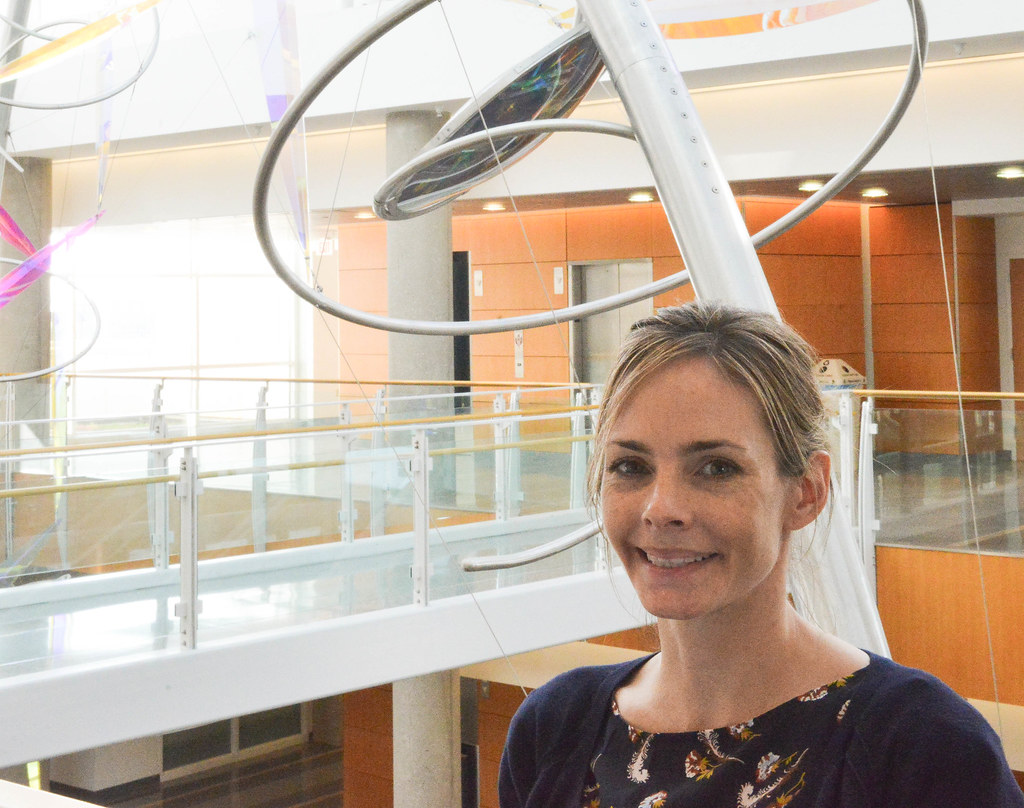
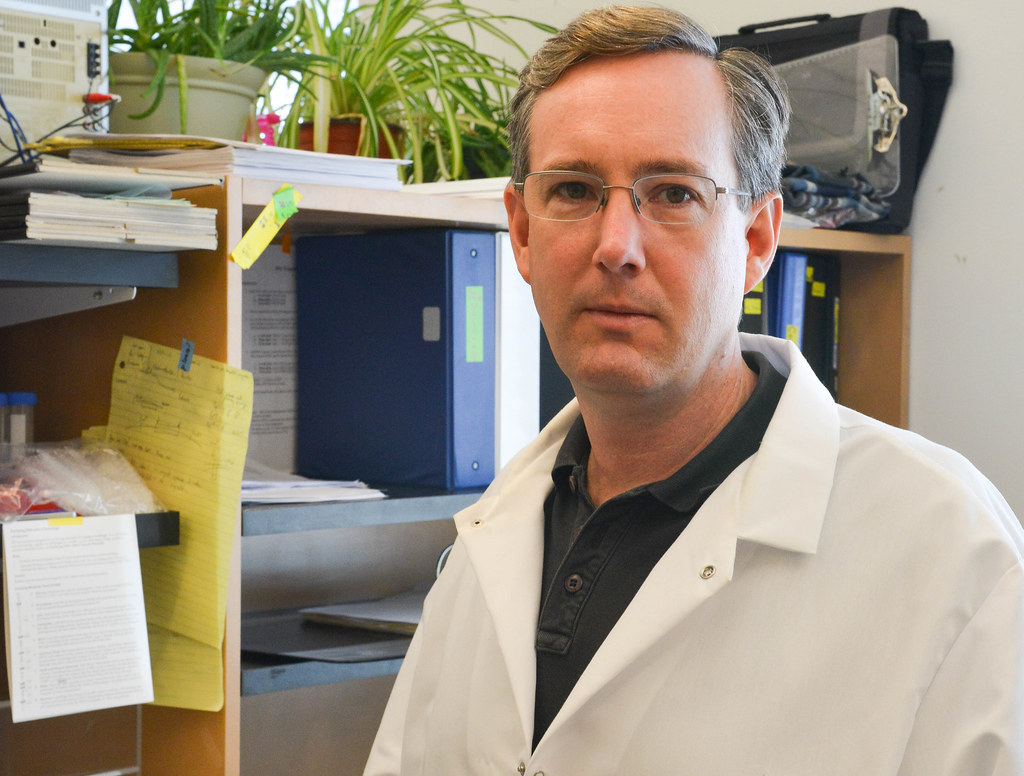
Dr. Carisa Petris stands in the McQuinn atrium of Bond Life Science Center. She and Bond LSC researcher Gary Weisman are using funding from a $100,000 Bond LSC grant to study the mechanisms of an auto-immune disease in the lacrimal glands of the eyes. They are hoping treatments for the disease in mice they study could be applied to humans. | photo by Phillip Sitter, Bond LSC
By Phillip Sitter | Bond LSC
They may not get much respect, but tears and spit are the products of a delicate secretive system that people would pay their respects to in mourning if they discovered that system was dying.
Gary Weisman and Dr. Carisa Petris are working together to help heal the damage caused by such a chronic lack of tears and saliva. The pair recently received a $100,000 Bond Life Sciences Center Grant for Innovative Collaborative Research to allow Bond LSC’s Weisman to partner with Petris, an eye surgeon working at MU Hospital.
They want to study the mechanism by which the auto-immune disease Sjögren’s syndrome cripples the glands of the eyes in mice. By comparing that mechanism to how it works in human eyes, they hope to examine if effective treatments for the mice could in turn help people.
“Dr. Weisman has characterized [Sjögren’s syndrome] in the salivary glands, and then there are similar glands in the eye called the lacrimal glands, and those are the tissues that we’re going to study,” she said of their collaboration.
Much of the grant money will go toward the costs of obtaining and housing new knockout mice for the study. These mice have a disabled, or knocked out, gene that causes them to express a certain trait like the dry eyes and development of Sjögren’s in this case.
“It takes a few weeks to a couple months for the disease to fully manifest itself, so we’ll house those mice for that time, and then of course, we’ll be treating them with the drug, and not with the drug, some for harvesting just the lacrimal glands and [studying] the surface of the eye,” Petris said.
Even though Sjögren’s syndrome and inflammation research are big topics, there’s just no good solution to the problems yet.
“There are a few [eye] drops that are used for Sjögren’s now, and they’re at best helpful, but they don’t cure the disease, so that would be the ultimate goal. They help decrease the inflammation that goes along with it and increase the tear production. The drops are also limited in their longevity too — you can only use them a certain length of time before they tend to not work so well anymore,” Petris said.
Petris referred to one drug that shows promise. The drug or another like it would interrupt the autoimmune response that causes the damaging inflammation that leads to Sjögren’s. It has already shown good results for reducing the symptom of dry mouth in mice, so Petris said she and Weisman will add it to some of the eyes of their mice and see if has any similar effect it reducing dryness there.
Dr. Peter Ostrum spoke at Bond LSC in celebration of World One Health Day
Dr. Peter Ostrum, who once played the character of Charlie Bucket in 1971’s “Willy Wonka and the Chocolate Factory” —also starring the late Gene Wilder — smiles after giving a lecture to an audience at Monsanto Auditorium in Bond LSC. After “Willy Wonka,” Ostrum did not pursue acting further, and went into a career in veterinary medicine. | photo by Phillip Sitter, Bond LSC
By Phillip Sitter |Bond LSC
The character of Charlie Bucket found his golden ticket to a happy life wrapped in a Willy Wonka chocolate bar. Peter Ostrum, who at the time was just a child actor playing Charlie, later found his in horse pastures.
After playing Charlie in 1971’s “Willy Wonka and the Chocolate Factory” alongside the late Gene Wilder starring in the titular role, Ostrum didn’t pursue acting any further. He spoke about life as a veterinarian Nov. 3 at Monsanto Auditorium in Bond Life Sciences Center.
“People are always curious about what happened to Charlie. Why wasn’t he in any other films? Did he survive Hollywood? I’m relieved to tell you that my life didn’t end up as a trainwreck,” Ostrum said, getting some laughs from the crowd gathered to listen to him speak.
“The film industry just wasn’t for me,” he explained, although he did enjoy working alongside Wilder and co-star Jack Albertson, who played Grandpa Joe. Ostrum said that every day on lunch break during filming in Munich, Germany, Wilder would share a chocolate bar with him.
Back at home in Ohio, Ostrum worked at a stable, and had several positive interactions with veterinarians. He admired the profession, and working with horses specifically. He even went on to be a groomer for the Japanese three-day equestrian event team at the 1976 Summer Olympics in Montreal.
He wanted to become an equine veterinarian after a year working at an equine veterinary clinic. However, Ostrum discovered that dairy cow care fell more in line with his dreams, and after getting his veterinary degree at Cornell, he’s been doing that ever since — in upstate New York where he is also a husband and father of two children.
Ostrum described how agriculture and veterinary medicine have changed over recent years, with changing numbers and sizes of farms, the rising power of animal welfare groups and an increased desire from consumers to know where their food comes from. People want to know whether animals are treated humanely and whether farms are negatively affecting the environment, he said.
All of these changes and others require increased transparency, education and community outreach efforts by everyone working in agriculture, Ostrum said. In candidates for veterinary associates, he said that he looks for “the intangible skills at the heart of who people are” — their character and their ability to connect with clients and patients.
Ostrum also mentioned the importance of mental health awareness among veterinarians and other health professionals. “We can’t help others if we can’t help and support ourselves,” he said.
Efforts to understand the genome of one plant through its many genetic varieties could lead to nutritional improvements in the staple crops billions of people depend on
By Phillip Sitter | Bond LSC
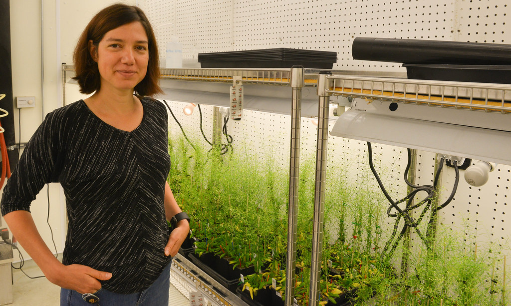
Ruthie Angelovici stands next to some Arabidopsis thaliana samples in the basement of Bond LSC. She is leading projects to study the relationships between genotypic and phenotypic variation in Arabidopsis and how this affects the amino acid content of the plants, and the resistance of their seeds to drought conditions. | Phillip Sitter, Bond LSC
It’s hard to avoid corn, rice or soybeans in your diet, and you’ve probably eaten or drank something today with at least one ingredient from them.
Unfortunately for the billions of people worldwide who depend on these crops as a staple, they aren’t actually all that nutritious. Specifically, they lack sufficient quantities of amino acids.
Twenty amino acids are required to build any protein, and within that about ten are considered essential, Bond Life Sciences researcher Ruthie Angelovici said. “Without amino acids, you can’t live.”
Amino acids might seem minor, but important parts and processes in our bodies from our muscles to enzymes are built from or work through them. That’s why Angelovici wants to enhance their availability in key foodcrops.
In the case of amino acids, “What we’re trying to understand is the basic question of how those accumulate in seeds, and then from that basic concept we’re going to try to improve that in grain,” Angelovici said.
The evolution of poor nutrition
No one really knows why so many of our most important crops that essentially sustain humanity lack sufficient essential amino acids.
Maybe plants don’t synthesize amino acids because the cost in energy for the plant is too high, or because higher levels of amino acids might make them more vulnerable to attacks from hungry insects. Maybe if plants produced higher levels of amino acids, the taste would be too strong for human palates, and so our ancestors long ago selectively bred those traits out of crop populations. Or, maybe in ancient farmers’ pursuits of other traits in their crops, like higher quantities of starch, humanity accidentally boosted one nutritional trait at another’s expense. There are a lot of unknowns when it comes to these theories, Angelovici said.
What is clear — and something Angelovici said she cannot stress enough — is how powerful a genetic tool she and her fellow researchers at Bond LSC have in the form of a collection of a vast amount of genetic variation of Arabidopsis thaliana.
“Arabidopsis thaliana is a model [plant] system that a lot of plant scientists use, although it is not a crop, or anything like that, but it’s a great model plant to start with, and then everything we learn from it, we can try and figure out if it’s the same in maize, rice, soybean, and translate it,” Angelovici explained.
Part of the mustard family, Arabidopsis grows quickly so researchers can study four or five generations in one year. As an added bonus, this huge genetic variety but can be grown in just one room instead of large fields. For Angelovici, that room is in Bond LSC’s basement and the basement of greenhouses nearby.
“We are growing right now 1,200 ecotypes of this Arabidopsis thaliana. So, what is an ecotype? It’s basically from the same species, but they have a slightly different genotypes. So, we’re looking at a vast genetic variation that represents genetic variation of this species across the world. Each ecotype comes from a different place,” she said.
For those of you wondering, a genotype is the specific sequence of information in an organism’s genetic code — its genetic identity. A phenotype is an observable physical trait controlled by the genetic sequence. For phenotype, think in terms of color, size, shape — just like in different breeds of dogs and cats, for example.
Even the smallest differences in genetics can produce the range of traits we observe, like the size difference between a Chihuahua and a St. Bernard — even though all the breeds are the same species. The same thing applies to plant species, too.
Angelovici said researchers can use all the genetic variation in their extensive Arapidopsis collection understand questions of how observable traits relate to genes, and vice versa.
Once that connection is established, “we basically have an address on the genome, and then we can go after the gene itself, understanding the function of the gene, and how that is affecting our variation of the phenotype, basically to help us understand the mechanism,” Angelovici explained.
“And if you understand the mechanism, we might be able to improve it, change it, either through genetic engineering or breeding. Basically, mining what Mother Nature has already done throughout many generations, and trying to figure out if we can utilize that in crops,” she added.
“We can measure the level of amino acid, but does the plant really care about the absolute level of amino acid, or relative level, and how they correlate with one another? It appears that these relationships are really important.”
All this algorithmic analysis can eventually improve results.
“When we get a candidate gene that we think affects one of the traits that we are interested in, we either knock it out or over-express it, and go back to the phenotype and figure out if it changes, and how,” Angelovici said.
“Along the way, we also try to understand if the phenotype is correlating with something that is larger, for example the plant’s growth, its development or the development of seeds.”
A plant under stress
An understanding of seed development might be especially important in understanding how drought affects the nutritional quality of future generations of water-stressed plants.
“Surprisingly, those are processes that are not well-understood — how the seed itself is adapting to water stress. A lot of people are working on water stress and drought at the plant level, in the yield [of a crop], but we’re trying to really understand what is happening on the level of the seeds, on the bio-chemical level, and then how that affects the next generation,” Angelovici explained.
If she and her fellow researchers find a super-resilient seed, they could learn to transfer its resiliency to drought to future generations of seeds.
Something they’ve seen already is that if you really water-stress a plant, while it may produce less seeds, seeds that it does produce are bigger.
“Right now the question is, are they bigger because they are trying to adapt for their harsher environment, or are they just trying to survive?” she said. Is the parent developing its offspring in a certain way to ensure the best possibility of success of that offspring, or just so it can survive to reproduce another day?
“We can only provide the data,” Angelovici said of her work in trying to answer questions like these, in order to improve the quality of human life by understanding and improving the quality of our food.
“This is the mechanism, and that is a tool we can provide,” Angelovici said of what the research can offer to people like farmers and other plant breeders. “Knowledge is power. What we do with this power is up to a lot of people.”
Ruthie Angelovici is an assistant professor in the Division of Biological Sciences, and is a researcher at Bond Life Sciences Center. She received her degrees in plant science from institutions in Israel — her B.S. and M.S. from Tel Aviv University, and her Ph.D. from the Weizmann Institute of Science in Rehovot. She was a postdoctoral fellow at the Weizmann Institute and at Michigan State University, and has been at MU since fall of 2015.
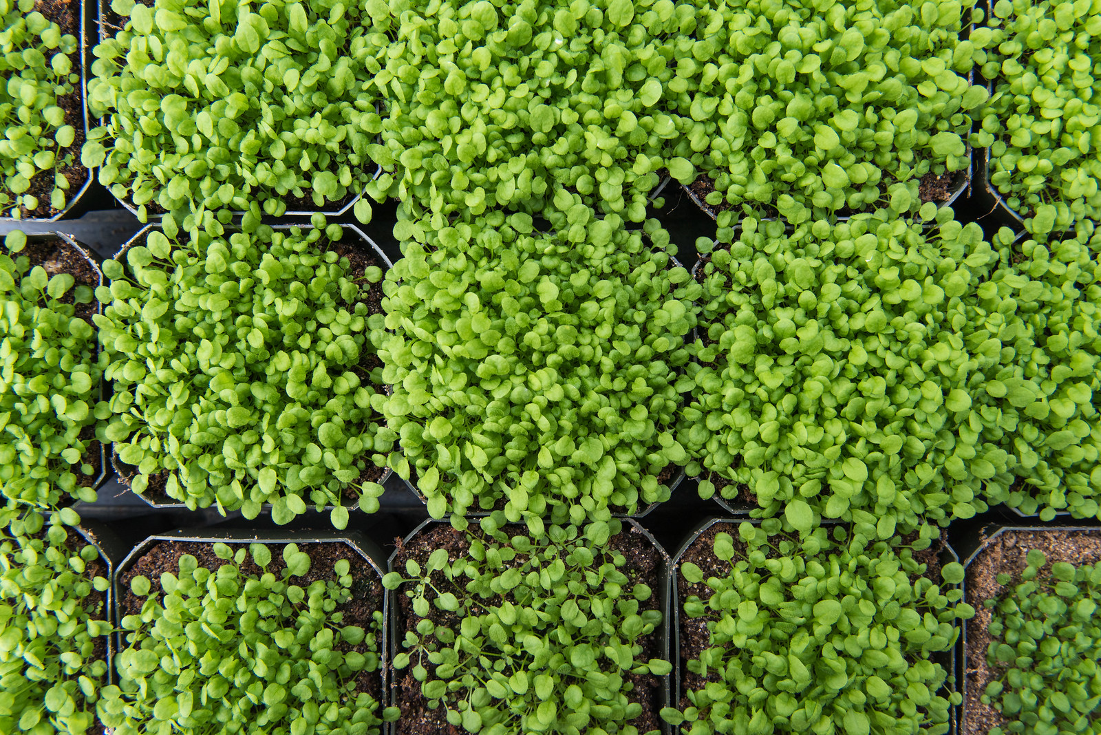
This file photo shows at least one other ecotype of Arabidopsis thaliana in a greenhouse in Bond LSC. Even small variations in the species’s genome can create the large number of observable varieties, sometimes with distinct sizes and shapes, other times with genetic differences that can only be observed on the microscopic level. | Roger Meissen, Bond LSC
Inter-departmental MU team aims to improve enzyme use and recovery for spectrum of industrial, medical and military applications
By Phillip Sitter | Bond LSC
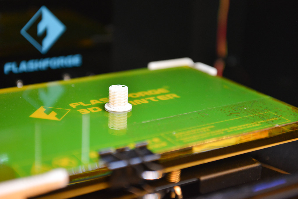
A mostly-finished cylindrical bio-reactor site sits in a 3D printer after the printing has stopped. With a 3D printer in-house, Chung-Ho Lin said that the inter-departmental team he is part of can generate four or five different prototypes a day to test in their bio-reactor model, instead of having to order from different fabrication companies. A basic printer like this used to cost $8,000, but within the last year prices dropped to only about $1,000. Lin is a research assistant professor at MU’s Center for Agroforestry, and the team and project are coordinated by Hsinyeh Hsieh, a veterinary pathobiology research scientist in George Stewart’s Bond LSC lab, where the team also does most of its work. | Phillip Sitter, Bond LSC
As Sagar Gupta watched a 3-D printer on a lab countertop construct a jumbo pencil eraser-sized, white plastic cylinder of what looked like a shell holding inter-woven letter Xs, he remarked that the only limitation to what you can print is the size of the printer.
“The timing is perfect, otherwise we wouldn’t have been able to afford it,” Chung-Ho Lin said of the availability of cheaper 3-D printers within the past couple years.
The two men were acutely aware, as the printer continued its methodical manufacture, that they may be architects of the first steps in a bio-chemical revolution.
It’s a revolution that could be hugely profitable financially and may help to save lives on battlefields, clean up some kinds of pollution and enable humans to venture further into space for a cheaper cost, among other things.
To understand how this cross-disciplinary team working in George Stewart’s lab at the Bond Life Sciences Center got there, we have to back up a little bit.
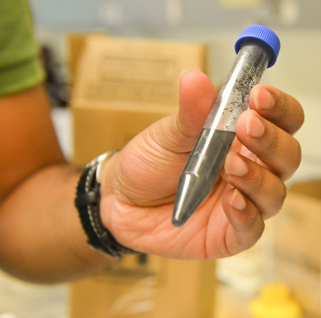
Sagar Gupta holds a vial of carbon solution. Most of the team’s prototype designs for bio-reaction sites are made of carbon, and some are even bio-degradable. | Phillip Sitter, Bond LSC
From a bottleneck to a bioreactor
Their work began three years ago with a project to develop technology to reduce the cost of converting cellulose into glucose for biofuels — essentially the process by which raw plant fiber from wood or leaves is turned into a sugar that can be more efficiently burned to produce energy.
“That has been the bottleneck for the biofuel industry,” said Lin.
The team — consisting of Lin, a research assistant professor at MU’s Center for Agroforestry; Stewart, Hsinyeh Hsieh and several undergraduate and recently graduated students including Gupta — already developed E. coli bacteria that can mass-produce engineered enzymes to convert cellulose into glucose.
These enzymes speed up the reactions and reduce the cost because they have linkers attached to them — protein hooks that let them be recovered after a single use as catalysts in biological reactions, rather than having to throw them out. Hsieh said she developed this with Stewart’s input, and the assistance of a recently graduated student, Che-Min Su.
However, the team needed a platform for the linkers to hook onto — something they could continuously use to reel in their catch.
The answer in their search for the correct platform arrived when affordable 3-D printing technology came onto the market. With their own 3-D printer in-house, they custom-designed different platforms for their experiments and completely bypassed having to shop around with different fabrication companies.
All of the ingredients were there with that plastic cylinder Gupta and Lin watched print. The team now had a cheap way to mass produce and repeatedly recover enzymes. With this capability, they could produce a more efficient bioreactor — a controlled, isolated system in which desired reactions can take place with higher outputs of quantity and quality of a desired product.
It’s much like the more familiar concept of a nuclear reactor, which controls and isolates a nuclear chain reaction to harvest the most energy possible. The catalysts in that reaction are radioactive particles that give off heat as they decay. In a physical reaction, the heat released boils liquid water into gaseous steam, and the steam turns a turbine generator that makes electricity.
But in the team’s bioreactors, catalysts are enzymes that chemically react with cellulose and transform it into glucose instead of electricity. The glucose can be fermented further into butanol that can ultimately be used for liquid fuels to power vehicles.
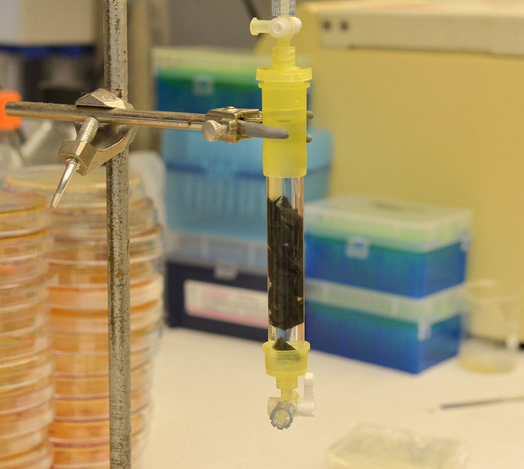
A bio-reactor column stands packed with carbon fibers submerged in enzymes. If the column were hooked up a continous flow system, substrate would be pumped through it to spur bio-chemical reactions on the surface of the carbon fibers, or whatever other type of site is packed inside. | Phillip Sitter, Bond LSC
Money and blood
While only at a bench-top, proof-of-concept scale, the team’s first bioreactor has lasted more than four months. With prospects to increase its size, they “could be saving at least $10 to $12 million per year on an industrial scale,” said Gupta. Gupta graduated in May from MU with an MBA, and now works for Lin.
That estimate is just for one individual bioreactor. Begin to multiply it, and the cost-savings add up very quick.
“Nowadays, probably a majority of pharmaceutical companies have already switched their manufacturing process into the enzymatic process. One thing nice about the enzymatic process is that it can eliminate [the need for] a lot of hazardous chemicals. They also tend to have a better yield,” Lin explained.
Lin added that there is a bonus of complexity within this kind of 3-D platform system. Individual enzymes have different linkers, and this allows for multiple enzymes to catalyze reactions and be recovered on the platform at the same time. This is especially cost-saving because the conversion of cellulose into glucose requires three different kinds of enzymes.
“Because of this high specificity, we don’t need any enzymatic purification process,” he said.
Once the enzymes hooked to a platform start to naturally decay, the team can simply remove the decayed enzymes by a hot water bath and soak it in a new batch of enzymes, just like swapping out an empty printer cartridge for a full one with fresh ink.
While their primary focus is on biofuels, they are very aware that more efficient and cheaper bioreactors could have huge implications for a broad spectrum of industries.
One use they are developing could effectively transform one blood type into another using enzymes.
“This is not a completely new technology, but in the past, I would say back in the 90’s, some people tried some clinical trials and they ran into a problem, because a lot of times after the conversion, [loose] enzymes would get into the recipients’ bloodstream and cause an auto-immune reaction,” said Lin.
However, by being able to immobilize enzymes with their linkers on this 3-D device, they should be able to get around that problem, he said.
“I think there’s great potential for the soldier on the battlefield,” Lin cited as an application for the technology. A field doctor or medic wouldn’t have to worry about waiting on a certain type of blood for a transfusion, because they could convert another batch of blood into a universal-donor type.
Another team member, Hien Huynh explained that the more enzyme you add in ratio to the substrate, in this instance blood, the faster the conversion process will go — “maybe just 30 minutes.”
Hsieh wrote that “Blood type conversion would be the ultimate challenge for our bioreactor, because it has so many clinical aspects to be concerned [about] and conquered. It is a challenge but our [multi-disciplinary] team is willing to take it on and make it work.”
Lin said that the team has already submitted a letter of intent to the U.S. Department of Defense, “hopefully to secure some support for the blood-conversion application.”
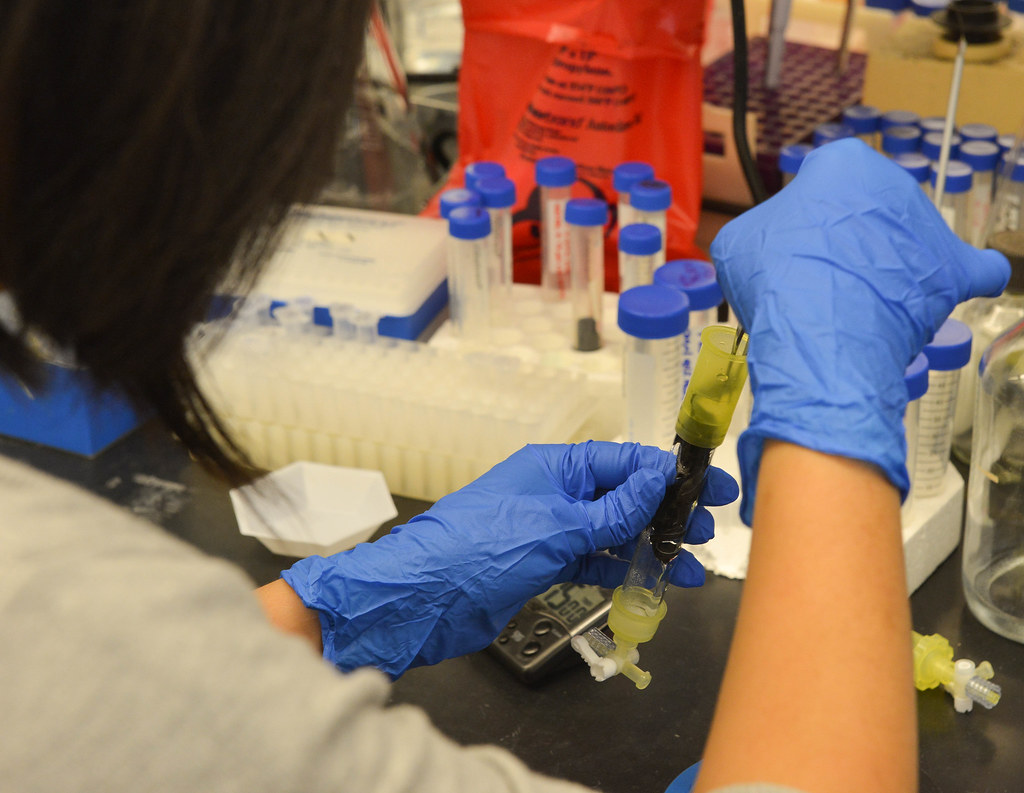
Hien Huynh packs a bio-reactor column with carbon fiber bio-reactor sites that look like feathers. The sites are coated in enzyme before being packed into the column. | Phillip Sitter, Bond LSC
Enzymes in action
There are other potentially massive implications for the battlefields of the future.
“You can immobilize anti-microbial, anti-fungal and anti-inflammatory enzymes on a surface to use as a wound-healing patch,” Lin said, noting that such a patch could be used on the battlefield, as well as for cosmetic surgery recovery.
But the applications don’t stop there. Other uses could use enzymes to clean up TNT residues leeching out of unexploded ordinance like cluster bomblets, mortars, rocket-propelled grenades and landmines buried in the ground before the toxic residues contaminate groundwater.
Even within the confines of biofuels, there’s a strong military market. By 2020, the Navy wants 50 percent of its total energy consumption to come from alternative sources as opposed to petroleum-based fuels — part of a broader strategy to go green. The U.S. military in the near future wants to reduce the cost of its energy consumption and secure a stable domestic supply of energy.
According to the U.S. Government Accountability Office, from fiscal years 2007 to 2014, the Department of Defense bought 32 billion gallons of petroleum-based fuels at a cost of $107.2 billion.
Away from the military sphere, Lin detailed other uses for cheaper, higher quality enzymes. It could purify and recycle urine into clean water on space flights on for astronauts or convert waste into energy with an ammonia fuel cell that’s already available.
Mass-produced enzymes can be used for water treatment on earth, too. Pollutants like dioxin and herbicides like atrazine that contaminate soil can be bio-remediated in the same way that TNT residues can be cleaned up.
The food industry already uses enzymes as flavor removers to remove strong tastes from products like beer.
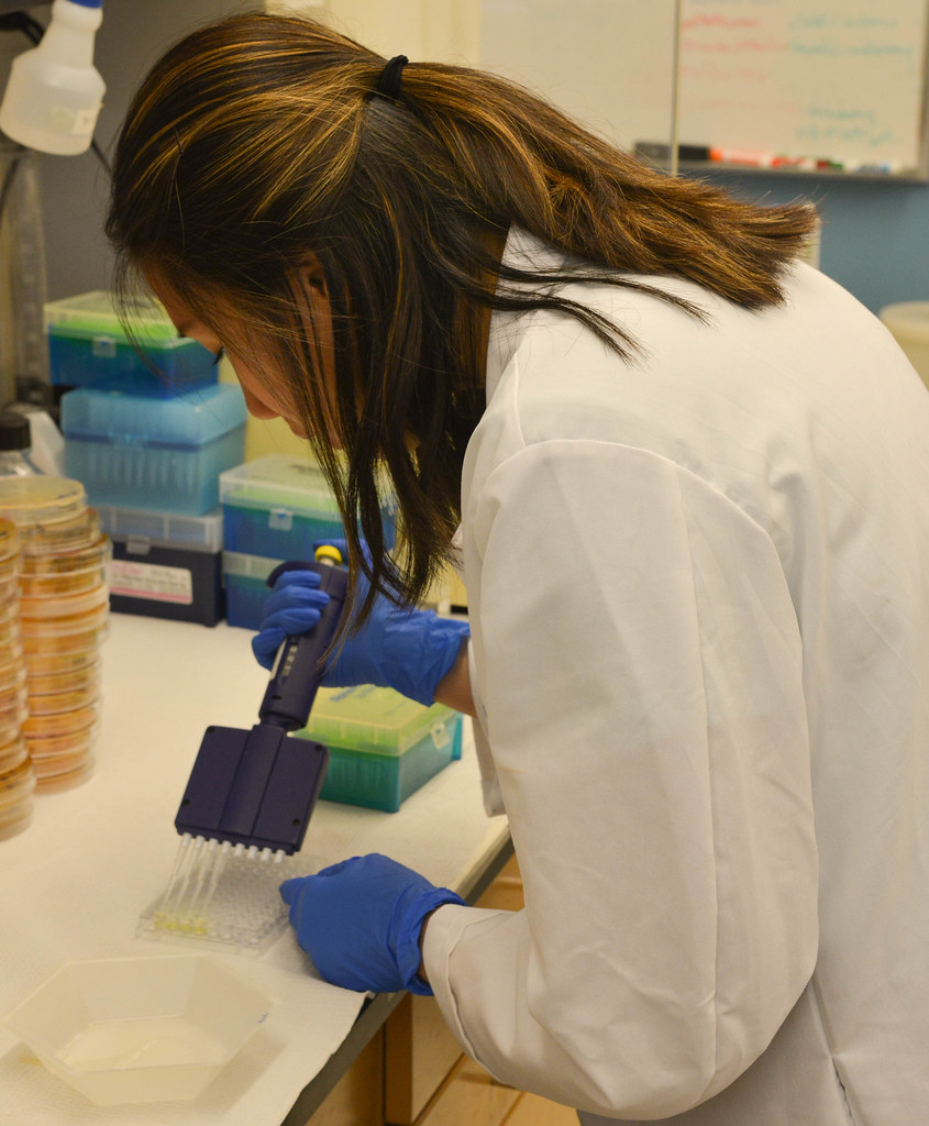
Minh Ma simulates the end result of a successful operation of the bio-reactor. She extracts and separates samples of real glucose product produced by the reactions in the column. A stronger yellow color in the solution indicates a higher concentration of glucose. | Phillip Sitter, Bond LSC
A bright bioreactor future
To call the team’s work revolutionary might be a bit premature.
There is a whole process ahead of them, including patent filing and university reviews, before the team can approach investors with the assurance their discoveries are legally protected. And, future investors will ultimately help determine how the technology is used.
But, Lin and the others might just have found themselves in the right place at the right time to make major breakthroughs, and that’s not all due to just advancements in technology.
“We have identified new directions and found a new niche to be competitive. I think the most important resource we have is people, and their brains,” Lin said.
Hsieh wrote that “To assemble a successful team is to put the right talent in the proper position and to inspire them to challenge themselves. I was lucky to come across so many young, talented students who are eager to learn and work hard for their bright future on MU’s campus.”
Hsinyeh Hsieh, a veterinary pathobiology research scientist in George Stewart’s Bond LSC lab, coordinates this project. Hsieh is an expert in gene fusion, enzyme production and characterization and enzymatic blood type conversion. Stewart is a medical bacteriologist, McKee Professor of Microbial Pathogenesis and chair of Veterinary Pathobiology at MU.
Lin works with Stewart and Hsieh to develop concepts, design prototypes and assemble the rest of the team — students and recent graduates — that optimizes the enzymatic reactions and the physical and chemical aspects of their bioreactor system. Minh Ma is a junior studying bio-chemistry. Mason Schellenberg studies bio-engineering, will be a senior and worked to find the most efficient platform design that the team’s 3-D printer could produce. Hien Huynh is a recent graduate who works on immobilizing enzymes. In addition to his MBA, Gupta also has a background that includes nano-technology, molecular engineering and financing. He concentrates on the feasibility and market potential of the team’s work.
Tiger Energy Solutions, LLC is the team’s industry partner — a spinoff startup from the team’s research project . Their focus in the development of a cheaper and higher quality method of converting cellulose into glucose for biofuels is to produce aviation biofuel. Tiger Energy serves as the interface between the team and industry while the team’s work is scaled-up for commercialization.
From left to right: Minh Ma, Hien Huynh and Sagar Gupta are three of the teams members, standing here in George Stewart’s lab. Ma is junior studying bio-chemistry. Huynh is a recent graduate who works on enzyme immobilization. Gupta is also a recent graduate — he obtained his MBA in May — and he focuses on the financial feasibility and market potential of the team’s work. | Phillip Sitter, Bond LSC
NASA, NIH-funded work seeks to understand bio-chemical mechanisms of life on Earth, and among the stars
Donald Burke-Agüero stands in his office in Bond LSC, holding a model of an RNA protein structure. Burke-Agüero studies the bio-chemical functions of RNA, and how those functions might be able to be artificially designed or replicated. | Phillip Sitter, Bond LSC
By Phillip Sitter | Bond LSC
Any child obsessed with Legos knows the fun of creation bound only by imagination and the size or variety of the blocks within their pile.
For some scientists, that spirit extends into adulthood, but instead of plastic parts they think about arranging blocks of nucleic acids.
Scientists may not be able to create dinosaurs, dragons or mythical sea creatures the way kids with Legos can. Through the manipulation of nucleic acid building blocks though, they may be better able to understand how the processes of life on Earth work, as well as out among the stars.
“I have a lot of fun asking what is possible,” said Donald Burke, a Bond Life Sciences Center investigator who spends his time researching the building blocks of life.
Burke said he has been interested in the origins of life for 40 years, and he has been associated with NASA for about 20 years.
NASA’s interest in understanding the origins of life is pretty straightforward. It wants to know what clues to look for on other worlds to figure out if those planets also support life.
Many of Burke’s previous discoveries at Bond LSC are funded by NASA’s exobiology and evolutionary biology program.
“No, I have not thought of an excuse to fly anything up there. I’ve tried to think ‘which of my experiments would make sense to do in a micro-gravity or zero gravity environment?’” he explained of the prospect of sending some of his work into orbit, with a wry smile.
But, there’s even more to understanding the building blocks of life than looking for bio-chemical signatures out among the stars. Knowing how these parts are put together allows scientists like Burke to understand the origins and processes of Earth’s biology, and, conceivably create chemical and biological processes or even organisms not found in nature in the near future.
A quadrillion arrangements of blocks, one arrangement at a time
“Many of the molecules of life are built from strings of amino acids, or nucleotides or other building blocks,” Burke explained. He also noted that these buildings blocks are not just strings, but fold up into three dimensional shapes.
RNA, or ribonucleic acid, stands out as an essential building blocks in the bio-chemical processes of life.
Put simply, RNA is a kind of molecular structure of nucleic acids similar to DNA (deoxyribonucleic acid) that comes in many combinations. These combinations are at the core of every cell, and play a role in coding, decoding, regulating and expressing the basic operating instructions for each cell — its genes.
The molecules we’re talking about are almost unimaginably small. In one test tube, Burke said there can be one quadrillion of them — that’s a one with 15 zeroes after it. Put another way, that’s roughly equivalent to the estimated number of ants that live on Earth.
Burke’s work focuses on the end goal of being able to artificially create original RNA combinations. In what’s known as experimental evolution, “the population of molecules in the tube is evolving as a result of us imposing experimental constraints upon it.”
This artificial synthesis of RNA molecules looks to create random sequences or variations on natural RNA to create new ones non-existent in nature. A second route aims to selectively choose molecules with certain properties, and use them to build altogether new combinations.
“Their string-like properties allow us to copy them, and make more copies, and make more copies, and make more copies. Their shape-like properties allow us to observe the bio-chemical behaviors they may have,” Burke explained how he and other scientists interact with RNA’s structure in the lab.
“I don’t think we know what those limitations are yet,” he said of the capabilities of RNA.
The motivation for wanting to be able to intentionally design RNA molecules is so that it “can do the things we want it to do under the conditions where we want it to do those things,” he explained of the process of the process of selecting RNA sequences for specific properties.
“I want the ones that will bind a tumor cell. I want the ones that will bind a viral protein. I want the ones that will catalyze useful chemical reactions.”
RNA’s path to the future following in biology’s footsteps
The National Institutes of Health and other organizations recognize that engineered forms of RNA have the potential to fight diseases, and they have funded Burke’s work.
He has studied RNA that instructs human cells on how to defend themselves from HIV and is now looking at other RNA that interferes with the proteins of the Ebola virus.
The expectation is that such therapeutics would work in conjunction with other treatments. In the future, they could be expanded to help fight other viruses, cancers and other diseases.
RNA could also be used to start, or catalyze, chemical reactions. As Burke explained, catalysts remove barriers to chemical reactions — “they don’t make things happen that wouldn’t otherwise happen, but they speed up the process.”
Synthetic RNA could be used to accelerate removal of toxins from soil or to get the bacteria in our guts to recognize cancerous tumor cells and kick-start an immune response.
But, the future of RNA research may soon reveal a few different Holy Grail moments on its horizon.
One such Holy Grail that Burke said will definitely happen will be observations consistent with the presence of life on other worlds, based on evidence like an atmosphere having certain chemical compositions.
Another likelihood could involve construction of a self-replicating, fully-artificial organism, either created from scratch or reverse-engineered from other organisms.
For those of you already anticipating the plot of a low-budget sci-fi thriller, Burke offered to assuage your fears.
“The notion of it escaping out in the world and taking over Los Angeles is [only] good 1950’s B-movie” material, because the conditions under which this artificial organism would survive would probably be difficult to maintain even in the controlled environment of lab, he said.
Instead of B-movie science, Burke explained that “really, I’m thinking about what kinds of chemistries we want to see take place, and then building the enzymes that would make it possible.”
“Biology has had a few billion years to work on this, but we’re just starting to figure it out.”
Donald Burke-Agüero is a professor of molecular microbiology and immunology and joint professor of biochemistry and biological engineering.
Turtles could help determine how exposure to harmful chemicals during development affects male and female brains Jeff Sossamon | MU News
Bisphenol A (BPA) is a chemical used in many consumer products including water bottles, metal food storage products and certain resins. Often, aquatic environments such as rivers and streams become reservoirs for BPA, affecting turtle habitats. Last year, a team of researchers led by the University of Missouri determined that BPA can disrupt sexual function in painted turtles, causing males to develop female sex organs. Now, the team has shown that BPA also can induce behavioral changes in turtles, reprogramming male turtle brains to show behavior common in females. Researchers worry this could lead to population declines in painted turtles.
“Previously, our research team found that BPA and ethinyl estradiol (EE2), a hormone found in birth control pills, could ‘sex-reverse’ turtles from males to females,” said Cheryl Rosenfeld, an associate professor of biomedical sciences in the MU College of Veterinary Medicine and an investigator in the Bond Life Sciences Center. “Painted turtles and other reptiles lack sex chromosomes. The gender of painted turtles and other reptiles is determined by the incubation temperature of the egg during development. Studies have shown that exposure to endocrine-disrupting chemicals (EDCs), such as BPA, can override incubation temperature and switch the sex of males to females. In our latest study, we found that BPA also affects how the male brain is ‘wired,’ potentially inducing males to show female type behavioral patterns.”
Researchers applied a liquid form of BPA and ethinyl estradiol to painted turtle eggs and incubated the eggs at a temperature that typically results in males. Five months after hatching, turtles were tested with a spatial navigation test that included four food containers, only one of which was baited with food. Each turtle was randomly assigned one food container that did not change over the trial period.
Researchers predicted that male turtles exposed to BPA and EE2 would exhibit improved navigational ability — similar to behaviors observed in female turtles. Results showed that developmental exposure to BPA and EE2 improved spatial navigational learning and memory in males, as evidenced by increased number of times spent in the correct target zone and greater likelihood of solving the maze compared to control turtles, who were male based on the lower incubation temperature.
“Previous studies have found that female turtles are much more adept at spatial navigation — think of female sea turtles that return many years later to the same beaches where they hatched to lay their own eggs,” Rosenfeld said. “We found that developmental exposure to BPA essentially overrides the brain development of male turtles as indicated by the enhanced navigational ability of the turtles we studied. While improved spatial navigation might be considered a good thing, it also may suggest that when they reach adulthood male turtles will not exhibit courtship behaviors needed to attract a mate and reproduce, which could result in dramatic population declines.”
Rosenfeld notes that this is the first study to show that these harmful chemicals not only reverse the physical sex-characteristics but also affect the brain in a turtle species. Turtles are known as an “indicator species” because they can be used as a barometer for the health of the entire ecosystem. By understanding the possible effects EDCs have on turtles, researchers might be able to understand the possible effects the chemicals have on other wildlife species and humans, Rosenfeld said.
“Effects of developmental exposure to bisphenol A and ethinyl estradiol on spatical navigational learning and memory in painted turtles (Chrysemys picta),” recently was published in the journal, Hormones and Behavior. Lindsey Manshack, a student at the time in Rosenfeld’s lab in MU’s Bond Life Sciences Center authored the study. Dawn Holliday, adjunct assistant professor of pathology and anatomical sciences in the MU School of Medicine and assistant professor of biology at Westminster College in Fulton, Mo., and Sharon Deem, director of the Saint Louis Zoo Institute for Conservation Medicine, contributed to the study. Funding was provided by Mizzou Advantage, the Office of Research and the Bond Life Sciences Center at the University of Missouri. The content is solely the responsibility of the authors and does not necessarily represent the official views of the funding agencies.
Grand opening highlights specialty of large-scale metabolite profiling
Dr. Zhentian Lei , assistant director and assistant research professor of the MU Metabolomics Center, provides an overview of an ultra high-pressure liquid chromatograph coupled to mass spectrometry for the large-scale profiling of metabolites at the University of Missouri Metabolomics Center open house on Aug. 12. | photo by Zivile Raskauskaite, Bond LSC
By Phillip Sitter | Bond LSC
You might think you’ve entered the inside of a pinball machine for a moment when you enter lab 243 at the Bond Life Sciences Center.
But the wires and tubes strung around the room, connected to large instruments that produce sounds of whirring fans, humming motors and hissing pumps, are just part of the University of Missouri’s newest core facility, the MU Metabolomics Center.
At its grand opening and open house Friday, August 12, there was even a counter-top half-pipe with metal ball bearings to shoot down it as a demonstration of time of flight mass spectrometry.
This new center will serve as home of high-tech chemical analysis services that scientists in Bond LSC, across campus and the country can use to better understand the organisms they work with on a molecular level.
Lloyd Sumner, director of the MU Metabolomics Center, and Assistant Professor Ruthie Angelovici discuss the use of NMR for metabolite identification during the University of Missouri Metabolomics Center open house on Aug. 12. | photo by Zivile Raskauskaite, Bond LSC
“We have a series of experiments that allow us to profile hundreds to thousands of different metabolites, and that gives people a large-scale, high resolution biochemical traits for whatever they’re looking at, whether it be plants, microbes or animals,” explained Lloyd Sumner, director of the center. “That is useful in understanding what is happening in response to stresses, disease, drug treatment or pest/pathogen interactions that occur in nature.”
Metabolites are the building blocks and energy sources that fuel your metabolism. In your body, what you eat and drink is processed and yields small molecules that are ready to become raw chemical material for construction processes and energy to fuel these processes, like energy stored in the form of fats and lipids, amino acids for the construction of proteins and enzymes. Metabolite are essentially the raw materials.
In order to be studied, complex metabolite mixtures are separated and observed as individual, uniquely identifiable molecules.
This separation can be accomplished in a couple different ways.
“We have instruments that couple chromatography with mass spectrometry. We use that for comparative profiling. Some of the instruments utilize gas chromatography, some of the instruments use liquid chromatography. Chromatography is the technology used to separate these complex mixtures into its individual components. Once we have the mixture’s components separated, we weigh them and that gives us an idea of their identification,” Sumner explained.
These internal components of a triple quadrupole mass spectrometry are used for explaining how the instrument helps identify the metabolites within a sample during the University of Missouri Metabolomics Center open house on Aug. 12. | photo by Zivile Raskauskaite, Bond LSC
Mass spectrometry works by bombarding molecules with electrons. This bombardment process generates charged molecules that can also fragment into smaller, electrically-charged pieces. These charged pieces can then be “weighed,” or separated, according to their mass-to-charge ratio and identified.
“Something that we find a lot of the time is that we see metabolic differences, but we can’t always identify all of the metabolites associated with those differences. In those cases, we also use the gold standard for chemical identification of unknown molecules,” Sumner said of the nuclear magnetic resonance (NMR) spectrometer in the corner of the lab.
A person points at a 600 MHz Nuclear Magnetic Resonance Spectrometer used for metabolite identification during the University of Missouri Metabolomics Center open house on Aug. 12. | photo by Zivile Raskauskaite, Bond LSC
Placards warn people that when NMR produces a magnetic field 235,000 times stronger than the Earth’s — by comparison, a typical refrigerator magnet’s field is about 83 times as strong as the Earth’s.
Sumner explained that most people at Bond LSC won’t use the equipment directly themselves. The center’s Assistant Director Dr. Zhentian Lei and other staff will perform most analyses and training users to prepare, process and understand their data.
Sumner said “we train our core users to do their own sample preparation, data processing and data interpretation. Most of the equipment we have in here [cost] hundreds of thousands of dollars, and so we actually have staff that will do the data acquisition, and we try to make it more cost-effective for users by training them to prep their own samples and process their own data.”
The training workshop in metabolomics will be August 15 through 19. The training Monday through Thursday will be hands-on, and Friday will be a symposium day highlighting current metabolomics research. We will likely offer another training workshop in the Spring of 2017, and then annually thereafter.
For more information on using the MU Metabolomics Core or future training, email Director Lloyd Sumner at sumnerlw@missouri.edu or Assistant DirectorZhentian Lei at leiz@missouri.edu.
Bond LSC scientist internationally recognized for work on salivary glands and autoimmune disorders
By Phillip Sitter | Bond LSC
You might not think too highly of spit, but you would quickly regret not having any.
People with Sjögren’s syndrome suffer chronic dry mouth and eyes from an overzealous immune system that attacks salivary and tear ducts, causing serious health issues.
Gary Weisman’s research might hold the key to understanding and managing this immune response, leading to effective treatment or even prevention of this ailment.
For this, the International Association of Dental Research, or IADR, awarded him the 2016 Distinguished Scientist Award for Salivary Research. Weisman accepted the award in June at the opening ceremonies of the IADR conference in Seoul, Republic of Korea.
Gary Weisman stands in his lab in Bond LSC where he studies the cellular mechanisms of auto-immune disease, specifically how the release of ATP from damaged cells signals receptors that trigger an immune response. | Phillip Sitter, Bond LSC
“We want the good, but not the bad,” said Weisman, a Bond Life Sciences Center investigator, of what we ideally want from our immune system’s functions.
Mice with un-checked autoimmune disease of their salivary glands have their glands destroyed. The disease can spread to other secretory organs next. An over-reactive immune system on a civil war-path can extend its damage to cause pancreatic failure and death.
The destruction wrought by Sjögren’s syndrome is self-inflicted, caused by an overreaction of our bodies’ defenses against infection and injury. This is what an autoimmune disease is.
But, our bodies’ immune cellular response team is complicated. Weisman said dozens of different cell types have been isolated and identified as part of the immune system, and he likens the immune system to fire, police and construction services in human society all working together.
While firefighters are meant to prevent further damage from an inferno, sometimes our bodies’ first responders start doing the equivalent of using dynamite to stop the spread of a fire.
In chronic inflammation, that autoimmune response can mean a burning, throbbing, constant pain. The key to a healthy immune response is balance. The balance has to be between containment and repair of damage caused by infection or injury and damage caused by chronic inflammation if that emergency response continues unabated.
Weisman has spent almost 30 years studying how to prevent our bodies’ immune system from over-reacting to threats and causing further harm.
Earlier in his career, Weisman studied how extracellular ATP plays a critical role in immune responses, and how too much of it can cause the over-reaction that leads to tissue destruction in autoimmune diseases. ATP, or adenosine 5’-triphosphate, is the main molecule used for energy in cellular activities inside cells. Weisman was one of the first scientists to study how damaged cells release ATP as a distress signal.
The released ATP signals receptors that “send out the alarm to the fire station” — the body’s immune cells, he said.
Once he understood this, Weisman began to manipulate the actions of released ATP to see how that would affect an immune response.
Mice with salivary gland autoimmune disease got healthy when the released ATP was prevented from activating their receptors on the surface of cells. Preventing the ATP receptors from being activated slowed down and even stopped the advance of autimmune disease.
Conversely, if you prevent the activation of the ATP receptors in lab mice with Alzheimer’s disease they die much more rapidly from the disease, Weisman said, suggesting that activation of immune cells by ATP is beneficial in slowing the progression of this disease.
Alzheimer’s disease and autoimmune diseases such as Sjögren’s syndrome are only some of the inflammatory diseases that Weisman has studied. With each of these diseases, the role of ATP receptors has to be investigated individually, suggesting that Weisman’s work may extend beyond salivary glands and the brain to other parts of the body.
“Our [ATP] receptor is also involved in heart disease,” Weisman said, and he added that other diseases like cystic fibrosis, cancer, lupus and arthritis have inflammatory components, too.
For now, we all fight a losing battle when it comes to our bodies’ management of the immune system. As we and our immune system age, it has the potential to destroy more than it protects and “eventually you could slip over to the dark side and die,” Weisman said.
In the meantime, Weisman said that a better understanding of the immune system could lead to more effective, targeted treatments of chronic inflammation and other autoimmune disorders. This could provide a new approach to control undesirable activation of the immune system beyond the use of with anti-histamines, anti-cytokines and ibuprofen.
Weisman is a Curator’s Distinguished Professor of Biochemistry. He began his salivary gland research at MU 27 years ago with Professor John Turner, before Turner’s retirement. Since then, his research has been continuously funded by the National Institutes of Health, where one of his recent grants was well scored and will likely be extended for another five years.
Lorson lab publishes research on a new therapeutic path to help treat spinal muscular atrophy
Erkan Osman shows iImages of neuro-muscular junctions. Osman, a post-doctoral fellow in Chris Lorson’s lab, co-authored research in the journal Molecular Therapy that details work in binding a synthetic nucleic acid to a normally useless motor neuron backup gene to help treat spinal muscular atrophy. | photo by Phillip Sitter, Bond LSC
By Phillip Sitter | Bond LSC
Imagine you are forced to jump out of an airplane.
Luckily, you find a parachute that even has a backup chute. You leap out of the plane and free-fall.
You pull the cord to open your parachute, but it doesn’t open. Don’t panic, though, you have a backup. But, you pull that cord and nothing happens. Now you face the reality of a death as firm and un-yielding as the ground rushing into your view.
This air disaster mirrors the mechanism and mortal threat posed for people born with the genetic problem that causes spinal muscular atrophy (SMA).
Chris Lorson’s lab at the Bond Life Sciences Center would like to change that situation by making an effective genetic backup to the defective gene that results in SMA. The journal Molecular Therapy, a publication of Nature, recently accepted their findings for publication.
The defect occurs in a specific gene called Survival Motor Neuron (SMN). If the SMN gene is defective because of mutation, this causes a deficiency of the SMN protein it is supposed to produce. Without this protein, the neurons that control muscle movement malfunction. Signals cease to stimulate muscles.
Muscles that are not stimulated atrophy, grow weak and waste away. At first this happens with the skeletal muscles, which leads to loss of motor function for simple activities like walking and swallowing. If it happens with the muscles that control breathing, you die.
News of the disease often presents a devastating prognosis. Infants have it worst; babies diagnosed with SMA only have a life expectancy of two to five years from birth.
Fortunately, our bodies have a sort of backup for the SMN gene, another one called SMN-2. But, like a useless backup parachute for an unlucky skydiver, SMN-2 isn’t actually very good at producing proteins of the quality needed to stave off SMA. It might just be a vestigial trait on its way down the evolutionary drain — it doesn’t even exist in the closest primate relatives of humans.
Discoveries in the Lorson lab look to make the SMN-2 gene an effective backup, and their recent publications indicate that this may be a viable possibility for future SMA treatments.
Christian Lorson studies the genes that cause SMA when they fail to adequately function. His team’s on a backup gene that greatly extended life expectancy in mouse studies. | photo by Phillip Sitter, Bond LSC
“What we’ve been working on in the lab is a potential therapeutic, and what it does, it’s a large small molecule that is called an antisense oligonucleotide, or ASO,” Lorson said. “And this is something that is essentially a synthetic piece of nucleic acid that is able to go in and bind to a specific sequence within a gene.”
Once bound to SMN-2, the ASO is designed to alter mRNA splicing, “essentially, the editing of a gene,” Lorson said. Speaking in terms akin to products leaving a factory, Lorson said that the attached ASO makes SMN-2 produce good quality proteins, the ones that it wasn’t able to produce before.
In other words, suddenly the backup protein-factory that was making poor-quality products is now pumping out top-of-the-line stuff that will work.
Previous research identified a strong ASO contender to experiment with, and Lorson said current research is about optimizing an ASO to extend survival times in mice with SMA — from just 13 days to five months after only one injection at birth.
Lorson stressed that his lab’s achievement doesn’t promise a fast cure for SMA. He said it is unlikely a single compound will address the full gambit of effects that people with SMA suffer, especially given that people can be identified as having SMA at any time from birth through later in life — often late onset SMA tends to be less severe than diagnosis as an infant.
There’s not yet any single compound treatment for SMA that has been approved by the Food and Drug Administration, Lorson said, so he cautions against getting hopes up of for a revolutionary treatment for SMA coming onto the market soon — “Near future but not tomorrow.”
He acknowledged, though, that “from a research perspective, things seem to be moving at lightning speed, but if you are a patient or a family member, things can never go fast enough, so I think there’s a realized sense of urgency, whether or not it’s for patients who don’t have the disease yet, are not born, or for patients who have had the disease for a decade and are wondering when their opportunity would come.”
Lorson’s work is funded in part by Cure SMA, FightSMA and the Gwendolyn Strong foundations. Erkan Osman, a post-doctoral fellow in Lorson’s lab and the first author on the most recent paper, won the emerging investigator award from FightSMA and Gwendolyn Strong in 2015.
Scientists use placental cells in lab to study virus
Megan Sheridan, an MU grad student, removes the base solution from a demonstrated sample of stem cells that will be grown into placental cells for study of Zika virus. Within four days of exposure to the correct hormones, the stem cells express genes of placental cells, and within another day start producing placental hormones. The cells are infected with Zika at day four to ensure maximum measurable interaction, as the stem cells naturally die in culture after about ten days. | photo by Phillip Sitter, Bond LSC
By Phillip Sitter | MU Bond Life Sciences Center
Scientists believe they have a better way to study how Zika virus can spread from a pregnant mother to her fetus — and their technique doesn’t even involve observations of babies in the womb or post-natal examinations.
“As soon as we heard about Zika, everybody’s light bulbs turned on,” said Megan Sheridan, a graduate student at the University of Missouri Bond Life Sciences Center.
Sheridan works in the lab of Toshihiko Ezashi at Bond LSC, and she, in turn, is part of a cross-campus team researching Zika with R. Michael Roberts, Alexander Franz, Danny Schust and Ezashi.
Roberts’ lab studies pluripotent stem cells — progenitor cells which can develop into any other type of cell in the body.
“We use the proper signals to drive stem cells to become like placental cells,” Sheridan explained. With this capability to stimulate stem cells with growth hormones and inhibitors at opportune moments, Roberts’ researchers realized they could create enough placental cells to create an environment similar to that of a womb in very early stages of pregnancy.
Megan Sheridan sits in front of a demonstration of her work with pluripotent stem cells. Sheridan is a graduate student who works in Toshihiko Ezashi’s lab, where she produces cells with placental characteristics from the stem cells in order to study placenta interaction with Zika virus. | photo by Phillip Sitter, Bond LSC
This is something which Sheridan thinks hasn’t been done before in regards to studying placental interaction with Zika. Their technique could give a look into the first trimester, when epidemiological studies say a fetus is most susceptible to infection.
Roberts’ lab is trying to understand the placental barrier’s vulnerability to Zika virus in its early stage of pregnancy. During this time, an infection could occur even before the mother is aware she is pregnant.
If the lab uses their technique to understand how Zika virus enters placental cells, then potentially they could also learn how to strengthen the placenta as a barrier to Zika and make it a first line of defense against infection of the fetus in the womb. If developing babies don’t get infected with Zika, then they won’t suffer the consequences of birth defects.
One such defect is microcephaly where a baby is born with a smaller than expected head, which may in turn be a sign that their brain has not fully developed. While infection with Zika virus is rarely fatal or otherwise severe in itself — many people don’t even develop symptoms — birth defects like microcephaly could cause further developmental problems like delays in learning how to speak and walk, intellectual disabilities, difficulty swallowing and problems with hearing and vision, according to global health organizations.
Microcephaly only became a widely documented effect of Zika after a particular strain surged across South and Central America with the infected mosquitoes that carry it, Sheridan explained, but this may be in part because previous Zika infections and outbreaks were themselves poorly documented.
While birth defects caused by Zika have drawn much media attention as the disease has spread northward through our hemisphere from Brazil, studies focusing on infection in the womb have only used placental material that has come to term. This may not be the most accurate way to see how the placenta gets infected in the first place early in pregnancy.
The pathway of Zika virus infection in lab mice isn’t really comparable to human infection, because mice aren’t infected with this virus naturally. Only lab mice that have had their genomes altered to be able to acquire the virus have susceptibility to the infection that can be modeled.
Roberts’ lab is currently working with the African strain of Zika and obtained strains from Southeast Asia and Central America recently. There’s about a 99 percent genetic similarity across strains, Sheridan said.
Zika virus was first discovered in Africa in Uganda in 1947, according to the Centers for Disease Control and Prevention. The first human case was documented in 1952, and subsequent outbreaks also occurred in Southeast Asia and the Pacific Islands. The Pan American Health Organization issued an alert about the confirmed arrival of the virus in Brazil in May 2015.
The lab has completed Zika infections of some of their stem cell-produced placental cells. Sheridan reassured that even though the lab works with live viruses, Zika is not airborne, and none of their work involves mosquitoes.
Roberts’ lab submitted one grant application earlier this year to the National Institutes of Health for funding for their research. While that application was denied, Sheridan said that they have a lot more preliminary data now and are hoping to submit a revised grant soon.
She said that their original work was “highly scored, but the funding level is still low,” meaning that obtaining funds for research into Zika virus is highly competitive nationally.
Legislation to fund more efforts into studying and preventing transmission of Zika virus is caught in congressional gridlock, according to The New York Times and other media outlets.
In the mean time, as the Roberts lab prepares its next grant application submission, Sheridan said of her efforts that she is “working hard to make progress on the project as quickly as possible.”
Please visit the CDC’s dedicated page for more information on Zika virus — including advice for travellers and pregnant women, description of symptoms and treatment, steps you can take to control mosquitoes and prevent other means of transmission of the virus and more background on the history and effects of the disease.