This past weekend not only ushered in Mizzou’s first home game of the season, but the return of Saturday Morning Science. The weekly lecture series connects the Columbia community with MU scientists and their research, from bio-engineering to volcanology to anthropology and linguistics.
Elizabeth G. Loboa, dean of the College of Engineering, kicked off the semester with her talk on tissue engineering in the age of drug-resistant bacteria.
Tissue engineering is about turning cells into tissues and organs, for example, fat-derived stem cells into muscle, bone and cartilage. The tissues take shape on tiny scaffolds that are bio-compatible and biodegradable.
The Loboa lab does this, but they’ve added an extra layer to their research: Loboa’s scaffolds also act as pipelines that deliver wound-healing and anti-bacterial compounds to cells as they grow into tissue. The idea is to reduce infection, inflammation and scarring as the wound heals.
“We’re trying to kill these bacteria while helping these stem cells become the cells we want to create,” Loboa said, about her research at the University of North Carolina-Chapel Hill and North Carolina State University.
Using a process called electrospinning, Loboa’s group makes scaffolds shaped like porous fibers, sheaths, or hollow sheaths. Depending on their structure, these scaffolds act like faucet taps that control the rate and timing at which anti-bacterial compounds are released.
“I look at our fibers as delivery platforms,” Loboa said.
Saturday Morning Science takes place 10:30 a.m. Saturday at the Bond LSC’s Monsanto Auditorium. Coffee and bagels are available preceding the talks. This semester’s schedule is as follows:
9/17: Carolyn Orbann, Assistant Teaching Professor, Department of Health Sciences, “Historical Epidemics, Novel Techniques: Using Historical and Ethnographic Materials to Build Computer Simulation Models”
9/24: Michael Marlo: Associate Professor of English, “Documenting linguistic diversity: a view from the East African Great Lakes”
10/1: Steve Keller, Associate Professor of Chemistry, “The 20 Greatest Hits in Science…In an Hour”
10/8: Manuel Leal, Associate Professor of Biological Sciences, “Are Lizards Smarter Than Those Who Study them?”
10/15: Stephan Kanne: Executive Director and Associate Professor, Thompson Center for Autism & Neurodevelopmental Disorders, “What Do We Look For When We Diagnose Autism?”
10/29: Libby Cowgill, Assistant Professor Anthropology, “Fitness for the Ages: How to Lift Like a Neanderthal?”
11/5: Arianna Soldati, Ph.D. Candidate, Department of Geological Sciences, “Living in a Viscous World: A Volcanologist’s Perspective”
11/12: Frank Schmidt and Gavin Conant, Professor of Biochemistry (Schmidt); Associate Professor of Bioinformatics, Department of Animal Science (Conant), “Networks in Biology and Beyond”
12/3: Elizabeth King, Assistant Professor, Division of Biological Sciences, “What’s the Best Way to Divide up the Pie: The Price of Long Life”
NASA, NIH-funded work seeks to understand bio-chemical mechanisms of life on Earth, and among the stars
Donald Burke-Agüero stands in his office in Bond LSC, holding a model of an RNA protein structure. Burke-Agüero studies the bio-chemical functions of RNA, and how those functions might be able to be artificially designed or replicated. | Phillip Sitter, Bond LSC
By Phillip Sitter | Bond LSC
Any child obsessed with Legos knows the fun of creation bound only by imagination and the size or variety of the blocks within their pile.
For some scientists, that spirit extends into adulthood, but instead of plastic parts they think about arranging blocks of nucleic acids.
Scientists may not be able to create dinosaurs, dragons or mythical sea creatures the way kids with Legos can. Through the manipulation of nucleic acid building blocks though, they may be better able to understand how the processes of life on Earth work, as well as out among the stars.
“I have a lot of fun asking what is possible,” said Donald Burke, a Bond Life Sciences Center investigator who spends his time researching the building blocks of life.
Burke said he has been interested in the origins of life for 40 years, and he has been associated with NASA for about 20 years.
NASA’s interest in understanding the origins of life is pretty straightforward. It wants to know what clues to look for on other worlds to figure out if those planets also support life.
Many of Burke’s previous discoveries at Bond LSC are funded by NASA’s exobiology and evolutionary biology program.
“No, I have not thought of an excuse to fly anything up there. I’ve tried to think ‘which of my experiments would make sense to do in a micro-gravity or zero gravity environment?’” he explained of the prospect of sending some of his work into orbit, with a wry smile.
But, there’s even more to understanding the building blocks of life than looking for bio-chemical signatures out among the stars. Knowing how these parts are put together allows scientists like Burke to understand the origins and processes of Earth’s biology, and, conceivably create chemical and biological processes or even organisms not found in nature in the near future.
A quadrillion arrangements of blocks, one arrangement at a time
“Many of the molecules of life are built from strings of amino acids, or nucleotides or other building blocks,” Burke explained. He also noted that these buildings blocks are not just strings, but fold up into three dimensional shapes.
RNA, or ribonucleic acid, stands out as an essential building blocks in the bio-chemical processes of life.
Put simply, RNA is a kind of molecular structure of nucleic acids similar to DNA (deoxyribonucleic acid) that comes in many combinations. These combinations are at the core of every cell, and play a role in coding, decoding, regulating and expressing the basic operating instructions for each cell — its genes.
The molecules we’re talking about are almost unimaginably small. In one test tube, Burke said there can be one quadrillion of them — that’s a one with 15 zeroes after it. Put another way, that’s roughly equivalent to the estimated number of ants that live on Earth.
Burke’s work focuses on the end goal of being able to artificially create original RNA combinations. In what’s known as experimental evolution, “the population of molecules in the tube is evolving as a result of us imposing experimental constraints upon it.”
This artificial synthesis of RNA molecules looks to create random sequences or variations on natural RNA to create new ones non-existent in nature. A second route aims to selectively choose molecules with certain properties, and use them to build altogether new combinations.
“Their string-like properties allow us to copy them, and make more copies, and make more copies, and make more copies. Their shape-like properties allow us to observe the bio-chemical behaviors they may have,” Burke explained how he and other scientists interact with RNA’s structure in the lab.
“I don’t think we know what those limitations are yet,” he said of the capabilities of RNA.
The motivation for wanting to be able to intentionally design RNA molecules is so that it “can do the things we want it to do under the conditions where we want it to do those things,” he explained of the process of the process of selecting RNA sequences for specific properties.
“I want the ones that will bind a tumor cell. I want the ones that will bind a viral protein. I want the ones that will catalyze useful chemical reactions.”
RNA’s path to the future following in biology’s footsteps
The National Institutes of Health and other organizations recognize that engineered forms of RNA have the potential to fight diseases, and they have funded Burke’s work.
He has studied RNA that instructs human cells on how to defend themselves from HIV and is now looking at other RNA that interferes with the proteins of the Ebola virus.
The expectation is that such therapeutics would work in conjunction with other treatments. In the future, they could be expanded to help fight other viruses, cancers and other diseases.
RNA could also be used to start, or catalyze, chemical reactions. As Burke explained, catalysts remove barriers to chemical reactions — “they don’t make things happen that wouldn’t otherwise happen, but they speed up the process.”
Synthetic RNA could be used to accelerate removal of toxins from soil or to get the bacteria in our guts to recognize cancerous tumor cells and kick-start an immune response.
But, the future of RNA research may soon reveal a few different Holy Grail moments on its horizon.
One such Holy Grail that Burke said will definitely happen will be observations consistent with the presence of life on other worlds, based on evidence like an atmosphere having certain chemical compositions.
Another likelihood could involve construction of a self-replicating, fully-artificial organism, either created from scratch or reverse-engineered from other organisms.
For those of you already anticipating the plot of a low-budget sci-fi thriller, Burke offered to assuage your fears.
“The notion of it escaping out in the world and taking over Los Angeles is [only] good 1950’s B-movie” material, because the conditions under which this artificial organism would survive would probably be difficult to maintain even in the controlled environment of lab, he said.
Instead of B-movie science, Burke explained that “really, I’m thinking about what kinds of chemistries we want to see take place, and then building the enzymes that would make it possible.”
“Biology has had a few billion years to work on this, but we’re just starting to figure it out.”
Donald Burke-Agüero is a professor of molecular microbiology and immunology and joint professor of biochemistry and biological engineering.
Turtles could help determine how exposure to harmful chemicals during development affects male and female brains Jeff Sossamon | MU News
Bisphenol A (BPA) is a chemical used in many consumer products including water bottles, metal food storage products and certain resins. Often, aquatic environments such as rivers and streams become reservoirs for BPA, affecting turtle habitats. Last year, a team of researchers led by the University of Missouri determined that BPA can disrupt sexual function in painted turtles, causing males to develop female sex organs. Now, the team has shown that BPA also can induce behavioral changes in turtles, reprogramming male turtle brains to show behavior common in females. Researchers worry this could lead to population declines in painted turtles.
“Previously, our research team found that BPA and ethinyl estradiol (EE2), a hormone found in birth control pills, could ‘sex-reverse’ turtles from males to females,” said Cheryl Rosenfeld, an associate professor of biomedical sciences in the MU College of Veterinary Medicine and an investigator in the Bond Life Sciences Center. “Painted turtles and other reptiles lack sex chromosomes. The gender of painted turtles and other reptiles is determined by the incubation temperature of the egg during development. Studies have shown that exposure to endocrine-disrupting chemicals (EDCs), such as BPA, can override incubation temperature and switch the sex of males to females. In our latest study, we found that BPA also affects how the male brain is ‘wired,’ potentially inducing males to show female type behavioral patterns.”
Researchers applied a liquid form of BPA and ethinyl estradiol to painted turtle eggs and incubated the eggs at a temperature that typically results in males. Five months after hatching, turtles were tested with a spatial navigation test that included four food containers, only one of which was baited with food. Each turtle was randomly assigned one food container that did not change over the trial period.
Researchers predicted that male turtles exposed to BPA and EE2 would exhibit improved navigational ability — similar to behaviors observed in female turtles. Results showed that developmental exposure to BPA and EE2 improved spatial navigational learning and memory in males, as evidenced by increased number of times spent in the correct target zone and greater likelihood of solving the maze compared to control turtles, who were male based on the lower incubation temperature.
“Previous studies have found that female turtles are much more adept at spatial navigation — think of female sea turtles that return many years later to the same beaches where they hatched to lay their own eggs,” Rosenfeld said. “We found that developmental exposure to BPA essentially overrides the brain development of male turtles as indicated by the enhanced navigational ability of the turtles we studied. While improved spatial navigation might be considered a good thing, it also may suggest that when they reach adulthood male turtles will not exhibit courtship behaviors needed to attract a mate and reproduce, which could result in dramatic population declines.”
Rosenfeld notes that this is the first study to show that these harmful chemicals not only reverse the physical sex-characteristics but also affect the brain in a turtle species. Turtles are known as an “indicator species” because they can be used as a barometer for the health of the entire ecosystem. By understanding the possible effects EDCs have on turtles, researchers might be able to understand the possible effects the chemicals have on other wildlife species and humans, Rosenfeld said.
“Effects of developmental exposure to bisphenol A and ethinyl estradiol on spatical navigational learning and memory in painted turtles (Chrysemys picta),” recently was published in the journal, Hormones and Behavior. Lindsey Manshack, a student at the time in Rosenfeld’s lab in MU’s Bond Life Sciences Center authored the study. Dawn Holliday, adjunct assistant professor of pathology and anatomical sciences in the MU School of Medicine and assistant professor of biology at Westminster College in Fulton, Mo., and Sharon Deem, director of the Saint Louis Zoo Institute for Conservation Medicine, contributed to the study. Funding was provided by Mizzou Advantage, the Office of Research and the Bond Life Sciences Center at the University of Missouri. The content is solely the responsibility of the authors and does not necessarily represent the official views of the funding agencies.
Grand opening highlights specialty of large-scale metabolite profiling
Dr. Zhentian Lei , assistant director and assistant research professor of the MU Metabolomics Center, provides an overview of an ultra high-pressure liquid chromatograph coupled to mass spectrometry for the large-scale profiling of metabolites at the University of Missouri Metabolomics Center open house on Aug. 12. | photo by Zivile Raskauskaite, Bond LSC
By Phillip Sitter | Bond LSC
You might think you’ve entered the inside of a pinball machine for a moment when you enter lab 243 at the Bond Life Sciences Center.
But the wires and tubes strung around the room, connected to large instruments that produce sounds of whirring fans, humming motors and hissing pumps, are just part of the University of Missouri’s newest core facility, the MU Metabolomics Center.
At its grand opening and open house Friday, August 12, there was even a counter-top half-pipe with metal ball bearings to shoot down it as a demonstration of time of flight mass spectrometry.
This new center will serve as home of high-tech chemical analysis services that scientists in Bond LSC, across campus and the country can use to better understand the organisms they work with on a molecular level.
Lloyd Sumner, director of the MU Metabolomics Center, and Assistant Professor Ruthie Angelovici discuss the use of NMR for metabolite identification during the University of Missouri Metabolomics Center open house on Aug. 12. | photo by Zivile Raskauskaite, Bond LSC
“We have a series of experiments that allow us to profile hundreds to thousands of different metabolites, and that gives people a large-scale, high resolution biochemical traits for whatever they’re looking at, whether it be plants, microbes or animals,” explained Lloyd Sumner, director of the center. “That is useful in understanding what is happening in response to stresses, disease, drug treatment or pest/pathogen interactions that occur in nature.”
Metabolites are the building blocks and energy sources that fuel your metabolism. In your body, what you eat and drink is processed and yields small molecules that are ready to become raw chemical material for construction processes and energy to fuel these processes, like energy stored in the form of fats and lipids, amino acids for the construction of proteins and enzymes. Metabolite are essentially the raw materials.
In order to be studied, complex metabolite mixtures are separated and observed as individual, uniquely identifiable molecules.
This separation can be accomplished in a couple different ways.
“We have instruments that couple chromatography with mass spectrometry. We use that for comparative profiling. Some of the instruments utilize gas chromatography, some of the instruments use liquid chromatography. Chromatography is the technology used to separate these complex mixtures into its individual components. Once we have the mixture’s components separated, we weigh them and that gives us an idea of their identification,” Sumner explained.
These internal components of a triple quadrupole mass spectrometry are used for explaining how the instrument helps identify the metabolites within a sample during the University of Missouri Metabolomics Center open house on Aug. 12. | photo by Zivile Raskauskaite, Bond LSC
Mass spectrometry works by bombarding molecules with electrons. This bombardment process generates charged molecules that can also fragment into smaller, electrically-charged pieces. These charged pieces can then be “weighed,” or separated, according to their mass-to-charge ratio and identified.
“Something that we find a lot of the time is that we see metabolic differences, but we can’t always identify all of the metabolites associated with those differences. In those cases, we also use the gold standard for chemical identification of unknown molecules,” Sumner said of the nuclear magnetic resonance (NMR) spectrometer in the corner of the lab.
A person points at a 600 MHz Nuclear Magnetic Resonance Spectrometer used for metabolite identification during the University of Missouri Metabolomics Center open house on Aug. 12. | photo by Zivile Raskauskaite, Bond LSC
Placards warn people that when NMR produces a magnetic field 235,000 times stronger than the Earth’s — by comparison, a typical refrigerator magnet’s field is about 83 times as strong as the Earth’s.
Sumner explained that most people at Bond LSC won’t use the equipment directly themselves. The center’s Assistant Director Dr. Zhentian Lei and other staff will perform most analyses and training users to prepare, process and understand their data.
Sumner said “we train our core users to do their own sample preparation, data processing and data interpretation. Most of the equipment we have in here [cost] hundreds of thousands of dollars, and so we actually have staff that will do the data acquisition, and we try to make it more cost-effective for users by training them to prep their own samples and process their own data.”
The training workshop in metabolomics will be August 15 through 19. The training Monday through Thursday will be hands-on, and Friday will be a symposium day highlighting current metabolomics research. We will likely offer another training workshop in the Spring of 2017, and then annually thereafter.
For more information on using the MU Metabolomics Core or future training, email Director Lloyd Sumner at sumnerlw@missouri.edu or Assistant DirectorZhentian Lei at leiz@missouri.edu.
Bond LSC scientist internationally recognized for work on salivary glands and autoimmune disorders
By Phillip Sitter | Bond LSC
You might not think too highly of spit, but you would quickly regret not having any.
People with Sjögren’s syndrome suffer chronic dry mouth and eyes from an overzealous immune system that attacks salivary and tear ducts, causing serious health issues.
Gary Weisman’s research might hold the key to understanding and managing this immune response, leading to effective treatment or even prevention of this ailment.
For this, the International Association of Dental Research, or IADR, awarded him the 2016 Distinguished Scientist Award for Salivary Research. Weisman accepted the award in June at the opening ceremonies of the IADR conference in Seoul, Republic of Korea.
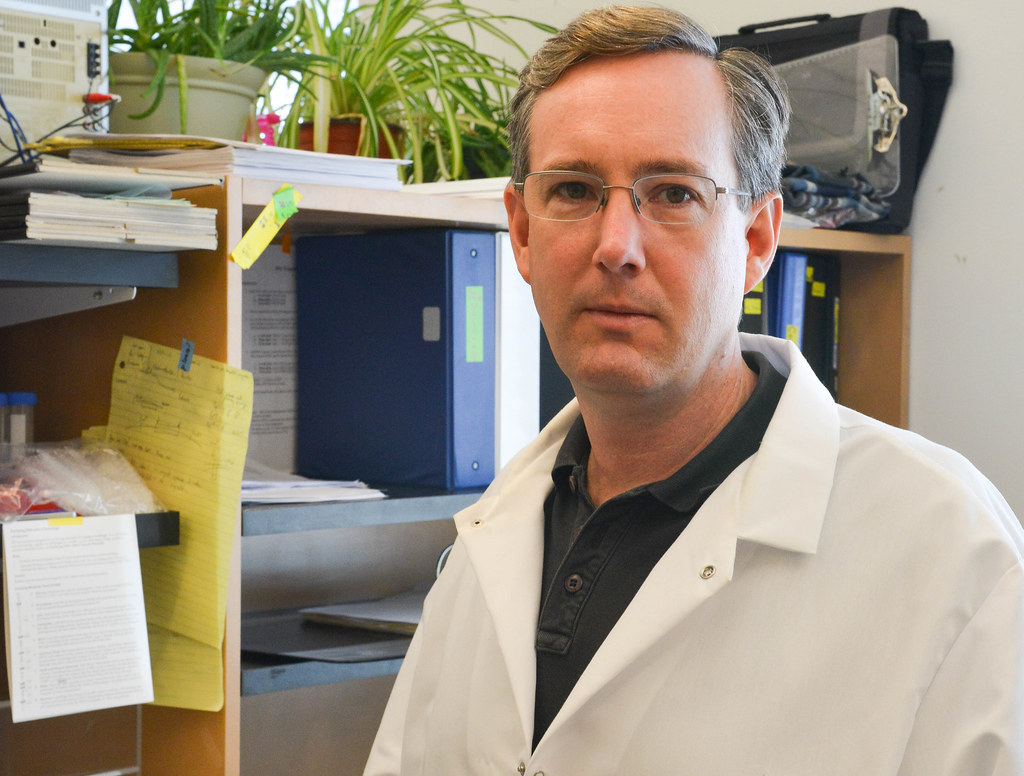
Gary Weisman stands in his lab in Bond LSC where he studies the cellular mechanisms of auto-immune disease, specifically how the release of ATP from damaged cells signals receptors that trigger an immune response. | Phillip Sitter, Bond LSC
“We want the good, but not the bad,” said Weisman, a Bond Life Sciences Center investigator, of what we ideally want from our immune system’s functions.
Mice with un-checked autoimmune disease of their salivary glands have their glands destroyed. The disease can spread to other secretory organs next. An over-reactive immune system on a civil war-path can extend its damage to cause pancreatic failure and death.
The destruction wrought by Sjögren’s syndrome is self-inflicted, caused by an overreaction of our bodies’ defenses against infection and injury. This is what an autoimmune disease is.
But, our bodies’ immune cellular response team is complicated. Weisman said dozens of different cell types have been isolated and identified as part of the immune system, and he likens the immune system to fire, police and construction services in human society all working together.
While firefighters are meant to prevent further damage from an inferno, sometimes our bodies’ first responders start doing the equivalent of using dynamite to stop the spread of a fire.
In chronic inflammation, that autoimmune response can mean a burning, throbbing, constant pain. The key to a healthy immune response is balance. The balance has to be between containment and repair of damage caused by infection or injury and damage caused by chronic inflammation if that emergency response continues unabated.
Weisman has spent almost 30 years studying how to prevent our bodies’ immune system from over-reacting to threats and causing further harm.
Earlier in his career, Weisman studied how extracellular ATP plays a critical role in immune responses, and how too much of it can cause the over-reaction that leads to tissue destruction in autoimmune diseases. ATP, or adenosine 5’-triphosphate, is the main molecule used for energy in cellular activities inside cells. Weisman was one of the first scientists to study how damaged cells release ATP as a distress signal.
The released ATP signals receptors that “send out the alarm to the fire station” — the body’s immune cells, he said.
Once he understood this, Weisman began to manipulate the actions of released ATP to see how that would affect an immune response.
Mice with salivary gland autoimmune disease got healthy when the released ATP was prevented from activating their receptors on the surface of cells. Preventing the ATP receptors from being activated slowed down and even stopped the advance of autimmune disease.
Conversely, if you prevent the activation of the ATP receptors in lab mice with Alzheimer’s disease they die much more rapidly from the disease, Weisman said, suggesting that activation of immune cells by ATP is beneficial in slowing the progression of this disease.
Alzheimer’s disease and autoimmune diseases such as Sjögren’s syndrome are only some of the inflammatory diseases that Weisman has studied. With each of these diseases, the role of ATP receptors has to be investigated individually, suggesting that Weisman’s work may extend beyond salivary glands and the brain to other parts of the body.
“Our [ATP] receptor is also involved in heart disease,” Weisman said, and he added that other diseases like cystic fibrosis, cancer, lupus and arthritis have inflammatory components, too.
For now, we all fight a losing battle when it comes to our bodies’ management of the immune system. As we and our immune system age, it has the potential to destroy more than it protects and “eventually you could slip over to the dark side and die,” Weisman said.
In the meantime, Weisman said that a better understanding of the immune system could lead to more effective, targeted treatments of chronic inflammation and other autoimmune disorders. This could provide a new approach to control undesirable activation of the immune system beyond the use of with anti-histamines, anti-cytokines and ibuprofen.
Weisman is a Curator’s Distinguished Professor of Biochemistry. He began his salivary gland research at MU 27 years ago with Professor John Turner, before Turner’s retirement. Since then, his research has been continuously funded by the National Institutes of Health, where one of his recent grants was well scored and will likely be extended for another five years.
Lorson lab publishes research on a new therapeutic path to help treat spinal muscular atrophy
Erkan Osman shows iImages of neuro-muscular junctions. Osman, a post-doctoral fellow in Chris Lorson’s lab, co-authored research in the journal Molecular Therapy that details work in binding a synthetic nucleic acid to a normally useless motor neuron backup gene to help treat spinal muscular atrophy. | photo by Phillip Sitter, Bond LSC
By Phillip Sitter | Bond LSC
Imagine you are forced to jump out of an airplane.
Luckily, you find a parachute that even has a backup chute. You leap out of the plane and free-fall.
You pull the cord to open your parachute, but it doesn’t open. Don’t panic, though, you have a backup. But, you pull that cord and nothing happens. Now you face the reality of a death as firm and un-yielding as the ground rushing into your view.
This air disaster mirrors the mechanism and mortal threat posed for people born with the genetic problem that causes spinal muscular atrophy (SMA).
Chris Lorson’s lab at the Bond Life Sciences Center would like to change that situation by making an effective genetic backup to the defective gene that results in SMA. The journal Molecular Therapy, a publication of Nature, recently accepted their findings for publication.
The defect occurs in a specific gene called Survival Motor Neuron (SMN). If the SMN gene is defective because of mutation, this causes a deficiency of the SMN protein it is supposed to produce. Without this protein, the neurons that control muscle movement malfunction. Signals cease to stimulate muscles.
Muscles that are not stimulated atrophy, grow weak and waste away. At first this happens with the skeletal muscles, which leads to loss of motor function for simple activities like walking and swallowing. If it happens with the muscles that control breathing, you die.
News of the disease often presents a devastating prognosis. Infants have it worst; babies diagnosed with SMA only have a life expectancy of two to five years from birth.
Fortunately, our bodies have a sort of backup for the SMN gene, another one called SMN-2. But, like a useless backup parachute for an unlucky skydiver, SMN-2 isn’t actually very good at producing proteins of the quality needed to stave off SMA. It might just be a vestigial trait on its way down the evolutionary drain — it doesn’t even exist in the closest primate relatives of humans.
Discoveries in the Lorson lab look to make the SMN-2 gene an effective backup, and their recent publications indicate that this may be a viable possibility for future SMA treatments.
Christian Lorson studies the genes that cause SMA when they fail to adequately function. His team’s on a backup gene that greatly extended life expectancy in mouse studies. | photo by Phillip Sitter, Bond LSC
“What we’ve been working on in the lab is a potential therapeutic, and what it does, it’s a large small molecule that is called an antisense oligonucleotide, or ASO,” Lorson said. “And this is something that is essentially a synthetic piece of nucleic acid that is able to go in and bind to a specific sequence within a gene.”
Once bound to SMN-2, the ASO is designed to alter mRNA splicing, “essentially, the editing of a gene,” Lorson said. Speaking in terms akin to products leaving a factory, Lorson said that the attached ASO makes SMN-2 produce good quality proteins, the ones that it wasn’t able to produce before.
In other words, suddenly the backup protein-factory that was making poor-quality products is now pumping out top-of-the-line stuff that will work.
Previous research identified a strong ASO contender to experiment with, and Lorson said current research is about optimizing an ASO to extend survival times in mice with SMA — from just 13 days to five months after only one injection at birth.
Lorson stressed that his lab’s achievement doesn’t promise a fast cure for SMA. He said it is unlikely a single compound will address the full gambit of effects that people with SMA suffer, especially given that people can be identified as having SMA at any time from birth through later in life — often late onset SMA tends to be less severe than diagnosis as an infant.
There’s not yet any single compound treatment for SMA that has been approved by the Food and Drug Administration, Lorson said, so he cautions against getting hopes up of for a revolutionary treatment for SMA coming onto the market soon — “Near future but not tomorrow.”
He acknowledged, though, that “from a research perspective, things seem to be moving at lightning speed, but if you are a patient or a family member, things can never go fast enough, so I think there’s a realized sense of urgency, whether or not it’s for patients who don’t have the disease yet, are not born, or for patients who have had the disease for a decade and are wondering when their opportunity would come.”
Lorson’s work is funded in part by Cure SMA, FightSMA and the Gwendolyn Strong foundations. Erkan Osman, a post-doctoral fellow in Lorson’s lab and the first author on the most recent paper, won the emerging investigator award from FightSMA and Gwendolyn Strong in 2015.
Scientists use placental cells in lab to study virus
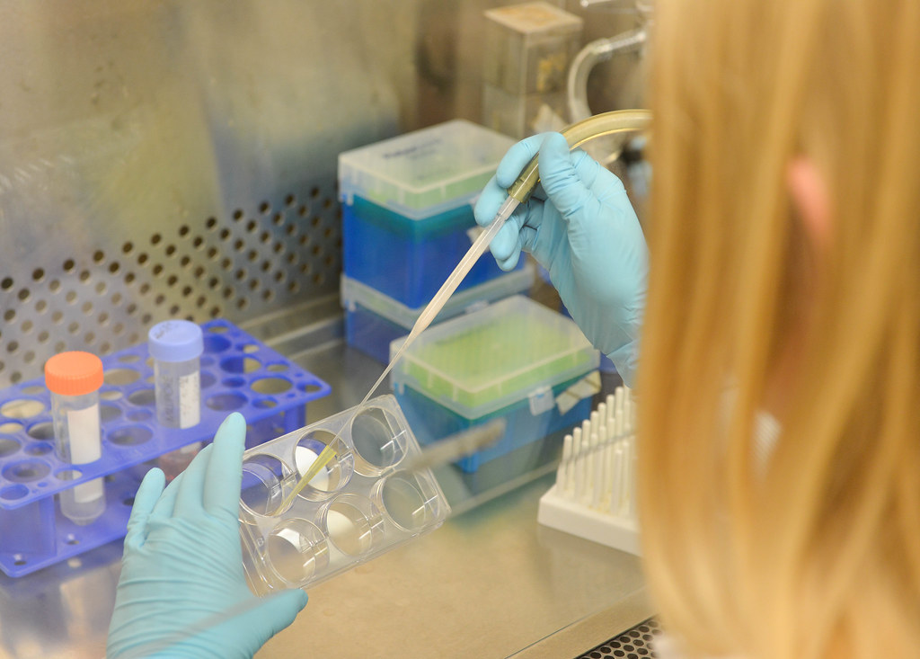
Megan Sheridan, an MU grad student, removes the base solution from a demonstrated sample of stem cells that will be grown into placental cells for study of Zika virus. Within four days of exposure to the correct hormones, the stem cells express genes of placental cells, and within another day start producing placental hormones. The cells are infected with Zika at day four to ensure maximum measurable interaction, as the stem cells naturally die in culture after about ten days. | photo by Phillip Sitter, Bond LSC
By Phillip Sitter | MU Bond Life Sciences Center
Scientists believe they have a better way to study how Zika virus can spread from a pregnant mother to her fetus — and their technique doesn’t even involve observations of babies in the womb or post-natal examinations.
“As soon as we heard about Zika, everybody’s light bulbs turned on,” said Megan Sheridan, a graduate student at the University of Missouri Bond Life Sciences Center.
Sheridan works in the lab of Toshihiko Ezashi at Bond LSC, and she, in turn, is part of a cross-campus team researching Zika with R. Michael Roberts, Alexander Franz, Danny Schust and Ezashi.
Roberts’ lab studies pluripotent stem cells — progenitor cells which can develop into any other type of cell in the body.
“We use the proper signals to drive stem cells to become like placental cells,” Sheridan explained. With this capability to stimulate stem cells with growth hormones and inhibitors at opportune moments, Roberts’ researchers realized they could create enough placental cells to create an environment similar to that of a womb in very early stages of pregnancy.
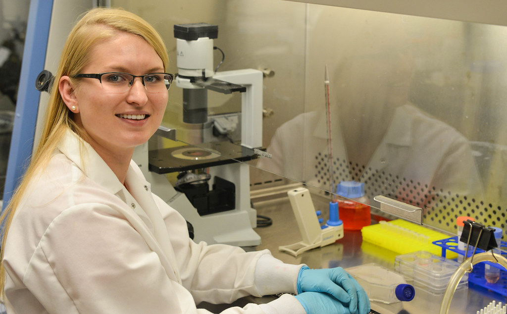
Megan Sheridan sits in front of a demonstration of her work with pluripotent stem cells. Sheridan is a graduate student who works in Toshihiko Ezashi’s lab, where she produces cells with placental characteristics from the stem cells in order to study placenta interaction with Zika virus. | photo by Phillip Sitter, Bond LSC
This is something which Sheridan thinks hasn’t been done before in regards to studying placental interaction with Zika. Their technique could give a look into the first trimester, when epidemiological studies say a fetus is most susceptible to infection.
Roberts’ lab is trying to understand the placental barrier’s vulnerability to Zika virus in its early stage of pregnancy. During this time, an infection could occur even before the mother is aware she is pregnant.
If the lab uses their technique to understand how Zika virus enters placental cells, then potentially they could also learn how to strengthen the placenta as a barrier to Zika and make it a first line of defense against infection of the fetus in the womb. If developing babies don’t get infected with Zika, then they won’t suffer the consequences of birth defects.
One such defect is microcephaly where a baby is born with a smaller than expected head, which may in turn be a sign that their brain has not fully developed. While infection with Zika virus is rarely fatal or otherwise severe in itself — many people don’t even develop symptoms — birth defects like microcephaly could cause further developmental problems like delays in learning how to speak and walk, intellectual disabilities, difficulty swallowing and problems with hearing and vision, according to global health organizations.
Microcephaly only became a widely documented effect of Zika after a particular strain surged across South and Central America with the infected mosquitoes that carry it, Sheridan explained, but this may be in part because previous Zika infections and outbreaks were themselves poorly documented.
While birth defects caused by Zika have drawn much media attention as the disease has spread northward through our hemisphere from Brazil, studies focusing on infection in the womb have only used placental material that has come to term. This may not be the most accurate way to see how the placenta gets infected in the first place early in pregnancy.
The pathway of Zika virus infection in lab mice isn’t really comparable to human infection, because mice aren’t infected with this virus naturally. Only lab mice that have had their genomes altered to be able to acquire the virus have susceptibility to the infection that can be modeled.
Roberts’ lab is currently working with the African strain of Zika and obtained strains from Southeast Asia and Central America recently. There’s about a 99 percent genetic similarity across strains, Sheridan said.
Zika virus was first discovered in Africa in Uganda in 1947, according to the Centers for Disease Control and Prevention. The first human case was documented in 1952, and subsequent outbreaks also occurred in Southeast Asia and the Pacific Islands. The Pan American Health Organization issued an alert about the confirmed arrival of the virus in Brazil in May 2015.
The lab has completed Zika infections of some of their stem cell-produced placental cells. Sheridan reassured that even though the lab works with live viruses, Zika is not airborne, and none of their work involves mosquitoes.
Roberts’ lab submitted one grant application earlier this year to the National Institutes of Health for funding for their research. While that application was denied, Sheridan said that they have a lot more preliminary data now and are hoping to submit a revised grant soon.
She said that their original work was “highly scored, but the funding level is still low,” meaning that obtaining funds for research into Zika virus is highly competitive nationally.
Legislation to fund more efforts into studying and preventing transmission of Zika virus is caught in congressional gridlock, according to The New York Times and other media outlets.
In the mean time, as the Roberts lab prepares its next grant application submission, Sheridan said of her efforts that she is “working hard to make progress on the project as quickly as possible.”
Please visit the CDC’s dedicated page for more information on Zika virus — including advice for travellers and pregnant women, description of symptoms and treatment, steps you can take to control mosquitoes and prevent other means of transmission of the virus and more background on the history and effects of the disease.
Professor Lloyd W. Sumner introduces University of Missouri Metabolomics Center. The new center provides leading-edge equipment to gain crucial information on the complex biology of health and disease in plants, animals and humans. The center, located on the second floor of the Christopher S. Bond Life Sciences Center on the MU campus, is one of a few in the country that encourages interdisciplinary research in metabolomics.
Stewart holds a different colony of anthrax in his lab. Stewart’s work with anthrax and other similar organisms focuses on understanding the tough protein shell of the bacteria’s spores that enable the pathogen to survive in soil for extended periods of time, even hundreds of years. | photo by Phillip Sitter, Bond LSC
By Phillip Sitter | MU Bond Life Sciences Center
For a tiny spore, anthrax holds a lot of danger and promise.
If you found yourself wondering about more than its safety in the lab, we have a answers to a few persistent questions.
What makes anthrax dangerous, and how does it spread? How common are infections of it in nature? If we have antibiotics that already treat it, beyond finding new and better ones why study the organism?
Bovine slayer and bio-weapon
Anthrax bacteria are highly resilient organisms. Like only a few other bacteria in nature, they produce spores with protein-shelled casings that lay dormant in soil for long periods of time, waiting to be taken in by grazing animals like cattle and sheep.
Once they enter the nutrient-rich environment of an animal’s bloodstream — exactly how the bacteria gets there after entering an animal’s mouth is unknown — spores germinate inside the white blood cells that absorb them. As anthrax reproduces rapidly inside its host, it releases toxins that quickly kill the infected animal. That sometimes happens in just a matter of hours according to farmers’ accounts, said George Stewart, Bond Life Sciences Center scientist, medical bacteriologist, McKee Professor of Microbial Pathogenesis and chair of Veterinary Pathobiology at MU.
When infected animals die the bacteria are exposed to oxygen in air that penetrates the decomposing body and new spores escape as the dead host decays. The newly-produced spores are deposited back into the soil, where they wait in a state of suspended animation for as long as it takes to be ingested by another grazing animal, sometimes decades to a hundred years.
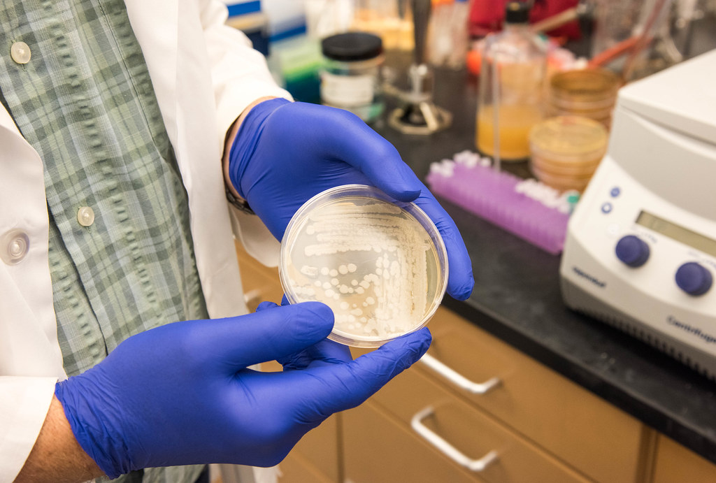
“Most of these plates are anthrax, but they’re all non-virulent,” Stewart says of the various bacterial colonies on the counter in his lab. Non-virulent strains are safe to handle with the precautions of a BSL-2 lab: gloves, a lab coat and eye protection. The Bond LSC does not have a BSL-3 or higher facility to study more dangerous pathogens like virulent strains of anthrax. That work is done at the Laboratory for Infectious Disease Research, or LIDR. | photo by Phillip Sitter, Bond LSC
Sporadic, natural outbreaks of anthrax can happen almost anywhere in the world except Antarctica, as the spores have been found to exist worldwide, Stewart said. In the United States, outbreaks in cattle and bison usually happen in the Plains and West in states like Colorado and Wyoming — anywhere that cattle or sheep have been raised or their wildlife equivalents graze, lending to the descriptor from Stewart of “anthrax belt” for the states stretching from Texas to the Dakotas.
During the Cold War, the U.S. and the Soviet Union, among others, were attracted to anthrax for their respective biological weapons programs because of the hardiness of the organism and high lethality rates of untreated gastrointestinal and, especially, pulmonary anthrax infections in humans. Today, Americans’ are probably most familiar with anthrax from the mailing attacks of 2001, when letters containing weaponized anthrax spores were delivered to the offices of media outlets and politicians, infecting 22 people and killing five.
Hero in a half-life shell?
Despite their grim reputation, anthrax spores hold a lot of potential for Stewart.
The outermost layer of the protein-shell structure of a spore holds particular interest — how it’s made, what proteins it’s composed of and the function of those proteins. Studying it could not only help find future anthrax vaccines and therapies, but also be used for other applications.
Scientists have coated spores of a close biological relative of anthrax, Bacillus thuringiensis, with plant growth-promoting and anti-insect enzymes and other proteins to treat crops. Its durable structure in the form of the spore’s protein shell attach to these enzymes and remain longer in the environment, making them more effective, Stewart said.
This long biological half-life even presents potential for bio-remediation work — using natural organisms to cleanse the environment of toxins and pollutants. In the case of B. thuringiensis, this might include cleaning up soil from the herbicide atrazine and the notorious pollutant dioxin, a by-product of various industrial processes including incineration, smelting and the production of paper pulp and some herbicides and pesticides.
According to collective findings of the National Institutes of Health and Environmental Protection Agency and others, atrazine is probably not a human carcinogen but can cause genetic damage in animals and is still acutely toxic to people at high enough levels. According to the World Health Organization (WHO), dioxin causes cancer and other ill human ailments.
Pollutants that contaminate soil can eventually leech into groundwater and also enter the food supply through grazing animals. That makes organisms adapted to living in soil like B. thuringiensis perfect candidates as potential carriers of agents used to clean up soil pollutants like atrazine and dioxin.
However, Stewart said that there is too much public stigma to use anthrax for similar applications, “which is too bad because Bacillus anthracis actually works a little better in these applications.”
Rethinking the anthrax image
So is anthrax a dastardly bio-villain or a misunderstood hero?
The truth is neither. Anthrax bacterium is just another living thing.
As a predator of other living things, needing to feed to survive and reproduce, it goes about its life cycle without any conscious agenda. It has no malice, and unlike a wolf, shark or hawk it doesn’t have a brain so it doesn’t even have instinct, only genetic code to mindlessly live by.
How we interact with anthrax is largely dependent upon us humans.
We have the vaccines to prevent it and antibiotics to treat it if it makes us sick. After that, we can harness and modify anthrax’s natural power for ill as a weapon, or maybe to do some good in the environment. So long as we have the technology to study and manipulate organisms like anthrax, the potential for both scenarios will always be there.
Meanwhile, at MU Stewart will continue to study anthrax, searching for its secrets in order to better understand it, to better serve the rest of us.
Next time you squirt mustard on a sandwich on enjoy wasabi with your sushi, you can thank a battle between broccoli and butterflies. Just ask Bond LSC biologist Chris Pires, our latest scientist in our Decoding Science audio series that runs on KBIA, 91.3 FM.
Pires has studied the process of co-evolution between plants in the order Brassica — including broccoli, caulifower and kale — and insects in the cabbage butterfly family to prove that this back and forth helped make both into what they are today.