New line of pigs do not reject transplants, will allow for future research on stem cell therapies
Story by Nathan Hurst/MU News Bureau
COLUMBIA, Mo. – One of the biggest challenges for medical researchers studying the effectiveness of stem cell therapies is that transplants or grafts of cells are often rejected by the hosts. This rejection can render experiments useless, making research into potentially life-saving treatments a long and difficult process. Now, researchers at the University of Missouri have shown that a new line of genetically modified pigs will host transplanted cells without the risk of rejection.
“The rejection of transplants and grafts by host bodies is a huge hurdle for medical researchers,” said R. Michael Roberts, Curators Professor of Animal Science and Biochemistry and a researcher in the Bond Life Sciences Center. “By establishing that these pigs will support transplants without the fear of rejection, we can move stem cell therapy research forward at a quicker pace.”
In a published study, the team of researchers implanted human pluripotent stem cells in a special line of pigs developed by Randall Prather, an MU Curators Professor of reproductive physiology. Prather specifically created the pigs with immune systems that allow the pigs to accept all transplants or grafts without rejection. Once the scientists implanted the cells, the pigs did not reject the stem cells and the cells thrived. Prather says achieving this success with pigs is notable because pigs are much closer to humans than many other test animals.
“Many medical researchers prefer conducting studies with pigs because they are more anatomically similar to humans than other animals, such as mice and rats,” Prather said. “Physically, pigs are much closer to the size and scale of humans than other animals, and they respond to health threats similarly. This means that research in pigs is more likely to have results similar to those in humans for many different tests and treatments.”
“Now that we know that human stem cells can thrive in these pigs, a door has been opened for new and exciting research by scientists around the world,” Roberts said. “Hopefully this means that we are one step closer to therapies and treatments for a number of debilitating human diseases.”
Roberts and Prather published their study, “Engraftment of human iPS cells and allogeneic porcine cells into pigs with inactivated RAG2 and accompanying severe combined immunodeficiency” in the Proceedings of the National Academy of Sciences.
This study was made possible through grants from Konkuk University in South Korea and the National Institutes of Health.
Roberts has appointments in the MU College of Food, Agriculture and Natural Resources (CAFNR) and the MU School of Medicine and is a member of the National Academy of Sciences. Prather has an appointment in CAFNR and is the director of the NIH-funded National Swine Resource and Research Center.
Resistance is the price of success when it comes to treating HIV.
Virologists at the Bond Life Sciences Center are helping to test the next generation of anti-AIDS medication to quell that resistance.
Stefan Sarafianos’ lab recently proved that EFdA, a compound that stops HIV from spreading, is 70 times more potent against some HIV that resists Tenofovir – one of the most used HIV drugs.
“HIV in patients treated with Tenofovir eventually develop a K65R RT mutation that causes a failure of this first line of defense,” said Sarafianos, virologist at Bond LSC. “Not only does EFdA work on resistant HIV, but it works 10 times better than on wild-type HIV that hasn’t become Tenofovir resistant.”
Sarafianos and a team of researchers found that EFdA (4′-ethynyl-2-fluoro-2′-deoxyadenosine) is activated by cells more readily and isn’t broken down by the liver and kidneys as quickly as similar existing drugs.
“These two reasons make it more potent than other drugs, and so our task is to look at the structural features that make it such a fantastic drug,” he said.
From soy sauce to virus killer
The path from EFdA’s discovery to current research is a bit unorthodox.
A Japanese soy sauce company named Yamasa patented this molecule, which falls into a family of compounds called nucleoside analogues that are very similar to existing drugs for HIV and other viruses. EFdA was designed and synthesized by Hiroshi Ohrui (Chem Rec. 2006; 6 (3), 133-143; Org. Lett. 2011; 13, 5264) and shown by Hiroaki Mitsuya, Eiichi Kodama, and Yamasa to have potential usefulness against HIV. Samples sent for further testing confirmed EFdA’s potential usefulness against HIV. This started more than a decade of research to pinpoint what makes the compound special.
EFdA joins a class of compounds called nucleoside reverse transcriptase inhibitors (NRTIs) that includes eight existing HIV drugs. Like all NRTIs, EFdA hijacks the process HIV uses to spread by tricking an enzyme called reverse transcriptase (RT). RT helps build new DNA from the RNA in HIV, assembling nucleoside building blocks into a chain. Since EFdA looks like those building blocks, RT is tricked into using the imposter. When this happens the virus’ code cannot be added to the DNA of white blood cells it attacks.
“NRTIs are called chain terminators because they stop the copying of the DNA chain, and once incorporated it’s like a dead end,” Sarafianos said.
A little help from some friends
Sarafianos isn’t alone in studying EFdA.
The virologist’s lab works closely with University of Pittsburgh biochemist Michael Parniak and the National Institutes of Health’s Hiroaki Mitsuya to explore the molecule’s potential. Mitsuya had a hand in discovering the first three drugs to treat HIV and Parniak has spent years evaluating HIV treatments using cultured white blood cells.
Sarafianos’ focus requires him to take a very close look at EFdA to define how it works on a molecular level. He uses virology, crystallography and nuclear magnetic resonance to piece together the exact structure, bonding angles and configuration of the compound.
By looking at subtle differences in EFdA’s sugar-like ring, his lab identified the best structure that looks the most like actual nucleosides, doesn’t break down easily and is activated readily by CD4+ T lymphocyte white blood cells.
“The structure of this compound is very important because it’s a lock and key kind of mechanism that can be recognized by the target,” Sarafianos said. “We’re looking at small changes and the ideal scenario is a compound bound very efficiently by the target and activating enzyme but not efficiently by the degrading enzymes.”
Treatment for the future
The research of Sarafianos, Parniak and Mitsuya continue to uncover the magic of EFdA. In 2012, they showed that the drug worked incredibly well to treat the HIV equivalent in monkeys.
“These animals were so lethargic, so ill, that they were scheduled to be euthanized when EFdA was administered,” said Parniak. “Within a month they were bouncing around in their cages, looking very happy and their virus load dropped to undetectable levels. That shows you the activity of the molecule; it’s so active that resistance doesn’t come in as much of a factor with it.”
HIV prevention is the newest focus in their collaboration.
By recruiting formulation expert Lisa Rohan at the University of Pittsburgh, they are now putting EFdA in a vaginal film with a consistency similar to Listerine breath strips.
“The only way we are going to make a difference with HIV is prevention,” Parniak said. “If we can prevent transmission, this approach could make a huge difference in minimizing the continued spread of the disease when combined with existing therapies for people already infected.”
While AIDS in the U.S. occurs mostly in men, the opposite is true in sub-Saharan Africa where more than 70 percent of HIV cases occur. Since a film has a better shelf life than creams or gels, it could benefit those at risk in extreme climates and third-world countries.
“We have nearly 30 drugs approved for treating HIV infected individuals, but only one approved for prevention,” Sarafianos said. “Women in Africa would benefit from a formulation like this as a means to protect themselves.”
Despite this success, Sarafianos and Parniak aren’t slowing down in figuring out how EFdA works so well.
“We want to understand how long EFdA stays in the bloodstream and cells,” Parniak said. “If we understand structurally why this drug is so potent it allows us to maybe develop additional molecules equally potent, and a combination of those molecules could be a blockbuster.”
Grants from the National Institutes of Health fund this research.
In 2013 and 2014, the journals Retrovirology, Antimicrobial Agents and Chemotherapy and The International Journal of Pharmaceutics published this group’s work on EFdA. Sarafianos is an associate professor of molecular microbiology and immunology and Chancellor’s Chair of Excellence in molecular virology with MU’s School of Medicine and a joint associate professor of biochemistry in the MU College of Agriculture, Food and Natural Resources.
Sometimes plants inadvertently roll out the red carpet for bacteria.
Researchers at the University of Missouri Bond Life Sciences Center recently discovered how a plant’s own chemicals act as a beacon to bacteria, triggering an infection. Proceedings of the National Academy of Sciencespublished their study April 21.
“When bacteria recognize these plant chemicals it builds a needle-like syringe that injects 20-30 proteins into its host, shutting down the plant’s immune system,” said Scott Peck, Bond LSC plant scientist and lead investigator on the study. “Without a proper defense response, bacteria can grow and continue to infect the plant. It looks like these chemical signals play a very large role in mediating these initial steps of infection.”
The question of how bacteria actually know they are in the presence of a plant has puzzled scientists for years. Being able to identify the difference between a plant cell and, say, a rock or a piece of dirt, means the bacteria saves energy by only turning on its infection machinery when near a plant cell.
“Our results show the bacteria needs to see both a sugar – which plants produce quite a bit of from photosynthesis – and five particular acids at the same time,” Peck said. “It’s sort of a fail-safe mechanism to be sure it’s around a host before it turns on this infection apparatus.”
Peck’s work started with one mutant plant called Arabidopsis mkp1.
Discovered several years ago by Peck’s lab, this little mustard plant acts differently than others by rebuffing the advances of bacteria. Lab tests confirmed that this mutant didn’t get infected by Pseudomonas syringae pv. tomato DC3000, a bacterial pathogen that causes brown spots on tomatoes and hurts the model plant Arabidopsis. Along with MU biochemistry research scientist Jeffrey Anderson and post doc Ying Wan, they showed that this mutant didn’t trigger the bacteria’s Type III Secretion System, the needle-like syringe and associated proteins that lead to infection.
Pacific Northwest National Laboratory (PNNL) worked with Peck’s team to compare levels of metabolites between the mutant Arabidopsis and normal plants. This comparison helped Peck identify a few of these chemicals – created from regular plant processes – that existed in much lower levels in their special little mutant.
Using the PNNL work as a guide, the team found five acids collectively had the biggest effect in turning on a bacteria’s infection: aspartic, citric, pyroglutamic, 4-hydrobenzoic and shikimic acid.
“The key experiment involved us simply adding these acids back into the mutant,” Peck said. “Suddenly we saw the mutant plant wasn’t resistant anymore and the bacteria were once again capable of injecting proteins to turn off the plant’s immune system.”
First contact and recognition means all the difference, whether bacteria or plant. Just a slight jump out of the starting blocks by one or the other could change who will win a battle of health or infection.
While low concentrations of these five acids trigger the bacteria’s attack, high levels blind it to the plant’s presence, leading Peck to believe it could be used to hinder bacterial growth. If this actually thwarts the bacteria’s head start, it could mean stopping disease in crops and could lead to a different approach in the field.
“A lot of the winning and losing occurs within the first 2-6 hours and it seems to be that if the microbe is too slow to turn off the immune system, the plant can actually fight off the infection,” Peck said. “In the future we could possibly make a new generation of anti-microbial compounds that don’t try to kill the bacteria, but rather just make them no longer virulent by blocking these chemical signals so the natural plant immune system can basically take over.”
Peck’s team believes at least some other bacteria will respond to these chemical signals, and he plans to test other bacterial pathogens to make certain. They also want to test bacteria to see if they are more virulent in humans once primed for attack by these plant chemical signals.
“In the long run the question is how far this extends. A lot of people get salmonella or listeria infections through a food source,” Peck said. “The question is do other bacteria that come in through plant food sources have similar perception systems and end up being more infectious in humans because they are already primed for infection.”
A $500,000 grant from the National Science Foundation supported this research.
A former Bond LSC graduate student is being recognized for a dissertation that stands out from the crowd.
Jeongmin Choi received the 2014 Distinguished Dissertation Award this month from MU’s Graduate Faculty Senate for her work identifying the first plant receptor for extracellular ATP. The journal Science published Choi’s “Identification of a plant receptor for extracellular ATP” Jan. 17, 2014.
Choi completed her dissertation working as a member of Gary Stacey’s lab team. Stacey, a Bond LSC researcher, nominated her work for this award. This is the second year work completed in Bond LSC garnered this award after Lefteris Michailidis won in 2013 for work on the HIV drug EFdA.
Choi has since received her Ph.D. and now resides in Cambridge, England.
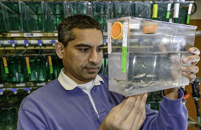
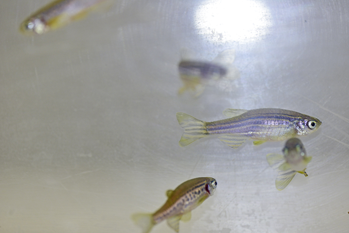
Three thousand zebrafish swim circles in tanks located on the ground floor of the Bond Life Sciences Center, content to mindlessly while away their existence by eating their fill and laying eggs.
Despite their very basic higher functions, Bond LSC researcher Anand Chandrasekhar wants to understand how their brains work. More importantly, he wants to know how individual neurons end up ordered, all in the right place to support the animal’s automatic functions like breathing, swallowing and jaw movement.
This could one day lead to better understanding of specific neuronal disorders in humans.
“We are studying how cells end up where they are, and in the nervous system that’s especially critical because these neurons are assembling circuits just like in computers,” Chandrasekhar said. “If those circuits don’t form properly, and if different types of neurons don’t end up where they are supposed to, the behavior of the animal is going to be compromised.”
Zebrafish are a perfect model to study for many scientists.
Given plentiful food, adult zebrafish lay thousands of eggs that fall through a screen in the bottom of fish tanks to be collected. These eggs turn into new embryos that are nearly transparent, allowing for easy observation of the 3-4 mm fish under a microscope.
Scientists have not only sequenced the zebrafish genome, but they’ve also inserted fluorescent jellyfish protein genes into the genome. This allows easy tracking of neurons.
“It’s called a reporter, and it’s used all the time to visualize a favorite group of cells. In our case, all of these green clusters of lighted up cells are motor neurons,” Chandrasekhar said. “Some groupings are shaped like sausages, some are more round, but each cluster of 50-150 cells sends out signals to different groups of muscles.”
These motor neurons are concentrated in the hindbrain. Akin to the brain stem, it controls the gills, breathing and jaw movement in these tiny fish. Genes controlling these functions are similar in zebrafish, higher vertebrates and humans despite their evolutionary paths diverging millions of years ago.
“The brain stem is the so-called reptilian brain that has changed very little, looking very similar in a lamprey or an eel all the way up to humans. All the elaborate brain structures you see in the cerebellum and the cortex are built up on top of that,” Chandrasekhar said. “Motor neurons from the brain stem send connections to muscles of the head including the gills, jaw and, in humans, the tongue, neck, voice box and muscles for breathing. We study those motor neurons and they undergo migration in many of these vertebrates systems.”
This migration and signals that control it are what Chandrasekhar’s research is all about.
For example, his recent studies suggest that a protein called Vangl2 plays an important role in regulating movement of neurons through the zebrafish embryo’s matrix of tissue. Proteins like this are present in many organisms, from flies to fish to mammals.
“When I say a neuron is migrating in its environment, it’s actually pushing its way in between all these other cells,” Chandrasekhar said. “Cells in the environment of this migrating neuron secrete proteins that may diffuse away from the cell and bind to a receptor on a migrating neuron and then kind of beckon this neuron to keep moving.”
Chandrasekhar’s work contributes to a better understanding of how basic neuronal networks are created in development. That sort of knowledge could one day help with understanding the mechanistic bases of diseases like spina bifida, a nervous system disorder that results in muscle control problems due to the spinal cord not completely closing. Versions of this defect affect 1 in every 2,000 births, according to the National Institutes of Health.
“The significance of the work that we are doing is quite high for development,” Chandrasekhar said. “It is clear that even for the process of closing of the neural tube in the spinal cord and the brain, those cells closing the neural tube actually know left side from right side. The same kinds of mechanisms are going to be important and required whether you are talking about zipping together the neural tube or about allowing cells to squeeze between other cells to migrate and end up in a target position.”
It’s the genetic equivalent to discovering a new sensory organ in plants.
A team at the University of Missouri Bond Life Sciences Center found a key gene that sniffs out extracellular ATP.
Scientists believe this is a vital way plants respond to dangers, such as insects chewing on its leaves. The journal Science published their research Jan. 17.
“Plants don’t have ears to hear, fingers to feel or eyes to see. They recognize these chemical signals as a way to tell themselves they are being preyed upon or there’s an environmental change that could be possibly detrimental, and they have ways to defend themselves,” said Gary Stacey, a Bond LSC biologist. “We have evidence that extracellular ATP is probably a central signal that controls the ability of plants to respond to a whole variety of stresses.”
ATP (adenosine triphosphate) is the main energy source inside any cell. All food converts to it before being used in a cell, and ATP is necessary to power many of the cell processes that create more energy. Its value as an energy reserve is squandered outside the cell.
Scientists spent years trying to figure out what this compound did while floating outside cell walls. Animal researchers found that answer in the 1990s. They identified the first ATP receptors, now seen to play roles in muscle control, neurotransmission, inflammation and development.
Plant scientists observe similar extracellular ATP responses in plant biochemistry, but until now could not identify the exact receptor for it or what it did.
“We call this new receptor P2K, and it’s unique to plants,” Stacey said. “Even though animals and plants hold some responses in common, they have evolved totally different mechanisms to recognize extracellular ATP.”
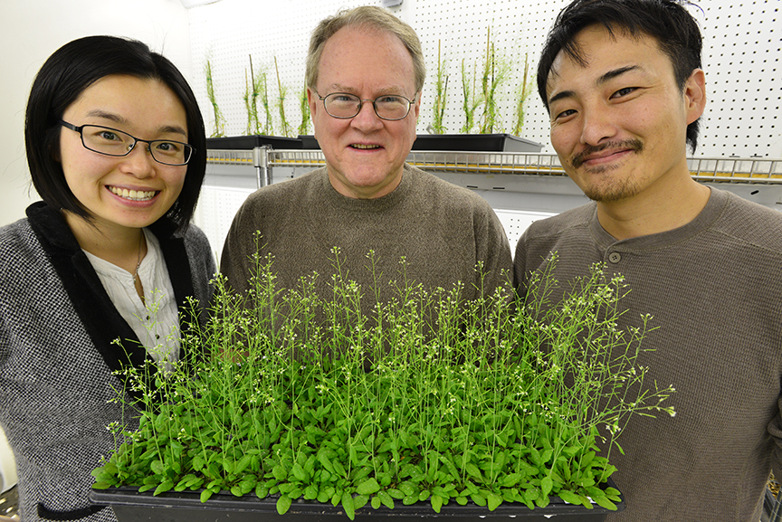
Led by Stacey, MU graduate student Jeongmin Choi and postdoc Kiwamu Tanaka screened 50,000 mutant Arabidopsis plants to find ones that didn’t respond to extracellular ATP. Using a protein called aequorin – which causes jellyfish to glow – the two-year process boiled down to whether a plant would produce light when ATP was added. Since aequorin only luminesces when it binds to calcium, those plants without extracellular ATP receptors stayed dark.
“If you add ATP to wild-type plants, calcium concentrations go up and the plants produce more blue light,” Choi said. “We found nine mutant plants with no increase in calcium and, therefore, no increase in light emission.”
By comparing the genetic sequences of these nine mutants, Stacey’s lab pinpointed the gene to chromosome 5 and labeled it DORN 1, since it doesn’t respond to the nucleotide ATP.
This discovery casts a different light on previous research.
“What we think is happening is that when you wound a plant, ATP comes out in the wound and that ATP triggers gene expression, not the wounding in and of itself,” Stacey said. “We think ATP is central to this kind of wound response and probably plays a role in development, in a lot of other kinds of things.”
Future research will focus on exactly how this receptor works with ATP. Tanaka plans to study its protein structure, how it reacts to pests in lab situations and possible co-receptors that could also play a role in recognizing ATP.
Grants from the U.S. Department of Energy Office of Basic Energy Sciences and the Republic of Korea supported this research.
You can’t see the resemblance, but cells in Michael Roberts’ lab share a family tree with some newborns.
Their common genetics may help explain severe, early-onset preeclampsia, an inherited disorder that leads to a placenta that is often small and inefficient and possibly due to the mother’s body not fully welcoming her pregnancy.
University of Missouri Health Center scientists such as Danny Schust and Laura Schulz, work with Roberts and Toshihiko Ezashi, both Bond Life Sciences Center reproductive biologists, to search for its complex cause.
That starts in the delivery room. OBGYN’s Danny Schust and his residents save small pieces of umbilical cord from preeclampsia pregnancies, allowing Roberts and Ezashi to grow cells with the disease.
“We’re essentially recreating the previous pregnancy, going back in time as far as that baby is concerned,” Roberts said. “That allows us to look at the disease in a Petri dish, look at the properties of these cells to try and figure out what’s wrong with them.”
Preeclampsia affects 3-7 percent of births worldwide, and leads to around 50,000 deaths annually. Symptoms like high blood pressure and protein in the urine tip off doctors to the disorder, but it can go unnoticed until late in the pregnancy unless the symptoms are severe. Left unchecked the disease can lead to seizures and death. The only cure for the serious early-onset form of the disease is to deliver the baby prematurely, usually between 28-33 weeks. This leaves the newborn underweight and with complications like underdeveloped lungs.
Creating useful cells from the collected umbilical cords takes a little bit of work. Cells grown from the umbilical cords are converted into induced pluripotent stem cells, with potential to become any type of cell in the body. Researchers then use a series of hormones, growth factors and other conditions to create placental cells mirroring the previous pregnancy.
Normally the placenta – containing genes from both mother and father – grows into the wall of the uterus to establish a supply of blood, nutrients and oxygen to support the embryo. But in preeclampsia, those cells encounter problems.
Roberts and Schulz focus on extravillous trophoblasts, placental cells that invade the wall of the womb, while collaborators Toshihiko Ezashi and Danny Schust study synctiotrophoblasts, placental cells responsible for uptake of oxygen and nutrients from the mother’s blood.
“The question becomes do these placental cells grow too much or too little, and it appears that in preeclampsia they don’t grow enough,” Roberts said. “We’re just beginning to look at their ability to move and grow through a jelly-like substance that impedes a cell’s mobility and ability to pass through small pores on a membrane. We’re also comparing the gene expression of the cells from preeclamptic patients with those from normal births..”
The Roberts/Ezashi/Schulz/Schust team still has several years left on two five-year National Institutes of Health grants and hopes to narrow the search for genes linked to preeclampsia. If they pinpoint the culprit genes, scientists could one day potentially correct the problem by developing drugs that restore normality to the placental cells or even by using an induced pluripotent stem cell approach.
“It might mean you could go back to correct the defect in those cells or take that patient’s cells, make some normal cells and perhaps substitute them back in to effect a cure,” Roberts said. “My own feelings are that we’re a very long way from doing that, but that is the thought.”
Michael Roberts is a Curators’ Professor of Animal Science, Biochemistry and Veterinary Pathobiology in the College of Agriculture, Food and Natural Resources (CAFNR) and the College of Veterinary Medicine, respectively. Toshihiko Ezashi is a research associate professor in CAFNR. Danny Schust is an associate professor and Laura Schulz an assistant professorof Obstetrics, Gynecology, & Women’s Health, at MU’s School of Medicine.
Positron emission tomography (PET) scans detect radioactive tracers and how they travel over time. In humans the scan tracks blood flow to find cancers, understand brain activity and show uptake of drugs in preclinical trials.
In plants, PET scans shine light on how plants divvy up sugars to protect against attackers.
“Doctors inject radioactive tracers into a patient, and it lights up a tumor on a PET scan because the tumor accumulates a lot of sugar and energy to grow,” said Jack Schultz, director of MU’s Bond Life Sciences Center. “The question in plants is how and where do sugars travel when young leaves are attacked by a pest.”
The sugar trip
Young leaves are of particular interest.
They attract pests because they are full of the nutrients while also being more vulnerable to attack. These youngsters can’t photosynthesize enough sugar to build chemical defenses for themselves, so they get sugars elsewhere through the phloem, part of the plant’s transportation system.
“Think of phloem as a tube with ports for the leaves,” Schultz said. “Older leaves are almost always making sugars, loading them into the phloem where they are transported to various places.”
Abbie Ferrieri, a former doctoral student in Schultz’ lab, wondered if these old leaves were supplying young leaves directly or otherwise. Using Arabidopsis thaliana, she worked with Brookhaven National Laboratory to investigate using PET scans. This mustard plant is commonly used as a model in research because of its relative simplicity and the breadth of knowledge about its inner workings.
First, she fed radioactive sugar – 2-[18F]fluoro-2-deoxy-d-glucose – to old leaves on the plant. Then she injured young leaves with a mechanical wheel and introduced methyl jasmonate to the wounds. Methyl jasmonate works as a chemical signal, telling other parts of the plant that an attack is happening.
The researchers took PET scans to determine where these sugars traveled. Ferrieri found that most sugars travel to roots and also to leaves in a line above and below them, a connection called orthostichy, in the injured plant.
But, three hours later, PET scans showed that radioactive sugars migrated to the attacked leaves regardless of whether they were in the same row on the stem. Damaged leaves then used those sugars to make phenolic glycosides, compounds that help defend the leaf.
Using the more short-lived radioactive tracers, Ferrieri saw leaves send sugar to the roots within minutes of an attack on nearby leaves. But 24 hours later, damaged leaves started receiving more sugar.
Root of the issue
This change in sugar flow and its control is a continuing curiosity that scientists like Schultz want to decipher.
Schultz’ lab knew from previous research that an insect chewing on a young leaf triggered an enzyme called invertase to break down sugars faster and call for more. But, Ferrieri thought roots might play a bigger role in controlling sugar.
To test her theory, she decided to shut the roots of her plants down using ice water.
“Plants are cold-blooded, essentially, so by submerging their roots in a lake of ice water, she found that the roots altered the rates all these things happened,” Schultz said. “You could say the roots are acting somewhat like a brain, controlling how fast carbon is moving from one place to another at any point in time.”
From human to plant
Plant scientists have been playing with radioactive particles for decades. Researchers like Melvin Calvin used radioactive carbon in the 1940s to figure out how plants capture carbon dioxide for photosynthesis.
But, this field focused more on medical applications until the past decade when plant research received renewed interest.
“The focus has changed a bit to move more into general biological and not just nuclear medicine,” said Silvia Jurrison, an MU professor of chemistry who works to find better uses of the technology.
Bond LSC labs – including those of Gary Stacey, Paula McSteen and Jack Schultz – collaborate with Jurrison to use radiotracers to explore plant processes. Jurrison hopes new compounds will make that easier.
“We’re working to develop other fluorinated compounds, such as sucrose, which could be more relevant and useful in studying plants since they use sucrose more readily.”
While research in Schultz’ lab works to explain basic plant processes, it one day could lead to new advancements.
It could help sequester carbon to slow global warming or be used to better protect future crops from pests.
“Plant defense’ effectiveness depends not only on what they are made of, but also how fast and how strongly they can respond. Anything you can understand about how a plant defends itself against pests is potentially useful,” Schultz said.
Forget fruits and vegetables, seeds provide a critical part of the average person’s diet. From beans to cereal grains, understanding how genes and soil types impact nutrition could one day help produce more nutritious food.
One University of Missouri researcher wants to know which genes control the elements in these nutrient-rich packages.
“Iron and zinc deficiencies are considered two major nutritional disorders in the world, so there’s a lot of interest in developing plants with enhanced amounts of these micronutrients,” said David Mendoza-Cozatl, a Bond Life Sciences Center plant scientist. “The question for labs like mine is how can you convince a plant to accumulate more of these metals even though high concentrations can be toxic to plants?”
In a five-year collaboration with researchers at the University of Nevada and UC San Diego, the group measured the amounts of 14 elements in both plant seeds and leaves of mutant Arabidopsis thaliana plants planted in different soil types (salty, alkali, heavy metal and normal).
These mutants were special. Each plant had a different gene disabled, allowing researchers to tell if the disabled gene affected uptake of minerals into the seeds or leaves.
The teams found that 11 percent of genes influence proteins relevant to the nutritional content in seeds. Soil types also played a role in the significance each gene’s impact.
The approach could be likened to understanding how a car works.
“What we are doing here is we have a car with different parts missing, and the question we are asking is what happens to the car without each of these parts,” Mendoza-Cozatl said. “In plants we ask how more or less elements or nutrients can accumulate without each of these parts, these genes. That’s how we are trying to assign the function of a gene to nutrient homeostasis.”
Mendoza-Cozatl’s work with the group focused on soil laced with non-essential heavy metals (e.g. cadmium and arsenic). They grew mutant plants from seeds and compared nutrient content of those plants’ leaves and seeds to a baseline. To do this, researchers “digested” leaves and seeds, separately, then quantified the amounts of elements.
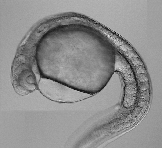
“We turn everything into basic elements by putting the seeds or leaves in a tube, adding nitric acid and boiling them,” Mendoza Cozatl said. “We found that the mineral makeup of the seed and leaves can be different.”
This surprised him since nutrients in seeds come through the leaves via the phloem. The phloem is living tissue that transports nutrients throughout a plant.
Nutrition isn’t the only area that could benefit from knowing what controls the transport of minerals in plants.
A newly engineered plant could be made to use less fertilizer or move particular types of minerals, like toxic heavy metals.
“Many former industrial areas contain fields contaminated with heavy metals like cadmium and arsenic,” Mendoza-Cozatl said. “Understanding genes important to nutrient transport could help both with bioremediation in soil and bio fortification in food.”
Genes identified through this study will lead to new research in the Mendoza lab as well as other labs involved in this large project.
“The mechanism underlying these changes in nutrient seed composition are not known, so we still need to find how these genes are affecting the seed composition,” Mendoza said. “That’s where the advance will be more significant, and we’re not there quite yet.”
Henry Nguyen‘s progress to identify the genes responsible for root-knot nematode is garnering some attention. The Bond LSC and CAFNR plant scientist helps find genetic candidates for the disease, as explained in this recent CAFNR story by Randy Mertens.
——-
A team of scientists from the University of Missouri, the University of Georgia and the Beijing Genome Institute have used next-generation sequencing to identify two genes — out of approximately 50,000 possibilities — that defend soybeans from damage caused by the root-knot nematode (RKN) parasite. This parasite causes millions of dollars in yield losses each year in the United States.
This is the first time the process has been used in soybean research. Using another genetic technique, the team is now working to identify the specific gene that prevents RKN from infecting the soybean. With this knowledge, resistant soybean varieties or cultivars can be bred for farmers.
Researchers believe that this process also can be applied to other crops to map genes important for traits, such as yield and stress responses, said Henry Nguyen, director of the National Center for Soybean Biotechnology (NCSB), housed at the MU College of Agriculture, Food and Natural Resources.
The root-knot nematode is a microscopic roundworm that can become a parasite on an enormous variety of crop species including the soybean, potato, sugar beet, rice, coconut palm, banana, pepper, tobacco, watermelon, tomato and peanut. It is one of the three most economically damaging plant parasites worldwide, causing an average worldwide yield loss of 5 percent, Nguyen said.
J. Grover Shannon, associate director of the NCSB and David M. Haggard Endowed Chair of Soybean Breeding at MU, estimates that RKN and other nematodes (excluding soybean cyst nematode) accounts for annual losses of more than $50 million in soybean yield in the United States. The U.S. is the world’s largest producer of soybeans, growing more than 3 billion bushels, or 33 percent of the world’s production. The value of the 2011 U.S. soybean crop exceeded $35.7 billion. U.S. soybean and soy product exports exceeded $21.5 billion in 2011.
Jinrong Wan, MU research scientist, said RKN larvae infect plant roots, causing the formation of root-knot galls that drain the plant’s photosynthate and other nutrients. Infection of young plants may be lethal, while infection of mature plants causes decreased yield.
RKN is a silent killer of profits, Wan continued. Often, it thrives as a parasite throughout a growing season in annuals, or over many years in perennial crops, without any above-ground signs or symptoms. Only when harvest is over and yield has been quantified is the parasite’s damage commonly seen.
No method currently used effectively controls RKN. Crop rotation is a typical technique, but with incomplete results because RKN can live for a long time and can infect an enormous variety of plants. Nematicides are another intervention, but are dangerous to the respiratory systems of animals. Biological control is practiced in some cropping systems, with two organisms showing only limited success. Clearly the most efficient method of control is by using resistant cultivars, said Nguyen.
A Faster and Better Assay
Nguyen said next-generation sequencing technology is a new and important tool for plant scientists.
Currently, scientists use single nucleotide polymorphisms to map genetic markers to determine what genes are responsible for important traits, such as disease resistance. That process, said Nguyen, is too slow considering the tens of thousands of genes that have to be surveyed.
To speed up the process, Nguyen’s team used Next-Generation Sequencing Technology to sequence the whole genome of more than 200 soybean inbred lines. Another advantage of using this method is that the mapping resolution can be significantly improved – the genes can be narrowed down to a very small chromosome region quickly, Tri Vuong, MU research scientist, added.
This story was originally posted on CAFNR News where you can find on MU agriculture research.