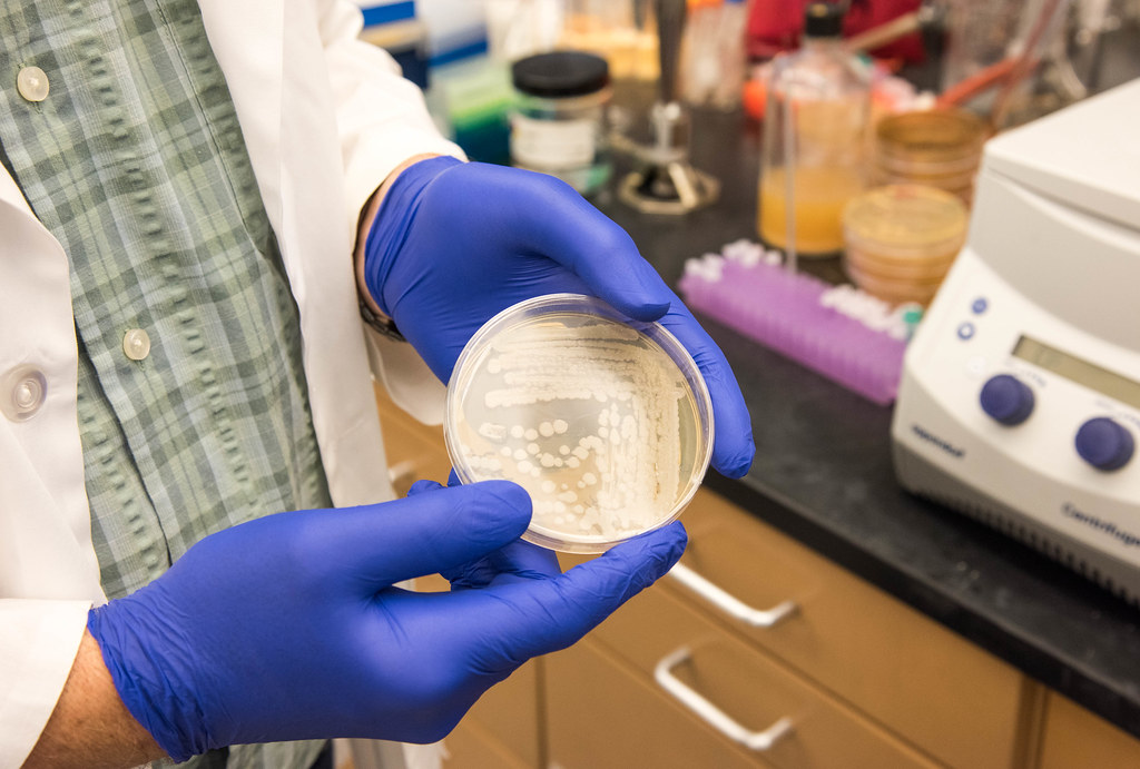
Stewart holds a different colony of anthrax in his lab. Stewart’s work with anthrax and other similar organisms focuses on understanding the tough protein shell of the bacteria’s spores that enable the pathogen to survive in soil for extended periods of time, even hundreds of years. | photo by Phillip Sitter, Bond LSC
By Phillip Sitter | MU Bond Life Sciences Center
For a tiny spore, anthrax holds a lot of danger and promise.
If you found yourself wondering about more than its safety in the lab, we have a answers to a few persistent questions.
What makes anthrax dangerous, and how does it spread? How common are infections of it in nature? If we have antibiotics that already treat it, beyond finding new and better ones why study the organism?
Bovine slayer and bio-weapon
Anthrax bacteria are highly resilient organisms. Like only a few other bacteria in nature, they produce spores with protein-shelled casings that lay dormant in soil for long periods of time, waiting to be taken in by grazing animals like cattle and sheep.
Once they enter the nutrient-rich environment of an animal’s bloodstream — exactly how the bacteria gets there after entering an animal’s mouth is unknown — spores germinate inside the white blood cells that absorb them. As anthrax reproduces rapidly inside its host, it releases toxins that quickly kill the infected animal. That sometimes happens in just a matter of hours according to farmers’ accounts, said George Stewart, Bond Life Sciences Center scientist, medical bacteriologist, McKee Professor of Microbial Pathogenesis and chair of Veterinary Pathobiology at MU.
When infected animals die the bacteria are exposed to oxygen in air that penetrates the decomposing body and new spores escape as the dead host decays. The newly-produced spores are deposited back into the soil, where they wait in a state of suspended animation for as long as it takes to be ingested by another grazing animal, sometimes decades to a hundred years.
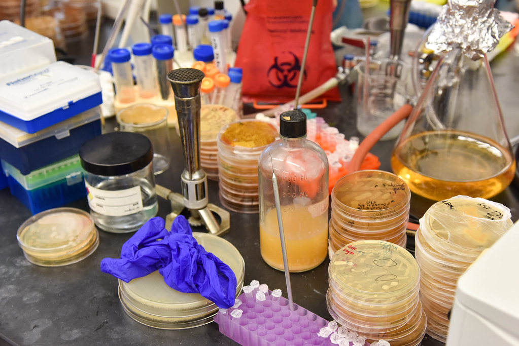
“Most of these plates are anthrax, but they’re all non-virulent,” Stewart says of the various bacterial colonies on the counter in his lab. Non-virulent strains are safe to handle with the precautions of a BSL-2 lab: gloves, a lab coat and eye protection. The Bond LSC does not have a BSL-3 or higher facility to study more dangerous pathogens like virulent strains of anthrax. That work is done at the Laboratory for Infectious Disease Research, or LIDR. | photo by Phillip Sitter, Bond LSC
Sporadic, natural outbreaks of anthrax can happen almost anywhere in the world except Antarctica, as the spores have been found to exist worldwide, Stewart said. In the United States, outbreaks in cattle and bison usually happen in the Plains and West in states like Colorado and Wyoming — anywhere that cattle or sheep have been raised or their wildlife equivalents graze, lending to the descriptor from Stewart of “anthrax belt” for the states stretching from Texas to the Dakotas.
During the Cold War, the U.S. and the Soviet Union, among others, were attracted to anthrax for their respective biological weapons programs because of the hardiness of the organism and high lethality rates of untreated gastrointestinal and, especially, pulmonary anthrax infections in humans. Today, Americans’ are probably most familiar with anthrax from the mailing attacks of 2001, when letters containing weaponized anthrax spores were delivered to the offices of media outlets and politicians, infecting 22 people and killing five.
Hero in a half-life shell?
Despite their grim reputation, anthrax spores hold a lot of potential for Stewart.
The outermost layer of the protein-shell structure of a spore holds particular interest — how it’s made, what proteins it’s composed of and the function of those proteins. Studying it could not only help find future anthrax vaccines and therapies, but also be used for other applications.
Scientists have coated spores of a close biological relative of anthrax, Bacillus thuringiensis, with plant growth-promoting and anti-insect enzymes and other proteins to treat crops. Its durable structure in the form of the spore’s protein shell attach to these enzymes and remain longer in the environment, making them more effective, Stewart said.
This long biological half-life even presents potential for bio-remediation work — using natural organisms to cleanse the environment of toxins and pollutants. In the case of B. thuringiensis, this might include cleaning up soil from the herbicide atrazine and the notorious pollutant dioxin, a by-product of various industrial processes including incineration, smelting and the production of paper pulp and some herbicides and pesticides.
According to collective findings of the National Institutes of Health and Environmental Protection Agency and others, atrazine is probably not a human carcinogen but can cause genetic damage in animals and is still acutely toxic to people at high enough levels. According to the World Health Organization (WHO), dioxin causes cancer and other ill human ailments.
Pollutants that contaminate soil can eventually leech into groundwater and also enter the food supply through grazing animals. That makes organisms adapted to living in soil like B. thuringiensis perfect candidates as potential carriers of agents used to clean up soil pollutants like atrazine and dioxin.
However, Stewart said that there is too much public stigma to use anthrax for similar applications, “which is too bad because Bacillus anthracis actually works a little better in these applications.”
Rethinking the anthrax image
So is anthrax a dastardly bio-villain or a misunderstood hero?
The truth is neither. Anthrax bacterium is just another living thing.
As a predator of other living things, needing to feed to survive and reproduce, it goes about its life cycle without any conscious agenda. It has no malice, and unlike a wolf, shark or hawk it doesn’t have a brain so it doesn’t even have instinct, only genetic code to mindlessly live by.
How we interact with anthrax is largely dependent upon us humans.
We have the vaccines to prevent it and antibiotics to treat it if it makes us sick. After that, we can harness and modify anthrax’s natural power for ill as a weapon, or maybe to do some good in the environment. So long as we have the technology to study and manipulate organisms like anthrax, the potential for both scenarios will always be there.
Meanwhile, at MU Stewart will continue to study anthrax, searching for its secrets in order to better understand it, to better serve the rest of us.
Next time you squirt mustard on a sandwich on enjoy wasabi with your sushi, you can thank a battle between broccoli and butterflies. Just ask Bond LSC biologist Chris Pires, our latest scientist in our Decoding Science audio series that runs on KBIA, 91.3 FM.
Pires has studied the process of co-evolution between plants in the order Brassica — including broccoli, caulifower and kale — and insects in the cabbage butterfly family to prove that this back and forth helped make both into what they are today.
Grad students present brain science, crop biology research in series kick-off
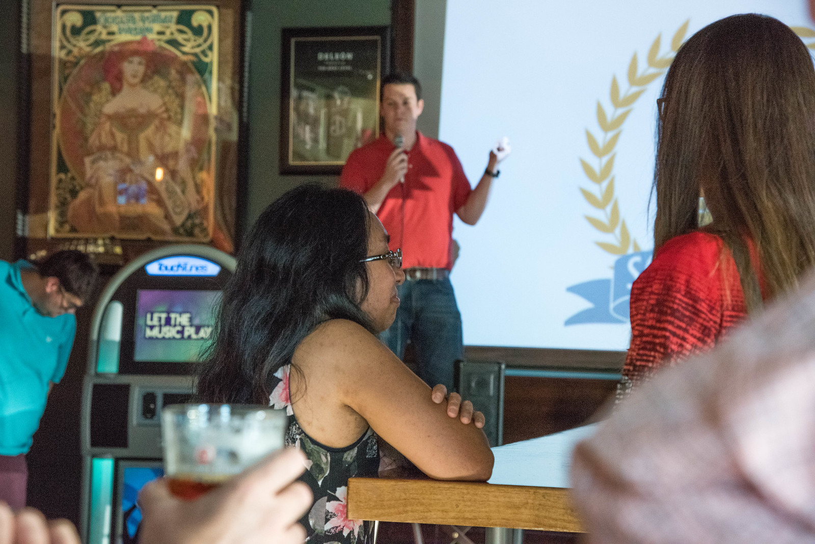
University of Missouri PhD student in biological sciences Nat Graham introduces the first Science on Tap CoMo event on the evening of Tuesday, June 28 at Ninth Street Public House. | photo by Phillip Sitter, Bond LSC
By Phillip Sitter | MU Bond Life Sciences Center
You never know what conversations you might overhear at a bar.
The talk centered on neural proteins and vitamin A-fortified bananas Tuesday night as about 40 science-minded people met at 9th Street Public House for the first Science on Tap CoMo.
Science on Tap is a monthly program scheduled for the fourth Tuesday of each month, and it gives Mizzou graduate students in science, technology, engineering and mathematics a chance to present research in their field to a casual audience.
Anahita Zare and Nat Graham at Public House, both graduate students at the University of Missouri carried the conversation.
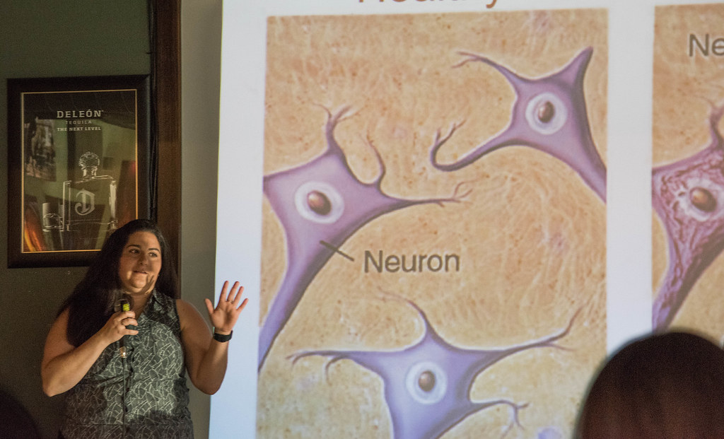
MU PhD student Anahita Zare shows the differences between healthy neural tissue and that affected by Alzheimer’s disease in her presentation at the first Science on Tap CoMo event Tuesday, June 28 at Ninth Street Public House. | photo by Phillip Sitter, Bond LSC
Zare, a Ph.D. candidate in chemistry at MU, spoke about her work on the development of a laser that, once completed, will allow her and other researchers to be better able to study neural proteins in their natural environment. With this ability to better scrutinize drug interactions with these proteins — as opposed to just before and after observations — the work could let researchers make advances in the race for cures to diseases of the brain like Alzheimer’s.
The incremental progress of work on the laser is all about the tiniest of details. Zare used an analogy of letters and words – “I’m changing letters and watching what happens to the words …” “I know what I’m changing and how I’m changing it, and then see its manifestations,” Zare said about the task.
Nat Graham, an MU PhD student in biological sciences, explains how many people around the world have nutrient deficiences in their diets, which genetically-engineered crops could help prevent. Graham spoke at the first Science on Tap CoMo event at Ninth Street Public House. | photo by Phillip Sitter, Bond LSC
Graham addressed the coming global food security crisis by offering a solution in the form of genetically-engineered crops with higher nutritional content. While the introduction of genetically-modified organisms (GMOs) into the global food supply have drawn criticism and protest, Graham was steadfast when he said “I believe lives would be saved if this were released.”
He specifically spoke of vitamin A-enriched bananas. It could be ideal for countries like Uganda, a very large exporter and consumer of bananas as a staple crop. Selective breeding techniques — including even exposure of test crops to radioactivity to promote genetic mutations that may prove to be useful — are options in the development of vitamin-enriched crops, but Graham said these other techniques are too unpredictable and time-consuming to guarantee the results needed.
Graham said he had no fear of GMOs, and in fact really wanted to try a vitamin A-enriched “super banana,” but obtaining one is difficult because of regulations that forbid crossing state lines with these bananas.
He also reminded the audience that although the study of plants often seems boring, it goes beyond gardens and forests. Crops are plants, too, and, among many other things, beer comes from crops. So, food security affects your drink.
George Stewart, McKee Professor of Microbial Pathogenesis and Chair of Veterinary Pathobiology holds up a colony of Bacillus anthracis in his lab. The strain of anthrax he holds is non-virulent, and is therefore safe to handle under BSL-2 precautions as opposed to BSL-3 for virulent strains that cause disease in humans. | photo by Phillip Sitter, Bond LSC
By Phillip Sitter | MU Bond Life Sciences Center
You’ve seen it before in the movies.
Sweaty scientists put on their full-body, spacesuit-like get-ups to stave off a potentially extinction-level outbreak and at least one scientist invariably gets infected with the deadly agent of disease.
While popular culture propagates this sense of peril, in reality bio-containment labs are designed with safety in mind.
“People tend to think bio-containment facilities are dangerous, mostly from movies I think, but the history is actually spectacular,” said George Stewart, a medical bacteriologist, a Bond Life Sciences Center scientist, McKee Professor of Microbial Pathogenesis and Chair of Veterinary Pathobiology.
For Stewart — whose lab works on the basic science behind anthrax — no one is “under the gun because of big outbreaks, not with the pressure you see in movies.”
But one type of pressure is an important part of bio-containment lab safety. Air pressure differences maintain certain labs at lower pressure compared to the rooms and hallways around them, ensuring that air will only flow in toward a lab and not out, keeping any airborne pathogens trapped inside.
“They know if everything is done properly, it’s perfectly safe for them and even safer for everyone else [outside a lab],” Stewart said of the safety features, procedures and systems of bio-containment lab safety in place at facilities like those at the Bond LSC and elsewhere at MU.
Stewart said he is vaccinated against anthrax. “Whether I have protective immunity or not, I don’t know.”
“Almost like you were working underwater”
The powerful capabilities of anthrax and other lethal pathogens call for particular safety precautions for scientists.
Stewart looks like he’s straight out of the movie Contagion when donning the full-body suit for his more dangerous research in a Bio-Safety Level (BSL) 3 facility at MU’s Laboratory for Infectious Disease Research (LIDR). In the trees on the eastern fringes of campus, the specialized building is where he and other researchers study diseases animals can transmit to humans, including plague, Brucella, tularemia and Q fever, and mosquito-borne diseases like dengue, chikungunya and now Zika virus. The safety protocols and systems at a BSL-3 lab like the LIDR facility Stewart described reflect the likely transmission by aerosols of the human pathogens inside.
After passing through security access to the building and the labs inside, Stewart enters an ante room off of a hallway. The air pressure in this room and the lab beyond it is such that air will only flow in toward the lab, and not out and away.
Anything that goes into the lab only leaves if it is autoclaved, disinfected in a steel machine using pressurized steam that “essentially kills everything, even heat-resistant spores,” so that means Stewart changes clothes, removes his watch, phone and any other personal items.
Next come layered scrubs and a water-proof Tyvek suit with booties and a hood that cover everything but his hands and face. Two layers of gloves take care of his hands, but shielding his face is a bit more technical.
A plastic face cover with a Tyvek hood shrouds over Stewart’s shoulders. Inside, a pump fills the hood with positively-pressured filtered air – this has the inverse effect of the negative pressure of the rooms and keeps air flowing out away from his face and not toward it.
Everything inside the lab and the building is about redundancies like that. A final safety measure is that all work on pathogens take place in bio-safety hoods – HEPA-filtered cabinets.
Stewart said it can be difficult to hear with the air filter systems blowing, so every move by researchers is calculated and announced. Colleagues take their time in handing off equipment to one another, so as to avoid torn gloves.
It’s “almost like you were working underwater as two divers,” Stewart said about working in the BSL-3 lab with a colleague.
“Everything is orchestrated in a very intentional way.”
Only dangerous when dry
Anthrax isn’t always lethal, so the scene is quite different inside a BSL-2 lab at Bond LSC where Stewart studies non-virulent strains.
BSL-2 labs study infectious agents that can cause disease in humans, but are usually treatable. Researchers only need lab coats, gloves and eye protection in these labs and all waste must be autoclaved. Here anthrax and other colonies of organisms are stacked in covered Petri dishes and handled without any Tyvek or air pumps.
The Anthrax bacterium researched here is missing a specific plasmid, a DNA molecule essential for virulence that protects the anthrax bacteria from white blood cells that attack them.
Stewart holds a colony of anthrax and says that it is impossible to see a visually recognizable colony like this naturally in soil. | photo by Phillip Sitter, Bond LSC
On top of that, all samples in this lab are wet, and anthrax spores are only dangerous as aerosols when dry. Before 2001, Stewart said virulent strains of anthrax were only labeled BSL-2 agents for this reason.
The anthrax letter attacks that year not only changed some of the organism’s lab classifications, but also interest in it. Prior to 2001, Stewart said there was not a lot of funding available for anthrax-focused researchers, a small and tight-knit community. Even though the attacks did spur an increased investment of government money into the field at the time for defense against anthrax as a bio-weapon agent, almost 15 years later Stewart said that funding is more or less back at pre-2001 levels, “perhaps marginally better.”
There are now only a handful of anthrax-dedicated labs in the U.S., Stewart said, trying to name them off as he counted his fingers. The Bond LSC has not had any lab higher than a BSL-2 plus for years now, not since the BSL-3 research moved to LIDR, Stewart said.
Several local residents called when LIDR was under construction and asked questions and voiced concerns about the facility and the work to be done there, Stewart recalled. However, Stewart said he and his colleagues gave the callers honest answers, and he has not heard of any pushback since.
Stewart sees bio-containment labs as positive technological achievements in the study of disease – without them, many advances in treatment would never have been possible. In terms of the work done at facilities at MU and in the Bond LSC, Stewart said “we have the facilities, we have the equipment, we have the training,” to ensure the safety of researchers inside the labs, and even more so everyone else on the outside.
Anthrax is not contagious and responds well to antibiotics, despite concerns in the scientific community Stewart shared that there is a possibility antibiotic resistance could be intentionally engineered into anthrax.
Stewart could only think of a couple of cases when lab workers got infected with the organism through mishaps, and those were at USAMRIID – the United States Army Medical Research Institute for Infectious Disease at Fort Detrick, Maryland – when anthrax was produced there in very large quantities for research of it as a bio-weapon during the Cold War.
When you spend a lot of your time working with potentially lethal pathogens though, what do you tell your doctor when you come in with flu-like symptoms? Stewart said that not only does he and any of his colleagues disclose to their doctor the organisms that they work with, but doctors at MU already know exactly what organisms are being researched inside the LIDR labs. As a precautionary measure for their own well-being in case accidental infection did occur in a lab after all, Stewart or another colleague working with anthrax who turned up sick would receive antibiotics just to be safe – “there are standard operating procedures for everything.”
He does not want to make light of the dangerous organisms he works with, but inside the BSL-3 facility at LIDR, Stewart said that breathing in HEPA-filtered air all day there does do wonders for his hay fever.
Stewart couldn’t help but share a chuckle with that one. Laughter might be the most un-containable thing in nature.
By Zivile Raskauskaite | MU Bond Life Sciences Center
The Mizzou Botanic Garden organized Native Pollinators Symposium in Columbia as a part of National Pollinators’ Week, which runs June 20-26. | photo by Zivile Raskauskaite, Bond LSC
While walking through the A.L. Gustin Golf Course in Columbia you might be surprised by blossoms of milkweed or wild bergamot.
While some golfers consider it a pests, golf course superintendent Isaac Breuer said properly managed wildflowers in the golf course turned into an important sanctuary for pollinators, such as bees, birds and butterflies.
A.L. Gustin Golf Course superintendent Isaac Breuer presents best practices for native habitats in the golf course that help maintain native pollinators in Native Pollinators Symposium in Columbia.| photo by Zivile Raskauskaite, Bond LSC
“A lot of our food comes from pollinators,” Breuer said during a panel at the Native Pollinators Symposium on Thursday, June 23, in Columbia. “If I can help pollinators through the work at the golf course, I am on board.”
About 90 percent of all plant species need the help of pollinating animals. It has been estimated that pollinators deliver one out of every three mouthfuls of food people eat. The population of pollinators is dwindling, so the human-made habitats of native wildflowers can help to maintain the number of pollinators.
The practice of planting native plants at the A.L. Gustin Golf Course was one example of local initiatives to maintain native pollinator populations. Mizzou Botanic Garden organized the Native Pollinators Symposium as a part of National Pollinators’ Week, which ran June 20-26. People gathered in Monsanto Auditorium at the University of Missouri’s Bond Life Sciences Center to learn more about the importance of pollinators.
People gathered at Native Pollinators Symposium on June 23 to learn more about the importance of pollinators in Missouri. | photo by Zivile Raskauskaite, Bond LSC
Breuer shared his experience enriching the environment and turning the 18-hole golf course into pollinator-friendly. His staff worked together with Missouri Department of Conservation to establish natural habitats in specific areas of the golf course.
Now, the mix of native grasses and wildflowers cover more than seven acres of the course. Breuer said they do not affect the pace of the game because native plants are located in the areas where golfers usually do not play.
Golfers can see asters, blazing star, coreopsis, wild bergamot, purple coneflower, rattlesnake master and black eyed Susan blooming in spring and summer. The habitat needs 2-3 years to mature.
That time commitment pays off. By then, it not only looks good and draws wildlife, but also serves as education tool on the importance of natural habitat and native pollinators.
“This golf course is my office, so I try to do things out there that can make the golfers and the environment happy,” Breuer said.
Advanced Light Microscopy magnifies scope of research
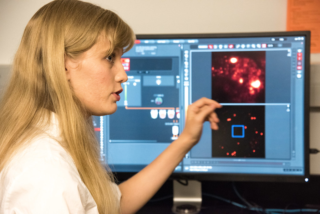
A sample is shown in the foreground that can be used in the digital light sheet microscope at MU’s Advanced Light Microscopy Core as Anand Chandrasekhar explains how he uses it to study neuronal development in zebrafish. | photo by Roger Meissen, Bond LSC
By Phillip Sitter | MU Bond Life Sciences Center
Microscopes have come a long way since Anton van Leeuwenhoek first looked at single-cell organisms in the 1600s.
Now, cutting-edge microscopes allow scientists a better look at how cells interact and work.
The results were easy to see Tuesday morning when a new digital light sheet illuminated all the cells in a zebrafish embryo of Anand Chandrasekhar, a Bond Life Sciences Center scientist and professor of biological sciences. The fish, with its two eyes, brain and spinal cord lit up like a green-colored digital ghost floating in invisible black waters of the monitor screen.
This new microscope joins an array in the Advanced Light Microscopy Core, or MCC, located at Bond LSC. The MCC is one of nine core facilities at MU that provide vital services across campus, from DNA sequencing to imaging.
Researchers and staff at the MCC showed on Tuesday how the new capabilities of the technology give them and MU a competitive edge in their research through better visualizations of their experiments.
The digital reconstruction produced by the light sheet combines “a whole bunch of images over an extended period of time,” Chandrasekhar said. Thousands of images of a cell or organism can be generated by the new equipment, at speeds of hundreds of frames per second, creating a picture that can easily take up a terabyte of hard drive space.
At those speeds, Chandrasekhar said he can “literally watch neurons in the brain light up” in real time. He observes how neuron changes occur in the fish’s brain as the animal goes about its different routine behaviors like avoiding possible predators and searching for food. With this capability, researchers like him can study how neuronal networks develop.
The new digital light sheet uses less damaging light than traditional microscopes and allows for clearer pictures, often with a 3-D look at their structure. More advanced imaging equipment also allow for faster, larger volume experiments that are gentler on the biological samples used in them.
Thomas Phillips, MCC Director and professor of biological sciences, explained how his center now can better serve scientists across campus.
In addition to the digital light sheet imaging system, the MCC also has two new super-resolution microscopy systems, what Phillips said researchers across campus expressed they needed most. The presence of these super-resolution systems in particular puts MU at the forefront of microscopic research.
“Whereas every school has a confocal, less than 10 percent have super-resolution capabilities,” Phillips said, and now MU has two super-resolution systems. “The new equipment adds totally new capabilities without interfering with the traditional confocal activities.”
Traditional confocal microscopy systems, while still useful, have limitations in their resolution which super-resolution systems overcome by how the specimens are illuminated.
In addition to having technology other institutions do not, MU has a higher quality version of super-resolution. Phillips explained that while systems at other institutions use two different lasers in the internal mechanisms of their imaging equipment, MU is one of less than 10 or 12 schools that has access to a super-resolution system that has a unique three laser combination that increases the resolution of the system.
“Super-resolution microscopy allows us to see how individual proteins are interacting inside cells in ways we haven’t been able to before,” he said.
Sergiy Sukhanov uses one of the super-resolution microscopes in cardiology research that looks at failure of organ systems. Sukhanov studies heart failure and atherosclerosis — the chronic, dangerous build-up of plaque in arteries that can create blockages that lead to heart attacks, strokes and death.
Sergiy Sukhanov explains how he uses a confocal microscope in MU’s Advanced Light Microscopy Core to study atherosclerosis and heart disease. | photo by Roger Meissen, Bond LSC
The associate research professor at MU’s School of Medicine showed on Tuesday images of protein interactions in the smooth muscle cells that line arteries. The goal of understanding these interactions is to help keep these smooth muscles in the healthy condition they need to be in to prevent catastrophic blockages.
With its capability of a resolution of up to 30 nanometers, Sukhanov said that the new equipment’s advantage for him is that he can actually see the cell he’s working on. Soon, he hopes to be able to work with live cells so he can observe changes in their protein structures in response to changes in their environment in real time.
Availability of the new equipment lured Sukhanov and his research team to MU over other institutions in 2014. He explained that he struggled to find the equipment he needed for his experiments at Tulane University in New Orleans or elsewhere in Louisiana. Other systems at other institutions usually only have a resolution of up to 50 nanometers.
Sukhanov’s decision is an example of the growth that the new imaging capabilities at the MCC can promote. As director Phillips explained, “you don’t plan experiments ahead of time if you don’t have the apparatus for it.”
MCC not only provides services across campus, but can give scientists insight into how to better look at their specimens.
First-year graduate student Jennifer Wolf displayed a super-resolution image of cancerous liver cells infected with Hepatitis C that been treated with a drug thought to prevent the virus from spreading. The drug aggregates viral capsid molecules – the outer part of a virus – within the infected cells to effectively contain them.
Jennifer Wolf, a first year grad student working in Stefan Sarafianos’ lab, explains an image of hepatitis C infected liver cancer cells captured by a super-resolution 3-D microscope housed at MU’s Advanced Light Microscopy Core. | photo by Roger Meissen, Bond LSC
Wolf said with the new equipment she is now able to see an individual molecule, and using that see the overlap of proteins, RNA and DNA fragments, which can help determine the effectiveness of drugs in treatment.
Associate director of the MCC Alexander Jurkevich explained that the super-resolution equipment also allows for the compilation of separate images into an even more detailed 3-D projection.
Wolf pulled up an image of green-colored mitochondria surrounded by red micro-tubules, green hubs of cellular activity connected by red highways that looked almost like a city from space at night. Wolf used the 3-D capability and rotated the image. She turned the biological intricacy on its side until it looked something like a galaxy on a cosmic horizon, only this view that maybe even fewer people have witnessed is microscopic.
“It’s important to use new technology like this to help the University of Missouri to stay on top,” Wolf said.
Bond LSC’s Matt Will explores the science behind food cravings in his research on mice.
Erik Potter from Mizzou Creative’s Eureka! Podcast tells us more in this month’s segment. Hear more of their science stories at news.missouri.edu/eureka
New outreach program teaches CAFNR students to make plant science knowledge accessible to a younger audience Written by Stephen Schmidt | Science Writer in the College of Agriculture, Food and Natural Resources
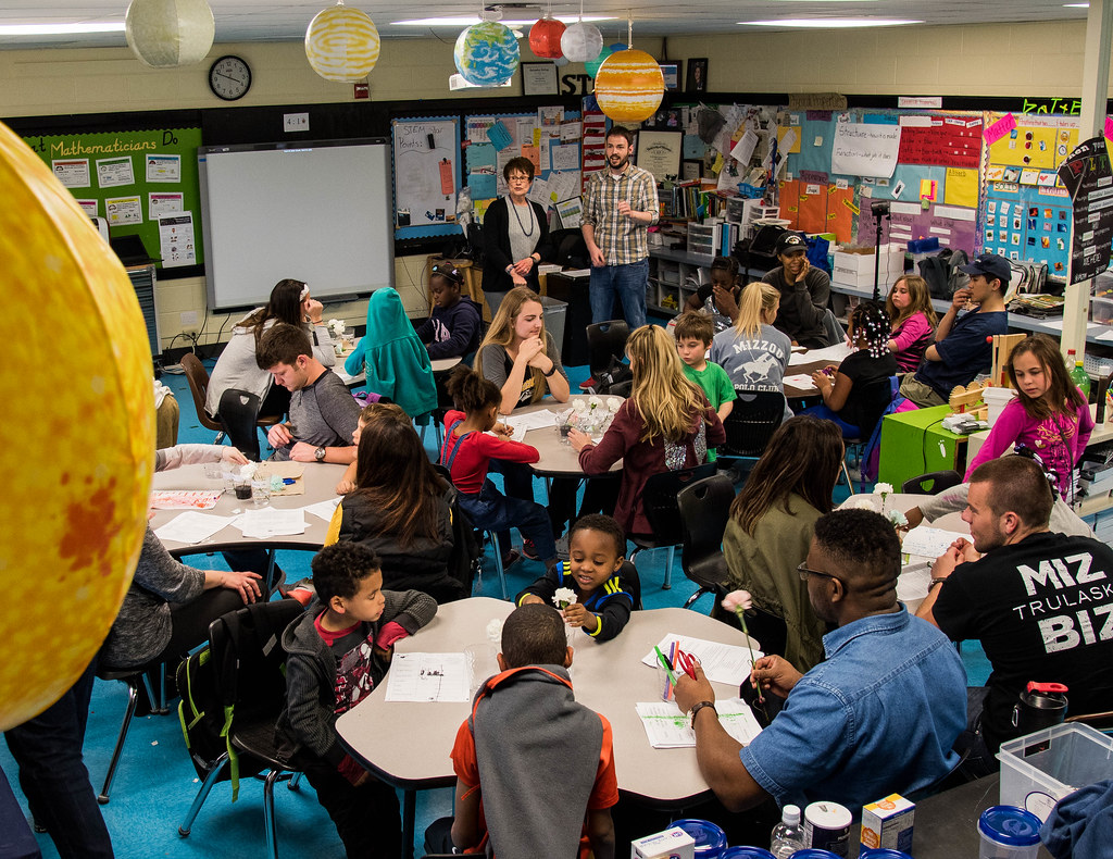
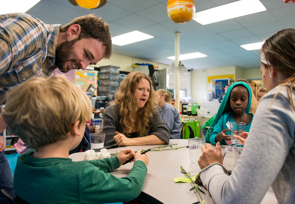
Although abundant light was shining through the windows, it was the quiet before the storm. Andrew Ludwig, a University of Missouri sophomore majoring in plant sciences, surveyed the small tables and chairs spread out before him in the laboratory of the Benton STEM Elementary School on a recent Monday afternoon. He sifted through his notes. He was ready, even though it was his first time stepping foot in the building — and he was about to talk to a crowd of students spanning from the first to the fifth grade.
MU graduate student Michael Gardner addresses the students gathered at a recent meeting of the Benton Elementary Science Club. Gardner co-presented a talk called “How Do Flowers Drink?” as part of the “It’s All About Plants” series that was launched earlier this spring. | Photo by Roger Meissen, Bond LSC
“I’m just going to try to stay enthused about everything because I’ve talked to some of my friends who are education majors and the big thing with conveying information to them is just being enthusiastic about it,” Ludwig said about his strategy on the “How Do Flowers Drink?” presentation he was about to give with fellow MU student Michael Gardner.
MU sophomore Andrew Ludwig talks with Leo Batchelder-Draper during an exercise exploring how plants absorb nutrients as his mother, Miriam Batchelder, looks on. | photo by Justin Stewart, Bond LSC
“I think the key is keeping them moving along, keeping them interested, because as soon as you lose their attention, they’re going to do whatever they want to do, so it’s going to be keeping things moving along at a pace that they’re still getting it, but that they’re not bored out of their minds,” added Gardner, who is a fifth-year graduate student specializing in plant stress biology. “We’re talking about water transport in a plant, which is a concept you can spend a semester on in a college-level course and still not be considered an expert on the topic.”
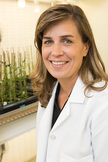
Both Ludwig and Gardner are a part of the team at the Bond Life Sciences Center lab of Melissa Mitchum, an associate professor in the Division of Plant Sciences. It was Mitchum who was able to launch the new “It’s All About Plants” series this spring with the Benton Elementary Science Club thanks to the science outreach portion of a new National Science Foundation grant that will further delve into a deeper understanding of the interactions between plants and parasitic nematodes she received in August and a partnership with the MU College of Education’s ReSTEM Institute, MU Office of Undergraduate Research and Columbia Public Schools.
The end result is a program that fosters learning of all varieties: The elementary students learn about plants, while the college students learn how to take complex ideas and break them down to a more accessible level. Furthermore, the program pairs undergraduate students such as Ludwig with graduate students such as Gardner and principal investigator mentors from across campus — involving the existing NSF-funded initiative Freshman Research in Plant Sciences (FRIPS) and the Students for the Advancement of Plant Pathology (SAPP) in the process.
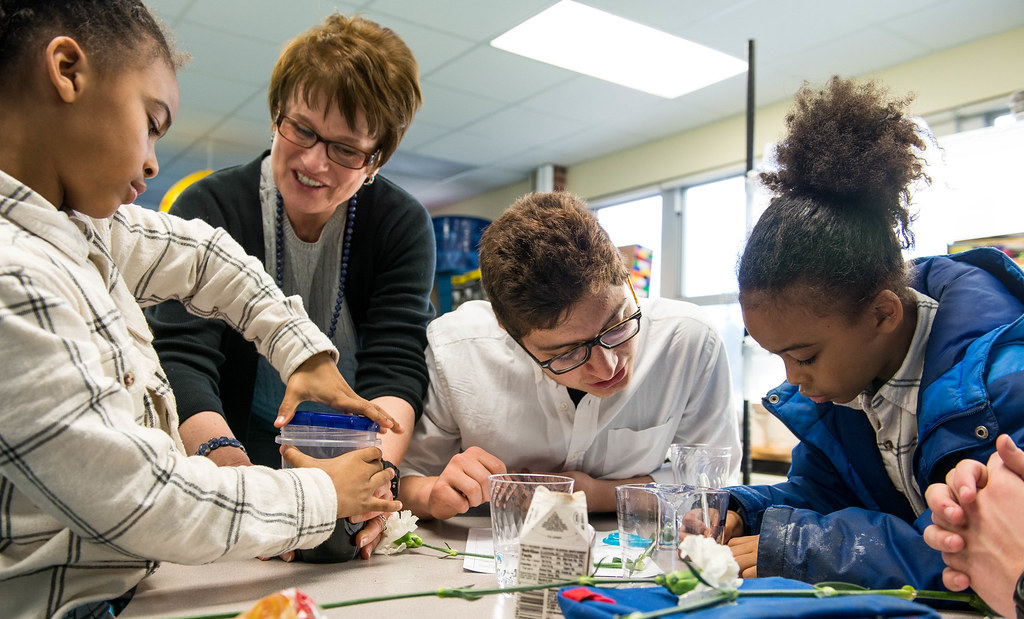
Deanna Lankford, a research associate from the MU ReSTEM Institute who helped found the Benton Elementary Science Club, helps Adrion Bradshaw open a container of blue food coloring as his twin brother Amahdrion fills writes out some observations with the help of volunteer Alp Kahveci. | photo by Justin Stewart, Bond LSC
“I think it’s a challenge for all of us who have advanced degrees to really think about where we were long ago and bring the concepts down to a basic level,” Mitchum said, “but I think it’s very important for us to be able to communicate our science at that level to really get the kids excited about research.”
Melissa Mitchum | photo by Roger Meissen, Bond LSC
She added that the program “really reinforces the concept of engagement and active learning.” In particular, it reinforces hands-on learning through a variety of activities. “I think that makes a big difference in learning,” said Mitchum, who will have sat with the children for five of the 10 presentations that started on Feb. 22. “I think that’s one of the reasons why I’m such a strong advocate of undergraduate research and getting in a lab and learning how to work in a lab. The same thing applies here. When the kids are doing experiments in the science club, they’re going to retain that information a lot more.”
School’s in session
“Class! Class! Class!”
“Yes! Yes! Yes!”
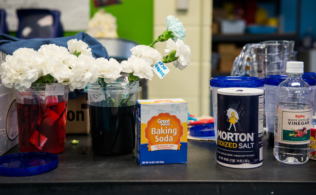
With those loud words reverberating through the room Ludwig had the attention of those before him — for a moment, anyway. The chatter had quieted down as he began to explain the main activity of the day, which would involve carnation flowers and six solutions of household items (sugar, salt, baking soda, vinegar, soda and food coloring) mixed with water in the same-sized cups – and one scenario with just water.
With the help of several volunteer MU students (whose hours are coordinated by Mitchum’s co-PI, Deanna Lankford, a research associate in the College of Education), the assignment was to pour 100 millimeters into each one of the cups.
The cups were then labeled with the name of the solution. This procedure was repeated three times. The white carnations were then cut to the height of the cup and placed in the water as a discussion ensued on what solutions would help the plant (besides plain water) and which ones would hurt.
Afterwards through the help of a video, the class was introduced to the idea of setting up controls and variables. The one variable, in this case, was the solution. All other factors (such as flower height, flower type, cup size and amount of liquid) were controlled, Ludwig explained. “We use the control to compare our other variables,” he said.
A moment later, Ludwig posed a question to the class: “Raise your hand if you think putting the flower in food coloring is going to change the color of the flower.” A collection of hands sprouted upward.
“We have some flowers that we put in the dye yesterday,” Ludwig continued, as he unveiled carnations that had white petals either tinged in red or blue to pass around to the class.
“Woah!”
“I was right!”
“I knew it!”
“Do we get to keep them?”
A white carnation begins to show traces of blue food coloring in its petals after sitting in a solution with food coloring and water for a short period of time. | photo by Justin Stewart, Bond LSC
A white carnation begins to show traces of blue food coloring in its petals after sitting in a solution with food coloring and water for a short period of time.
Following a brief video showing a similar experiment relating to celery and food coloring, Gardner explained the phenomenon to the class: “The plant is basically a big straw, so as the water evaporates up the plant, it pulls more water with it and then the food coloring too, so it gets to the very top of the plant, either the leaves of a tree or the top of a flower, and when it gets there the water will be able to evaporate, but the food dye can’t so that’s why your flowers are turning blue or red, OK?”
The food coloring portion of the afternoon turned out to be the top highlight for many of the participants. When asked about all of the projects he had worked on during the spring, Amahdrion Bradshaw, a second grader, proclaimed that “the funnest one was about dying the plants.”
Haily Korn, a fellow second grader, agreed, saying that her favorite part of the program is when you “do fun experiments” and that her favorite experiment was “when you get to dye stuff.”
A perfect fit
Lankford and the ReSTEM Institute formed the Benton Elementary Science Club, which meets one afternoon every week during the school year, seven years ago — the same time Benton officially became a part of the STEM (Science, Technology, Engineering and Mathematics) program.
“The science club has allowed our students to continue to expand their understanding of a variety of science topics through hands-on experiences after school,” said Heather McCullar, a STEM specialist who works at Benton. “The kids always leave club excited to share what they have learned with their families.”
Over the years, Lankford has helped many principal investigators with the education portion of their grants. When Mitchum asked her about getting involved with an existing partnership, Lankford immediately thought about the science club with its previously established learning format.
“Melissa said ‘I need help with this.’ And I said ‘Great, let’s talk,’” Lankford said, “And we did and we came up with the idea and it has been wonderful. I really like the fact that we have mentors and mentees doing the presentations.”
Under Mitchum’s direction, a series of meetings were set up with mentors and mentees last fall to develop the curriculum and lesson plans that would form the backbone of the course. Given that the grant is funded for a total of three years, the plan is to continue to teach the “It’s All About Plants” program, which has been well-received by students and school administrators alike, at Benton next spring.
Besides Mitchum, the other PI mentors from CAFNR who have taken part in the program are Gary Stacey, Curators Professor of Plant Sciences; Lee Miller, assistant professor of plant sciences; Xi Xiong, assistant professor of plant sciences; Walter Gassmann, professor of plant sciences; John Boyer, Distinguished Research Professor of Plant Sciences; Harley Naumann, assistant professor of plant sciences; Kevin Bradley, associate professor of plant sciences; Heidi Appel, senior research scientist, plant sciences; Jack Schultz, professor of plant sciences and director of the Bond Life Sciences Center; and Scott Peck, associate professor of biochemistry. PI mentors from the College of Arts and Science Division of Biological Sciences included Paula McSteen, Chris Pires, and Mannie Liscum.
The CAFNR students who took part in the science club series planning also recently hosted an outreach booth on t the Science Sleuth event on “Plants and Microbes” the MU campus April 16. In addition, Mitchum gave a talk at the Exploring Life Sciences symposium at the Bond Life Science’s Center recent Missouri Life Sciences Week.
Furthermore, the end goal is to take all of the lessons that are being created and turn them into a booklet that is easily accessible for teachers in Columbia Public Schools, and beyond, by posting the material online.
In the meantime, the lessons are being molded by the interactions of CAFNR students and those at Benton.
At a recent session of the “It’s All About Plants” series at the Benton Elementary Science Club, students learned about how plants transport water by conducting tests with a variety of solutions made up of household products, including, from left, baking soda, salt and vinegar. | photo by Justin Stewart, Bond LSC
“You have some students who can’t write their names yet, and other people who know everything we’re trying to tell them before we start,” Gardner said, afterwards, referring to the beginning of the class when a student at the front of the room recited a brief and succinct view of a plant’s water transport system. “Apparently his grandma told him all of this stuff already. So yeah that was definitely a challenge that I think we did OK at it.”
“I think it’s really great for everyone involved from the elementary school students who are getting to learn about specific topics from people who are very well informed about it to undergraduate and graduate students who are getting a wider arrange of presentation skills,” Ludwig added. “You can get locked in your head about your research. It’s good to be reminded that the knowledge we gain through research also has a real-world application and that part of the scientific process is sharing.”
It’s a challenge that Mitchum said could serve as a benefit to anyone in the scientific community.
“It’s not easy for us to do many times,” Mitchum said of breaking complex idea down into something easy enough for a child to understand, or even an average adult. “It takes practice. So. It’s something we have to learn. Science communication is very important these days. Especially when we talk about what we’re doing in a lab, it’s very molecular, very cellular, but we have to find a way to make it relevant.”
She added that programs such as this one show children that not all scientists have “crazy hair with the goggles and lab coat. They see ‘Oh, these guys are just like everyday people.’ This is a realistic career for them.”
Would Amahdrion be interested in such a career path?
“Nope,” he said. “I want to be a basketball player.”
Still, when given the choice of attending the science club sessions or just going home after school, he has a quick reply: “Spending extra time at school because you learn more.”
Work on HIV capsid proteins earns prestigious retrivology award
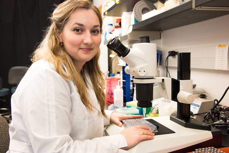
Anna Gres studies HIV capsid protein using X-ray crystallography. She recently won the 2016 von Schwedler Prize, which awards her $1,200 and gives her the oppportunity to speak this spring at the Cold Spring Harbor Retrovirus Meeting, one of the largest retroviral research conferences in the world. | photo by Roger Meissen, Bond LSC
Science is all about structure in the work of Anna Gres.
For the past four years, she’s looked closely at one HIV protein to figure out its shape in order to stop the virus.
“Capsid protein is extremely important during the HIV life cycle. About 1,500 copies of it come together to form the protective core around the viral genome,” said Gres, a graduate student in the lab of Bond Life Sciences Center’s Stefan Sarafianos and a Mizzou Ph.D. candidate in chemistry. “So, if you are able to somehow disrupt the interactions between the proteins or make them different, the virus loses its infectivity.”
Gres takes her work on this protein to a national stage next month when she speaks at the Retroviruses meeting at Cold Spring Harbor Laboratory — one of the most prestigious international conference on retroviruses — as the recipient of the 2016 Uta Von Schwedler prize. The prize recognizes the accomplishments of one distinguished graduate student as they complete their thesis.
HIV capsid protein has been studied for almost 30 years, but it’s been tricky to get a precise depiction of what it looks like. Gres uses X-ray crystallography to essentially capture the protein in all its 3-D glory. This method gives scientists the higher resolution picture to study the molecular structure of capsid protein. Her work allows the Sarafianos lab and others to study how it interacts and connects with other capsid proteins and the host protein factors of the cell HIV is trying to take over.
“In the past scientists had been splitting the capsid protein in two halves and crystallizing them separately. Another approach was to introduce several mutations to make it more stable,” Gres said. “You would think that it shouldn’t really matter if we have a few mutations, but the protein behaves in such a way that even slight changes result in subtly different interactions that are enough for the virus to lose its infectivity. We were able to crystallize the native protein without any mutations and that should give us more accurate picture.”
Now that the Sarafianos lab and Gres have a good idea of what that native protein looks like, they’ve moved on to other mutated versions of the protein that impair virus infectivity. This could give them insight into how scientists can stop HIV.
“Many labs reported numerous mutations in the capsid protein over the past 25 years that either increase or decrease the stability of the core, which often results in a noninfectious virus,” she said. “Right now we are interested in seeing what structural changes accompany these mutations and how they can affect the overall stability of the core.”
It’s been almost a week since the 2016 MU Life Sciences and Society Program Symposium, “Confronting Climate Change” wrapped up. If you missed the event, check out our Flickr gallery to see a little bit of the excitement.
We also had the chance to chat a few minutes with most of the LSSP speakers who graciously shared their insight on climate change in our lives. Check out these conversations on our YouTube page from the link at the top of the page.
Last but not least, we put together a radio piece for KBIA giving some speaker highlights. Visit our SoundCloud to see more.