Bond LSC principal investigators Bing Yang (left) and Ron Mittler were once again recognized on the list of Highly Cited Researchers. | photos by Roger Meissen, Bond LSC
By Roger Meissen | Bond LSC
Discoveries in research come with time, so the incremental accumulation of knowledge toward breakthroughs is fundamental to science and the future.
While many contribute to this understanding, a few scientists consistently produce research that others note more often in their own experiments. Two University of Missouri Bond Life Sciences Center researchers once again landed on that list of most cited scientists.
Bing Yang and Ron Mittler are included on the list of Highly Cited Researchers for 2023. The inclusion stems from authoring multiple highly cited publications in 2023 that rank in the top 1% by citations for their field.
This year marks the fifth straight year of inclusion in the list for Yang and the fourth year running for Mittler.
Only one in every 1,000 scientists receive this honor, and they join 6,849 researchers globally who made the Clarivate list. Clarivate runs Web of Science, a database aggregating published, peer-reviewed research from academic journals, conference proceedings and other citations.
Yang, a Bond LSC scientist and MU professor of Plant Science and Technology, works on targeted genome editing in plant species, including rice, maize, wheat, sorghum and soybeans. His lab aims to understand interactions between plants and pathogens like bacterial blight in rice so they can work toward varieties that can better withstand the disease, among other projects.
Mittler, a Bond LSC scientist and MU Curators’ Distinguished Professor of Plant Science and Technology, researches the role of reactive oxygen species (ROS) and its function in regulating biological processes in plants.
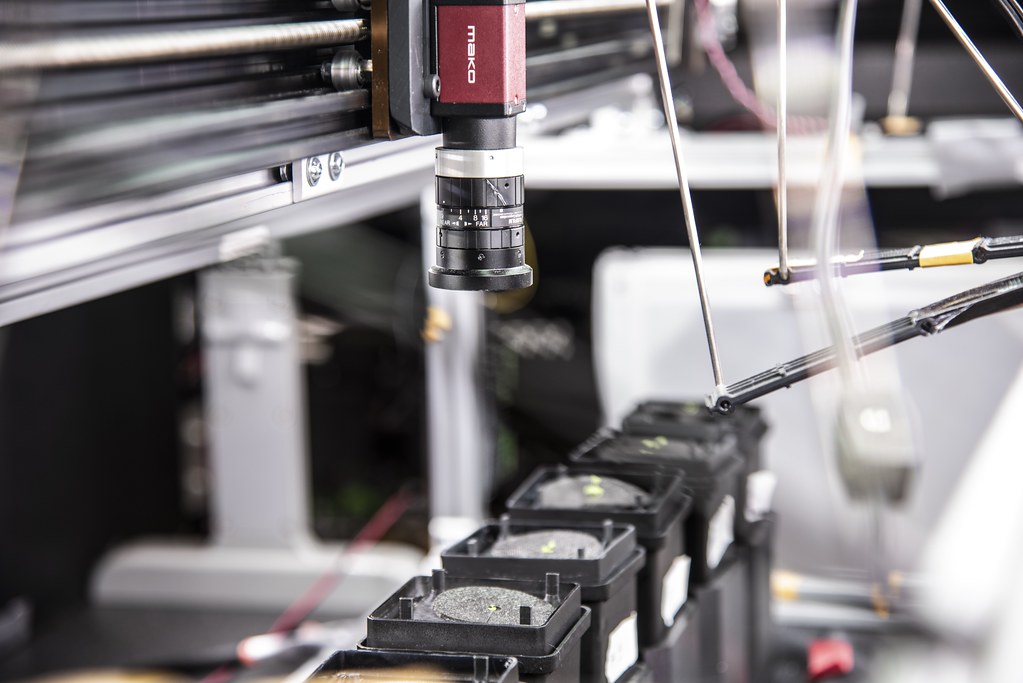
Landon Swartz, a graduate research student at the University of Missouri in the David Mendoza-Cózatl lab at Bond LSC, looks at the camera on the OPEN leaf machine and makes adjustments to the plant the robot is photographing. | Photo by Sarah Kiefer, Bond LSC
By Sarah Kiefer
The LED lights danced as the OPEN leaf system powered up. Quickly, the robot zips down the track to its preplanned destination, hovering above each plant sitting atop a 3D-printed mechanism, then the camera snaps a shot as it conducts this same routine every 30 minutes.
This open source, data driven tool is one way scientists like David Mendoza-Cózatllab at Bond LSC and work to simplify the tedious data collection required in plant research.
The OPEN leaf system — described in The Plant Journal in September 2023 — uses modern technology to do its job better and gather results faster than other machines of the same kind. But this National Science Foundation funded project is more than just a robot taking a picture of a plant.
The camera for the OPEN leaf system travels across its track, snapping photos of the growing plants along the way. | Photo by Sarah Kiefer, Bond LSC
“Phenotypes are a change in a plant that you can visualize. Our goal is to characterize phenotypes through time,” said Mendoza-Cózatl, a Bond LSC researcher, director of graduate studies and associate professor of Plant Science and Technology at the University of Missouri. “In my mind, time is one of the black boxes that we have not been able to solve because it is variable.”
Color plays a big role in measuring change in a plant. Using color quantization techniques that simplify colors into numbers, researchers can identify four basic colors to analyze. The machine captures RGB (red, green, blue) images using a camera and converts it into a LAB color space to do the heavy lifting. L stands for lightness, whereas A and B represent the green to magenta and blue to yellow color spectrums detected in something like a leaf of the plant.
“Color is a subjective thing to a lot of people and it’s a lot easier to look at a number and say this number is more than this number, but saying this color is more yellow than this one is a harder thing to recognize,” said Landon Swartz, the Mizzou computer science graduate research assistant who engineered the system. “Our process correlates the human perception of color which is actually very different from the way a computer sees color.”
Landon Swartz is a graduate research student at the University of Missouri who engineered and worked on the OPEN leaf system in the David Mendoza-Cózatl lab. | Photo by Sarah Kiefer, Bond LSC
When the colors are quantified, then the real work begins — plotting the points.
This procedure morphs a primarily visual measurement into a mathematical one. Just like with numerical data, Swartz can take colors and observe how they change over time by using coordinates from his simplified combinations.
“It’s a really dynamic process and something that’s not thought about a lot, but because no one’s ever done it before, it’s very hard to convince people it’s accurate and try to visualize or explain the process to others,” said.
Mendoza-Cózatl said this way of quantifying and recording takes the system to the next level.
“We thought this idea of describing color through time was solved a long time ago. Turns out, it was not,” Mendoza-Cózatlsaid. “Extracting the color of a plant has been done, but representing the color to viewers over time is something that has not been done.”
The team differentiated their system from others by considering a hybrid work environment to allow many collaborators through the Slack communication app. The app can be used to chat directly with the robot itself.
“We are trying to advance the field of phenotyping as a group,” Mendoza- Cózatlsaid. “It’s not just my machine, it can be your machine or anyone’s machine.”
Slack was first suggested by Drew Dahlquist, a former undergraduate in the Mendoza-Cózatl lab, during the COVID-19 pandemic when research labs needed to cut out the middleman on projects that require collaborators from afar.
“Let’s say I’m sitting in my basement, the experiment is running and I want to know how it’s going. I can message the machine ‘hey, can you send me the last couple of pictures that you took’ and the machine will send them,” Mendoza-Cózatl said. “The element that sets this system apart is you can then share it with colleagues, students, or anyone.”
Swartz and his collaborators worked to incorporate the details of the plant, not just the entire rosette — the circular arrangement of leaves on the plant. Their system recognizes each individual leaf without bias.
But how can observation of a leaf be biased in the first place?
Traditionally, these systems prompt the user to draw a line around each leaf for it to be recognized, which is a subjective observation based on how each person draws their boundaries.
The OPEN leaf system’s computer algorithm automatically recognizes each leaf to be analyzed separately.
The Arabidopsis thaliana, a model organism plant that Swartz works with sits on top of 3D printed plastic floaters that create a hydraulic watering system while the camera continues to take photos when prompted. | Photo by Sarah Kiefer, Bond LSC
“To me it’s really exciting because I was trained as a biochemist then I decided to work with plants. I never thought I would be publishing papers about computer vision and phenotyping,” Mendoza-Cozatl said. “It gives a fantastic example that science is really open. We saw a need, so we tackled the problem and came up with a solution that is very rewarding.”
Their new machine also makes the database more accessible and cheaper than other similar machines. Costing less than $3,000 to build, the blueprints and instructions to build the OPEN leaf system are accessible to anyone who wants to put it together.
“This type of project shows that you can do a lot with a little because you don’t need to pay thousands of dollars to get the same amount of insight into your research,” Swartz said. “This project has become a tool that we are going to use for papers to come, and we are hoping that other people will start to use it for their papers as well.”
The collaboration between engineers and biologists helped them address the evolving nature and nuanced challenges of the project.
“The most interesting part about this project is that there is a novelty to working in an interdisciplinary manner like this,” Swartz said. “Usually, how it works is someone comes to you with a problem and you present a solution. But here we are coming up with solutions to find the problems, which I think is a very fruitful approach.”
Swartz is already working on his next big project — an OPEN root system. This machine will accomplish similar goals but apply to the root system of the plants instead of the leaves.
The paper titled, “OPEN leaf: an open-source cloud-based phenotyping system for tracking dynamic changes at leaf-specific resolution in Arabidopsis” by Landon Swartz, Suxing Liu, Drew Dahlquist, Skyler T. Kramer, Emily S. Walter, Samuel A. McInturf, Alexander Bucksch, and David G. Mendoza-Cózatl was first published in The Plant Journal on September 21, 2023.
Marc Libault, a new Bond LSC principal investigator and professor of professor of plant science and technology in the College of Agriculture, Food and Natural Resources at the University of Missouri. | Photo by Sarah Kiefer, Bond LSC
By Sarah Kiefer | Bond LSC
Marc Libault only ended up one floor up from his old stomping ground in his recent move.
Libault — the most recent MizzouForward hire at Bond Life Sciences Center — returned this fall to bring his expertise in plant single cell biology to MU.
“What is exciting about this research is its innovation to generate a new knowledge in crop science; we’re the first group in the world to develop a very broad application of single cell biology in plants,” said Libault, Bond LSC principal investigator and professor of plant science and technology in the College of Agriculture, Food and Natural Resources. “I was excited to come back here to continue to work on solutions at the single cell level notably by working in collaboration with experts in proteomics, metabolomics, and phenomics at MU.”
Single cell biology is exactly what it sounds like: singling out individual cells in a plant in order to reveal their unique molecular features and mechanisms. This field can help scientists better understand the functions of different genes and proteins.
“If you want to engineer a complex machine such as a car, to enhance its performance you need to first understand the nature and the contribution of each part and how they work together,” Libault said. “In plants, you need to understand how each gene contributes to the biology of the cell and how each cell plays a part in the entire plant before creating meaningful genetic strategies to enhance crop biology. Having this more accurate picture allows us to enhance our current knowledge in plant and cell biology.”
While cutting and chopping are more common descriptors in cooking, researchers in Libault’s lab chop a plant sample in order to isolate the nucleus of the cells to study gene activity. His lab also focuses on spatial transcriptomics, a process that looks at how cells use their genes within the organs they inhabit. If they can understand the contribution of each cell in the plant and how they are interacting with one another, they can grasp how they work together to respond to environmental stressors.
“In society, each individual has unique competences and expertise. They work together to accomplish one goal. That’s similar to cells with unique functions working together in an organs to support its function,” he said.
By pooling this information together, they hope to identify important regulators that control how other genes are expressed then manipulate those molecular chains to see how the plant improves.
Libault is not exactly a newcomer at Mizzou.
Back in 2005, Libault came to MU from Paris, where he grew up. He worked as a postdoc in the Gary Stacey lab until 2011 when he became an assistant professor at the University of Oklahoma. He stayed there until 2017 when he moved into an associate professor position at the University of Nebraska. He got his bachelor’s degree from Denis Diderot University in molecular and cellular plant physiology and his master’s degree from Pierre et Marie Curie University in cellular biology and physiology. He went on to earn his Ph.D. in molecular and cellular plant physiology from Paris-Saclay University.
But, Libault’s last 20 years of research have been focused on understanding the mechanisms controlling gene activity including the role of epigenetics — the study of how environment and behavior change the way your genes work — helping him tackle questions like “how does the expression or repression of genes change from cell to cell?” This gives him a unique perspective as he learns to recognize the systems behind gene activity and apply that to his work on a single cell level.
The interaction between roots and bacteria is one of the areas of exploration of the Libault lab at the single cell level. In legumes like soybeans, bumps called nodules grow on the plant roots to facilitate the relationship between plant and bacteria. This nodulation gives the bacteria a place to live while it converts atmospheric nitrogen into food for itself and the plant.
“The bacteria capture the atmospheric nitrogen and convert it into a source of nitrogen supply for the plant,” Libault said. “In return, the plant will give sugar to the bacteria, which is why the two help each other.”
Outside of the lab, Libault enjoys watching Formula 1 racing or movies from the ‘70s and ‘80s in his free time. He is also interested in keeping up with French and European news. Although his balance between work and home life can become challenging at times, when things get hectic Libault prioritizes family time with his wife and three teenage boys as they continue to settle into Columbia after a few years apart.
Libault has been able to hit the ground running in his lab because much of his team made the trek south with him from Nebraska in September.
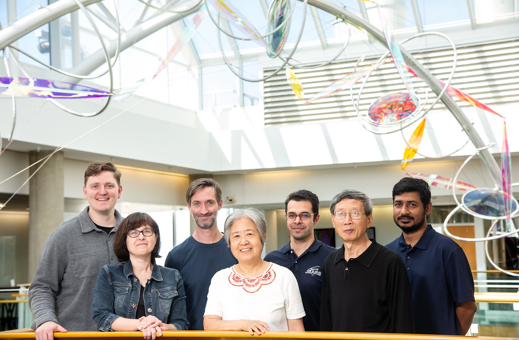
The Marc Libault lab at Bond LSC. From top left to bottom right, Sutton Tennant, master’s student; Marc Libault, Bond LSC principal investigator and professor of plant science and technology in the College of Agriculture, Food and Natural Resources; Sahand Amini, postdoctoral fellow; Md Sabbir Hossain, Ph.D. student; Sandra Thibivilliers, research specialist II; Hong Fu, lab technician; and Hengping Xu, postdoctoral fellow. | Photo by Sarah Kiefer, Bond LSC
Libault’s work necessitates collaboration to bring all the pieces together, and that’s what drew him back to MU.
“I know how collaborative the university and the researchers working here are, so that was a strong motivation for me to come back here and continue that kind of collective work,” Libault said. “What we have developed has been highly successful and that’s fun, but what’s next? What more can we learn? This is just the beginning of my next 25 years of research.”
MizzouForward is a transformative, $1.5 billion long-term investment strategy in the continued research excellence of the University of Missouri. Over 10 years, MizzouForward will use existing and new resources to recruit up to 150 new tenure and tenure-track faculty to address some of society’s greatest challenges. Investments also will enhance staff to support the research mission, build and upgrade research facilities and instruments, augment support for student academic success, and retain faculty and staff through additional salary support.
The Bengal tiger was one of five feline lineages compared to gives a comprehensive look at genome sequence structures that could have driven the evolution of distinct cat species. This new reference genome is comparable to the human genome in terms of its completeness, and could be used to for feline veterinary precision health. Denise Allison Coyle/Shutterstock
First-ever analysis compares nearly gapless genome across cat species and with humans to shine light on evolution
By Roger Meissen | Bond LSC
The tiger doesn’t know it, but a difference deep in its genome sets it apart from other cats.
This big cat preserved a distinct sense of smell thanks to a few chromosomes it uniquely retained over millennia of evolution that other feline species did not.
A study published in the journal Nature Genetics details this finding where scientists from the University of Missouri partnered with Texas A&M University and others to compare, for the first time, nearly gapless cat genomes across multiple species and with humans. This genome map correctly strings together nearly all chromosomes of these felines — from one end to the other without missing segments — to give an animal genetic reference that rivals the human genome.
Wes Warren, Bond LSC principal investigator and professor of genomics | photo by Roger Meissen, Bond LSC
“This study is a comprehensive look at the genome sequence structures that could be driving cat speciation, the evolutionary process by which populations became distinct species,” said Wes Warren, a Bond Life Sciences Center researcher and MU professor of genomics. “We found distinct differences between how great apes, including humans, and cat sequences evolved in a similar period of time. There were many interesting differences to consider, such as why certain chromosome regions have similar
but novel sequence landscapes even across species of cats.”
In tigers, scientists previously identified genes associated with the chemosensory system, perhaps lending them heightened senses of smell key to their survival.
“We see cats have olfactory receptors for sensing smell that are greater than most mammals but, among cats, the tiger stands out with the largest repertoire,” Warren said. “We know tigers have a solitary lifestyle, and that acute sense of smell helps males detect females for mating and lets them avoid the territory of other males to enhance their chances of survival.”
When thinking about other unique sensory features to explore, researchers also looked to the fishing cat, a rare nocturnal feline native to marshy areas in Southeast Asia. They searched for molecular signatures that would explain its sleek body, partially webbed feet and prowess for hunting fish, characteristics tailored to its life around water.
Scientists looked at the fishing cat, a rare nocturnal feline native to marshy areas in Southeast Asia. They identified molecular signatures that explain its prowess for hunting fish and characteristics tailored to its life around water. They found more complete genes to detect water-borne smells than in other feline species. | Attribution: Kelinahandbasket, Prionailurus viverrinus 01, CC BY 2.0
“There are specific olfactory receptors that detect waterborne odorants as well as a much larger number of receptors tailored for terrestrial smell detection; we asked if fishing cats would have more functional receptors for waterborne odorants than other cats, and that’s exactly what we saw,” Warren said. “In fishing cats, all gene copies were complete while other cats had broken copies, suggesting natural selection was at work because of their aquatic hunting behavior.”
While these illustrations are notable, scientists also learned from their extensive analysis of sequence structural variation.
What stood out overall were fewer differences in cat chromosomes — coding for traits like hair length and color, bone structure and size, fertility and senses — compared to great apes despite both families diverging into distinct species over a period of 13 million years.
Using the latest assembly techniques, the scientists pieced together the genomes of multiple cat hybrid species — the Bengal cat, the safari cat and the liger — and then compared the five lineages. New techniques severely limited genomic dark matter, which is genetic information researchers previously were unable to characterize and assign meaning to, thus enabling them to shine new light on this novel sequence structure.
Some of those pieces came from Leslie Lyons, the Gilbreath-McLorn Endowed Professor of Comparative Medicine in veterinary medicine and surgery at the MU College of Veterinary Medicine. She sequenced two of the cat hybrids nearly 25 years ago.
“I was at the National Cancer Institute as a postdoc when I sequenced the Geoffroy’s cat and Asian leopard hybrids, and I had no idea they would ever be reused for a project like this, but fortunately they were,” Lyons said. “Now, we have a cat genome that is comparable to the human genome, which is one of the best in the scientific world, so the cat has really leapt forward in terms of genomic resources.”
Structural differences in the X chromosome, in particular a region that displays features of a supergene, stood out to the team. This presumed supergene in cats, a complex interaction where a chromosomal region of neighboring genes are inherited to fix a trait with fitness consequences, could be the process of evolution that led to hybrid sterility if certain feline species cross. It is evolving faster than most of the cat genome. Much like the mule — a cross of a male donkey and a female horse — cannot produce offspring, the liger bears no offspring when lions and tigers mate. The team hypothesizes this is due to a failure in meiosis, the process that produces egg and sperm cells.
“We found multiple inversions on X chromosomes of interest in cats that include the complex DXZ4 region,” Warren said. “In these cats, we identified entire copies of this complex, repetitive region that harbors the only X-linked speciation gene identified in mammals.”
While these advances in genetics push our understanding of evolution in exciting new ways, Lyons has much more pragmatic uses for this work.
“I’m an end user,” Lyons said. “These good genetic maps allow us to find mutations that cause inherited diseases in cats, so this genome allows us to bring precision medicine to feline veterinary health care.”
“Our findings will open doors for people studying feline diseases, behavior and conservation,” he said. “They’ll be working with a more complete understanding of the genetic differences that make each type of cat unique.”
Nature Genetics published “Single-haplotype comparative genomics provides insights into lineage-specific structural variation during cat evolution” Nov. 2, 2023. The study was conceptualized by Bill Murphy — VMBS professor of veterinary integrative biosciences at Texas A&M and Wes Warren — principal investigator in the Bond Life Sciences Center and professor of genomics in the College of Agriculture, Food and Natural Resources at the University of Missouri. Additional collaborations involved researchers from the University of Washington, University College Dublin, the Institute for Systems Biology in Seattle, Louisiana State University and the Guy Harvey Oceanographic Center.
This work was funded through a grant from the Morris Animal Foundation, which works to improve and protect the health of animals through scientific innovation, education and inspiration.
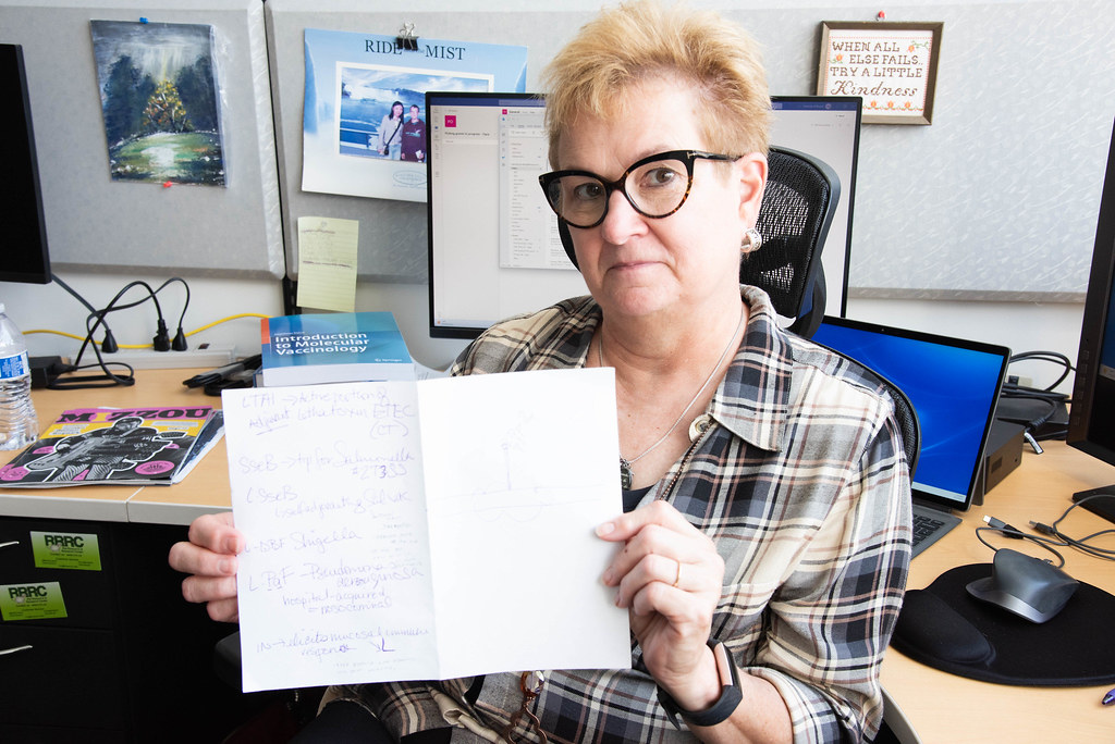
Wendy Picking, professor of veterinary pathobiology and principal investigator at Bond Life Sciences Center sits in her office and shows off some noteså she jotted down about the vaccines she’s working on. | Photo by Beni Adelstein, Bond LSC
By Beni Adelstein
Wendy Picking uses the power of proteins to fight pesky pathogens like Pseudomonas aeruginosa.
Picking and her team are one step closer to completing their mission to develop weapons like vaccines to fight against this bacterium. In March, they published these findings in Nature’s Journal, npj Vaccines.
“If your immune system is weakened and you get Pseudomonas in the hospital, you’re probably going to die,” said Picking, a principal investigator at Bond Life Sciences Center and professor of Veterinary Pathobiology. “We’re doing the basic research, so one day we can make a vaccine to prevent Pseudomonas, so you don’t die in the ER.”
Wendy has spent years developing vaccines against Pseudomonas aeruginosa and other bacteria like Shigellaflexneri with husband, Bill Picking, who focuses on the inner workings of the bacteria.
Pseudomonasaeruginosa causes pneumonia — a lung infection — while Shigella species causes diarrhea. Neither of these are any fun. These and other bacterial infections grow increasingly resistant to drug treatment in hospitals and beyond, so scientists need new ways to combat these pathogens.
A vaccine prepares the body to fight those bothersome bacteria.
By introducing a protein called an antigen into the body, it mimics an infection to trigger a defensive response. It’s like that antigen is a mug shot, so the body’s immune system recognizes what it’s up against and builds antibodies — a weapon tailor-made to fight it. The next time that pathogen tries to infect, the body will have had a way to fight it off.
For Wendy, making proteins that exist in the bacteria work toward this purpose is key.
In 2020, she earned her claim to fame — the discovery that fusing proteins together from the tip of the bacteria’s needle-like syringe amplifies an immune response to a vaccine to help fight off infection. Her lab was the first to discover this method works for these specific pathogens of Pseudomonas and Shigella.
“Producing proteins is expensive so we fuse the two tips to create a self-adjuvating protein, which combines two proteins to trigger and amplify a response,” Picking said.
When she says self-adjuvating, Wendy just means they created a new molecule that both stimulates the immune system and enhances that immune response. It’s like a go signal attaches itself to the vaccine’s proteins. The go signal tells the body to be more aggressive with fighting off those pathogens. Ideally, this will help fight off nasty symptoms of pneumonia or diarrhea.
These fused proteins originally helped the bacteria infect humans. They sit on the tip of a needle-like structure called a type 3 secretion system, which injects specialized proteins into cells to avoid being detected and attacked.
Wendy notes that developing the right animal models is one important part of creating a new vaccine. She has used mice for the Pseudomonas vaccine and rabbits for the Shigella vaccine.
“What you’re trying to do is mimic a human disease in a non-human model system, and you have to be sure that you’re stimulating the immune response in the right way,” she said.
Researchers observe how vaccines affect the immune systems of those animals and apply that knowledge to humans.
In their experiments, mice treated with these fused proteins survived being infected with Pseudomonas unlike the mice that received the placebo.
Now, Wendy’s team continues research on Pseudomonas and is shifting to research a Shigella vaccine. They will continue to test out the protein fusion method, refining vaccine candidates to fight off even more pesky pathogens.
Her most valuable findings are not limited to the experiments, and she said the most rewarding part of her work is seeing former students be successful.
“The most important lesson is picking your staff. Counting on your team, getting along with each other and working together is crucial,” she said. “I can now crash and burn; if these kids we work with are successful, then we’ve left the legacy that we wanted.”
“I want these vaccines to go to market, but we may be retired or dead when that happens.”
Wendy is looking forward to seeing what her group can come up with now.
New technologies like transcriptomics, RNA sequencing, and bioinformatics — available through MU’s research Core facilities in Bond LSC — will help them move their vaccine work forward to achieve this mission. Those pesky pathogens don’t stand a chance.
Wendy Picking’s most recent publication is “A protein subunit vaccine elicits a balanced immune response that protects against Pseudomonas pulmonary infection.” It appeared in the journal npj Vaccines in March 2023.
Lab of Olga Baker, a Bond LSC researcher and professor of otolaryngology at the University of Missouri. From left to right, Harim Tavares dos Santos, Olga Baker, Kihoon Nam, and Frank Maslow. | Photo by Sarah Kiefer, Bond LSC
By Sarah Kiefer
A digital declutter is a way to get rid of the seemingly endless files of old photos and documents, but when Harim Tavares dos Santos started sorting through computer files from the Baker Lab at Bond LSC, one image stood out and led him down a rabbit hole.
The picture showed tuft cells, a rare type of cell on his screen that seemed to wave hello with their finger-like structures.
“They looked different from other cells, so I looked into it more and I found that they actually had a name, but not much other than the basic research had been done on them,” said Tavares dos Santos – a senior research scientist in the Baker lab at Bond LSC.
Those tuft cells may be an important link in his study of Sjögren’s disease, a chronic autoimmune disease that destroys cells that make saliva and tears. He recently identified these cells in ducts – responsible for expelling saliva – of the submandibular glands across species in mice, pigs and humans using transmission electron microscopy, a process that can magnify a sample up to 2 million times its size.
Tuft cells — named after their tuft-like microvilli — serve as sentries on the surface of organs to detect chemicals then signal immune and nerve cells. In the gut, these specialized epithelial cells can sense chemicals from parasites and microorganisms to alert the body of the invaders. While first found in the intestines, they also line airways, nasal cavities and other hollow organs. For Tavares dos Santos, their presence in salivary glands provides a possible link to Sjögren’s.
Looking like a bottle-shaped base topped with a latex glove, these cells were first discovered in 1956, but have been vastly understudied. They use receptors similar to those that detect sweet and bitter taste to regulate inflammation in several organs, including the intestine.
Tuft cells are currently an enigma in many tissues, leaving more questions than answers for researchers. After establishing their presence in salivary glands across species, Tavares dos Santos wants to pinpoint what they do there. He hypothesizes that tuft cells are involved in Sjögren’s pathogenesis. Still, ongoing studies are being conducted to confirm or refute this notion. Tavares dos Santos recently obtained a NIH K99/R00 grant and a Sjögren’s Foundation grant to work on this project, in which Baker will mentor Tavares dos Santos during the training phase of both of these grants.
“These types of cells were just forgotten in time and there is now a huge gap between the discovery of tuft cells and the first reports of their function,” Tavares dos Santos said. “I hope to work towards closing that gap and determining their specific role in the salivary glands and how that impacts clinical treatments for Sjögren’s patients.”
Harim Tavares dos Santos looks at the fluorescent tuft cells that appear green on the monitor. Identifying how tuft cells work in salivary glands is essential in understanding the causes of Sjögren’s disease. | Photo by Sarah Kiefer, Bond LSC
He wants to understand the molecular, morphological, and functional roles of tuft cells in salivary glands health and disease. Once Tavares dos Santos deciphers the code for the role tuft cells play, he plans to expand this knowledge to other conditions affecting salivary gland function such as irradiated salivary glands from patients treated for head and neck cancers.
But for now, Tavares dos Santos will focus on Sjögren’s.
“This work makes me feel challenged because tuft cells are poorly understood, so everything we discover about the role of tuft cells in salivary glands is new information,” Tavares dos Santos said. “I am excited about the idea that this research could help people in the future.”
Harim recently received an NIH K99/R00: Pathway to Independence Award as well as a Sjögren’sFoundation grant that gives him the resources and the support to become a future faculty principal investigator.
Lab of Roman Ganta, Bond LSC principal investigator and McKee endowed professor of veterinary pathobiology. | Photo by Sarah Kiefer, Bond LSC
By Sarah Kiefer
It only takes a quiet walk through the Missouri woods to encounter ticks. As they crawl from the rich vegetation among the bushes and grass onto humans and animals alike, they wreak havoc on their hosts by passing on disease causing bacterial pathogens.
One of those pathogens known to cause a 100-year-old disease is Rocky Mountain Spotted Fever (RMSF). University of Missouri scientist, Roman Ganta, hopes to understand its inner workings to one day develop a vaccine against it.
Like several tick-borne pathogens belong to rickettsial bacteria, such as Ehrlichia Anaplasma, and Rickettsia species that cause severe diseases in various vertebrate animals, including people. Ganta has been investigating and developing vaccines against important tick-borne diseases that cause Anaplasmosis, Ehrlichiosis and RMSF in people, companion animals and agricultural animals.
“We are doing basic research first because it has to be translational, so we cannot continue without first understanding the fundamentals of the root cause of a disease,” said Ganta, a Bond Life Sciences Center principal investigator and McKee endowed professor of veterinary pathobiology. “Then we can apply that understanding to develop prevention methods to make the environment healthier and improve lives.”
RMSF is one of the most dangerous tick-borne diseases and without treatment, it can lead to death in a portion of the infected. RMSF gets its name from the red spots that appear on a patient’s skin due to damaged blood vessels. These spots can swell the arms, legs, face, and body, causing difficulty breathing and other complications. The bacterium Rickettsia rickettsii is primarily transmitted from an infected tick, although person-to-person and animal-to-animal transmission is possible.
Several tick species are known to harbor the pathogen, including the Lone Star tick (Amblyomma americanum) which has widespread distribution in Missouri and several neighboring and southeastern states.
Ganta’s research builds toward vaccines to protect against a number of tick-borne diseases.
The Ganta lab picks apart each individual gene of pathogenic rickettsial bacteria transmitted by ticks in causing diseases to identify whether it is essential for pathogens’ survival in ticks, vertebrate animals, or both.
Ph.D. graduate student Jonathan Ferm (right) prepares a pipette with cultures to inject into a sample while Dominica Genda (left) takes notes on the process. | Photo by Sarah Kiefer, Bond LSC
“We expected that all the genes for the vertebrate host would be equally essential for the tick, but that was not the case,” Ganta said. “Only a small group of genes were identified as equally essential for a pathogen’s growth in a tick which was surprising.”
Armed with this knowledge, the team can build a better defense against tick-borne rickettsial diseases.
“Because we now know what proteins are essential, we can create enhanced strategies for drug and vaccine development in promoting the health of people, companion and agricultural animals,” he said.
National Institutes of Health (NIH) funding helps him work on multiple vector-borne disease vaccine projects such as RMSF, Ehrlichiosis and Anaplasmosis. A vector refers to an organism—often a blood-sucking insect or tick—that carries a pathogen from one animal to another. Those pathogens are responsible in causing diseases like RMSF, Ehrlichiosis, Anaplasmosis, and Lyme disease.
To successfully infect a vertebrate host, the pathogens have developed ways to avoid rejection and hang on to their hosts. The pathogens derive nutrients from the hosts to support their survival. While scientists don’t entirely understand what benefit ticks get out of a pathogen, Ganta looks for clues in how the genes in a pathogen change their protein expression in vertebrate hosts and ticks. He wants to understand the gene expression of pathogens during their growth in ticks and vertebrate hosts so that he can identify what the pathogen needs to survive and use that knowledge to craft a vaccine specific to each pathogen.
Roman Ganta inspects a sample of cultures that is grown in order to generate a pellet that is then put under a microscope for closer observation. | Photo by Sarah Kiefer, Bond LSC
“How the proteins are expressed differently provide us with the whole story of what is essential for a pathogen in a tick and what is essential for it in a vertebrate host. Once we know that, the next step is to see what happens if you take those essential proteins out of those pathogens,” Ganta said. “Do the bacteria die or do they survive and grow differently in one host versus another? That’s what we investigate.”
Ganta has been pursuing this line of research for over 15 years with the continuous NIH grant support that began in 2007. His work on the RMSF vaccine project started as part of a $3.7 million NIH grant in August 2021 when he was a professor at Kansas State University (KSU). He has since continued it after his move to MU in early 2023.
Ganta’s vaccine projects on Ehrlichia and Anaplasma species pathogens started with an additional $3.2 million NIH grant that began in June of 2020 at KSU and has since been transferred to MU. Ganta’s research success has been creation of vaccines that are 100% effective in protecting dogs from the devastating diseases;RMSF and Ehrlichiosis, and Anaplasmosis in cattle. RMSF vaccine results were published in a paper in 2019, while Ehrlichiosis and Anaplasmosis vaccine work were published in 2015 and 2022, but this kind of vaccine research is yet to be extended to humans.
Ganta feels that improving the health of companion and agricultural animals will have a positive impact on the health and well-being of people. Ganta’s current focus is to test how long a vaccine protection will last and if the vaccines protect against infection in all areas of the world where the diseases are widespread.
“If these vaccines don’t protect for a long period of time, what do we do next? We have to find a better solution to extend the immunity, such as offering a booster vaccination or modify the vaccines to offer protection against distinct pathogen strains,” Ganta said.
Ganta’s RMSF vaccine is a whole-cell antigen inactivated vaccine, while Ehrlichiosis and Anaplasmosis vaccines are based on modified live attenuated bacteria. These approaches take the entire “cocktail” of proteins from the bacteria to trigger immune responses in patients without causing diseases.
“What do you do when you go to Alaska in the wintertime? You put on a coat. What do you do when you go to the Caribbean in the summertime? You wear something more comfortable for the hot weather. That process is called adaptation, and pathogens in ticks and vertebrate hosts do the exact same thing, adapting to different host environments” Ganta said.
Because pathogens constantly evolve, the vaccines must be able to handle those changes. Currently, Ganta and his team are fine-tuning vaccine variations for RMSF, so that the vaccine works against different strains of the bacteria and to define the length of protection in animals. Similarly, his team has been investigating and improving the vaccines against Ehrlichiosis and Anaplasmosis.
The team’s active modified live vaccines against tick-borne infections from Ehrlichia and Anaplasma pathogens are also effective in preventing the diseases such as Ganta’s modified live vaccine that has been effective in preventing canine ehrlichiosis and bovine anaplasmosis.
His research with the USDA grant support has been attempting to define pathogenesis with a far-reaching goal to develop a vaccine against a foreign animal tick-borne disease caused by Ehrlichia ruminantium. This pathogen results in heartwater disease in sub-Saharan Africa and parts of the Caribbean in both domestic and wild ruminants and can cause up to 80% fatalities in livestock population if introduced into the U.S. accidentally.
Ganta hopes that his continued vaccine research will one day help minimize several tick-borne diseases impacting people, companion animals and food animals.
William Picking standing next to a poster from his work in the Journal of Molecular Biology. This diagram depicts the structure of protein PscK from the pathogen Pseudomonas Lanuginose, which is used in a system to inject toxins into immune cells. Images A and C depict the protein’s structure and image B shows how the protein is used with a secretion apparatus.
Salmonella is one bacterium everyone’s heard of. It’s the scourge of meatpacking plants and involved in spinach recalls every year, causing unwelcome intestinal unrest and dangerous disease when encountered in high enough numbers.
William Picking wants to better understand how to combat this and other food and water system hazards. The Bond Life Sciences Center scientist reached down to take a piece of paper to quickly jot down a drawing of a nifty way some bacteria have evolved to avoid the defenses of the animal cells it infects. One of the focuses of his research is to determine how the bacterial type 3 secretion works. Understanding this secretion apparatus may help scientists create better, more targeted drugs to fight pesky bacterial infections and outbreaks.
His drawing showed the type 3 secretion apparatus (T3SS), the bacterial structure that injects specialized proteins into cells to avoid being detected and attacked by those cells. In some cases, the apparatus actually invades those cells so the bacteria can grow inside them. Scientists like Picking aim to use proteins from this needle-like structure to develop new strategies to prevent disease
This “injectosome” is basically a syringe. Composed of a base with a channel through it connected to a needle, this apparatus injects into and through the membrane of its host cell to get its proteins inside.
Scientists have a general idea of what the T3SS secretion apparatus looks like, but Picking’s current research focuses on understanding exactly how the injectosome works as a microscopic machine at a deeper level.
Picking and his team zoom in on the specific way that it selects the proteins to be secreted and energizes that process so they can deliver their specialized proteins.
This process must be coordinated so that the proteins are properly injected into the cell, so they invade those cells or, in some cases, kill the host cells trying to harm the bacteria.
The power behind this process comes from the ability of bacteria to recognize the surface of the organism to form a hole in the host cell membrane. The needle then passes those proteins through the hole.
The parts of the injectosome that are inside the bacteria are involved in determining what proteins are secreted and in what order. This overall internal segment is called the sorting platform and it also provides the actual power that drives the secretion process, causing the secreted protein to be unfolded and then forcing it through the needle and ultimately into the host cell. It makes the injection of this process easier and speeds it up.
“We have a general idea of what the sorting platform looks like but our research focuses on improving our understanding of this structure to get at the mechanisms guiding secretion,” Picking said. “We even want to understand the movements within this structure that are part of the secretion process. “
Picking’s main focuses are bacteria like Salmonella and Shigella, but other bacteria like Pseudomonas aeruginosa can shut down the communication some immune system cells use to combat an attacking microbe.
“By understanding this apparatus at a high resolution, at a detailed structural level, we can potentially design drugs to inhibit the process of infection.” Picking said.
The trick is to reverse engineer a drug that operates like an antibiotic from the knowledge of T33s. That small drug could block the process of infection, and the body’s immune system can kill off the invader.
The problem that arises for scientists is the antibiotic often kills the bacteria. This leads to antibiotic resistance, a process called selective pressure. That’s not good news. The drug engineered from reverse T33s shuts off the bacteria instead of killing it. But, by shutting off the bacteria, your immune system will naturally recognize the pathogen or foreign invader and then destroy it itself. This is an ingenious idea, gaining popularity with bacterial scientists meant to avoid the often-inevitable bacterial resistance.
Bacterial resistance is a chronic problem in our world where antibiotics become less effective as the bacteria evolve around its protection. This causes problems the problems you hear in hospital systems where MRSA bacteria threaten life after antibiotics fail to fight them off or even how low levels of antibacterial agents in hand soap create more persistent colonies on surfaces.
Zooming in on the T3SS injectosome may give clues to better fight infection and reduce the number of people affected by dysentery and other common bacterial infections.
“It’s a great goal, not a goal our laboratory will accomplish in the immediate future, but we hope to provide the information for someone else to create these drugs to get around the problems of antibiotic resistance.” Picking said.
Cynthia Tang is pursuing an MD/Ph.D. and has researched in the labs of Donald Burke and Henry Wan.
By Beni Adelstein | Bond LSC
Research skills aren’t built in a day, but Cynthia Tang’s diligence brought those skills to bear as she recently received a National Institutes of Health fellowship to further her budding career in science.
“Receiving the F30 fellowship means that the NIH sees value in my research proposal, in my training environment at the University of Missouri, and in my potential to become an independent physician-scientist,” said Tang, who works in Henry Wan’s lab at Bond Life Sciences Center.
This F30 predoctoral fellowship supports the research of students pursuing M.D.-Ph.D.’s. These awards help lighten the financial burden of a degree path that takes the better part of a decade for Tang and others pursuing their passions in research.
Tang came to the University of Missouri in 2018, started her journey as an M.D. student, then took a break to pursue her Ph.D. In the dual M.D.-Ph.D., the first two years are spent in pre-clinical studies followed by a full-time Ph.D. for three to five years and then students finish up with clinical training.
So, what project captured NIH’s attention?
Tang focused on how Covid-19 spreads and who gets it in rural areas. The aim is to identify people who have a higher chance of getting sick or being hospitalized so preventive measures can be offered earlier.
Lots of data must be gathered to accomplish this. Scientists sequence the genome of coronavirus variants to get a detailed profile and compare how that genetic makeup evolves over time. They do this by testing nasopharyngeal swabs—the Q-tips that are commonly used for covid tests—of people suspected of coronavirus infection. They use electronic health records to see who is getting sick, and add demographic information on age and location to the clues researchers analyze.
Tang focuses on rural populations in the U.S., especially in Missouri, because there is a gap in data from those communities. Once those at highest risk are identified, scientists can prioritize early prevention of COVID-19.
“Our biggest motivation for studying rural populations is so that we can better understand the way the virus changes and how to better serve those communities,” she said.
The process of planning for the grant itself enabled Tang to finetune the quality of her work.
“I feel like it helped speed up my Ph.D.,” she said. “We had to break down the research study into all the little pieces of what needs to be done and what to do when things go wrong. Everything must be so well thought out to put the grant together.”
After Tang’s initial fellowship application was rejected, she challenged herself to put together a completely new study design in under two months for her second proposal.
“It was the best thing that ever happened” said Tang, smiling. “I am extremely grateful to Dr. Wan and my thesis committee.”
This fellowship has provided many opportunities to Tang. She traveled overseas to present her findings at international conferences, which connected Bond LSC research with an international science community. Last year she was in Belfast, Ireland, and this month she will be in Valencia, Spain.
Where is Tang now?
She’s working remotely as she wraps up her Ph.D. and will graduate from Mizzou this upcoming December. Once she secures that Ph.D., she will head out to the University of North Carolina at Chapel Hill in January 2023 to finish her clinical training. Then she will enter the homestretch of her dual degree journey, finishing her M.D. in May of 2026.
Cynthia Tang currently serves as president-elect of the American Physician Scientists Association and will continue in this role next spring.
Kamal Singh (right), a principal investigator at Bond Life Sciences Center, assistant professor in the MU College of Veterinary Medicine, and the director of the Molecular Interactions Core stands next to Saathvik Kannan (left), a senior at Hickman High School and a computer programmer and researcher for the Singh lab. | Photo by Roger Meissen, Bond LSC
By Sarah Kiefer
The spikes that protrude from SARS-CoV-2 present a topography of peaks that drive one MU researcher to ask more questions.
To Kamal Singh, a principal investigator at Bond Life Sciences Center, assistant professor in the MU College of Veterinary Medicine, and the director of the Molecular Interactions Core those spikes are a changing map with every new variant of coronavirus, and they lead his lab to study its constant evolution of mutations and proteins.
The Singh lab recently found an unexpectedly high number of APOBEC-mediated mutations among a patient cohort from South India, who were vaccinated and have taken remdesivir to treat COVID-19. Using data from patients from South India, Saathvik Kannan, a senior at Hickman High School and a computer programmer and researcher for the Singh lab, tracked changes to the virus’ proteins.
“Viruses are known to evolve under pressure, so what’s happening in this case is that antibodies recognize the virus in a cell, and the virus sees that, ‘okay, there is someone to stop me,’ so it makes mutations,” Sing said.
The virus’ genome often alters, by mutating, inserting, or deleting the building blocks within RNA and DNA known as nucleotides, depending upon the pressure that viruses experience. Only some of these sugar-containing nucleotides are mutated, but these small changes can form a new virus variant.
“A few months ago, we didn’t know the reason behind all of these different mutations in patients because we did not look for APOBEC-mediated changes in viral genome,” Singh said.
Kannan found that between October 2022 to January 2023, the mutations among the patient cohort varied between 114 to 83. With that information, he identified 50% of patients who have diabetes and that when they take insulin the APOBEC protein is also induced. This new discovery leads to more questions about those with underlying conditions like diabetes or hypertension, and how they are affected differently by the drug.
“Ideally if a virus were to evolve you would see a linear progression, but you don’t see that here,” Kannan said. “This shows us that APOBEC proteins are likely one of the causes of this evolution in the virus and the makers of these variants.”
When the number of mutations vary significantly, like in the cohort, it re-affirms that the COVID-19 variants evolve independently.
Viruses don’t stop to wait for researchers to find the root of the problem. They constantly change, and in cases where the patients are vaccinated and take remdesivir, the virus evolves by taking advantage of the APOBEC protein.
Singh started down this path of research early on in the pandemic. Through PCR tests — which converts tiny amounts of RNA into DNA then copies it to a measurable level — he learned more about each mutation by comparing new samples to the original viral strain. Until this point, there had only been around 40 identified mutations in the spike protein in the different XBB sub-variant, but Singh’s lab has identified two more. The details have been recently published in The Lancet Journal. The two mutations found in this process, A27S and T747I, were unique to this cohort.
“For the first time we’re able to get more clues on what is actually happening because we were able to compare it to patient samples in the XBB subvariant and we had a wealth of data to work with,” Kannan said.
The Omicron variant yields new mutations constantly — currently at 43 and counting — and presents plenty of opportunity to look for differences. Singh directs his team to comb through variant sequences to identify differences in their genetic code. Soon they begin to identify trends from one month to another.
“The main difference here was that we have high quality patient data this time, from a patient cohort in Southern India, as opposed to getting it from a database,” Singh said. “The patient cohort tells us that the mutations are more than what was reported before.”
From January to June of 2020, Kannan found mutations that were present in one hundred percent of the sequences. When the Alpha Beta variant arrived in November 2020, they immediately knew what the mutated changes were. Their findings were published in the paper, ‘Clinical characteristics and novel mutations of omicron subvariant XBB in Tamil Nadu, India – a cohort study,’ in April 2023 in the Lancet Regional Health – Southeast Asia Journal.
After new variants — such as the XBB sub-variant of Omicron — materialized, Singh’s lab identified existing drugs and new drug compounds that match structural weaknesses presented by mutations.
One way Singh’s lab keeps abreast of new variants is by utilizing the tools they have available to their advantage with in-house programs either in Python or in R programming languages. Their lab can construct non-infectious, virus-like particles that contain all the same characteristics as the virus minus its genome, or its genetic material makeup.
A collaboration between University of Missouri and University of Nebraska Medical Center helped identify the new drug compound, MU-UNMC-2 in addition to the MU-UNMC-1, previously found in late 2021, as potential coronavirus treatments.
Singh and his lab use 3D visualization and computational tools to show the relationship between the mutations and their effect. They combine this knowledge with the 100% vaccinated and remdesivir patient cohort. | Photo by Sarah Kiefer, Bond LSC
Remdesivir, ribavirin, favipiravir, and molnupiravir were among existing drugs that can be used to treat COVID-19 under certain conditions. Remdesivir — previously ineffective as an Ebola virus treatment — initially proved somewhat useful for COVID-19. Molnupavir only showed moderate promise, and the two drugs were approved for emergency use by the FDA for the treatment of COVID-19. Remdesivir now has full FDA approval.
“To address the problem, you have to find the problem. Where is the virus? What can we address? And then we go after it,” Singh said. “My lab was the first one in the world who suggested when the pandemic came that these are a few drugs that can be used and remdesivir was one of them. I’m very proud that I could contribute to human health in that way.”
Singh views two drugs found in 2021 with a collaboration between the University of Missouri and the University of Nebraska Medical Center. The figure shows the two structures of the drugs and how they bind in between the virus and its entry point into the cell. | Photo by Sarah Kiefer, Bond LSC
With knowledge of this new protein in hand, Singh’s lab now is moving forward to focus most of their attention on the APOBEC protein mediated mutations and how underlying health conditions can affect an individual’s reaction to certain drug compounds.
“The virus has been outsmarting us, outfoxing us, until now. As soon as we get something, it changes itself,” Singh said. “That keeps us motivated as we chase it.”
The Singh lab’s research on the XBB sub-variant mutations was published in the paper ‘Omicron SARS-CoV-2 variant: Unique features and their impact on pre-existing anitbodies’ in the “Computational and Structural Biotechnology Journal” in June 2021 and their updated findings can be found in the paper ‘Clinical characteristics and novel mutations of omicron subvariant XBB in Tamil Nadu, India – a cohort study,’ published in “The Lancet Regional Health – Southeast Asia Journal” in April 2023.
Funding provided by: Bond LSC, Swedish research Council, American Lung Association, National Institute of Dental and Craniofacial Research in collaboration with Prof. Gary Weisman of the Bond Life Sciences center.