Rachel Carroll, research specialist in the Wes Warren lab at Bond LSC, stands with magazine cover for her study on fishing cats. | Photo by Braiden Wade
As animals face increasing pressure from habitat loss and changing climate in the wild, zoos worldwide strive to reinforce diversity for at-risk populations among their inhabitants.
So, when Rachel Carroll took charge of creating a genetic blueprint for the fishing cat — a feline native to Southeast Asia — she had one eye toward helping the often-overlooked “grumpy, overgrown house cat.”
“We’re hoping that we can really showcase the power that genetics can play in understanding not just the organism but diseases that could be occurring within them as well,” said Carroll, research specialist and lab manager in the Wes Warren lab at Bond Life Sciences Center.
Fishing cats were the heart of Carroll’s masters project she started in 2020. This species stands out from other cats with its affinity for water, unique swan diving technique used to catch food and moody personality. The International Union for Conservation of Nature lists these cats’ conservation status as “vulnerable,” and zoos have found that older fishing cats have a higher prevalence of bladder cancer.
Published in Nature’s Scientific Reports, Carroll’s effort to create a reference genome partly aimed to find gene mutations in fishing cats potentially linked to bladder cancer. Using genes from 11 cats at zoos across the country — some with bladder cancer and some cancer free — she looked at the cat equivalent of 152 genes known to increase cancer risk in humans, with 10 of those specifically linked to bladder cancer. That allowed her to identify a specific cat gene mutation that could lead to higher rates of bladder cancer. Since only 25 fishing cats are captive in U.S. zoos, many share a genetic pedigree that may result in higher rates of inherited disease.
“The work includes taking genetic sequences from affected cats — cats that are cancer free — and comparing those sequences to a reference genome for that species to see the differences and similarities within the sequences of these cats,” she said.
Creating the genome required mastering bioinformatics, a challenge Carroll welcomed. After learning to code, she operated Mizzou’s supercomputer to synthesize the complete genome.
She used a variety of bioinformatic programs to analyze if the fishing cat genome contains similar gene sequences to the known cancer risk genes in humans, and then she identified these mutations within the 11-cat dataset. Next, she used another program to determine if these mutations have a harmful effect on the organism.
Carroll found the gene BRCA2 to be more prevalent in cats with cancer compared to cats without, however, since her sample size was small, she cannot definitively say that this gene causes cat bladder cancer.
Carroll noted this study is just a jumping off point for exploring the genome of this species and, ultimately, promoting conservation. By understanding the genetic basis of diseases like bladder cancer, conservationists can develop better strategies to protect fishing cats and other endangered species. Zoological institutions will also have information to guide better management strategies to help decrease the chances of disease occurrence in a smaller population.
Carroll noted other major factors impact animal populations that are important to consider. Heavy deforestation, aquaculture development and farming development all lead to habitat loss, which is a danger to these cats as well.
“Habitat loss is really the biggest driver and risk factor for all species on Earth right now,” she said. “And, unfortunately, until we can get a really good hold on that, it’s really hard to find ways to promote conservation efforts since that’s happening so rapidly.”
For now, she’s happy to increase interest in these lesser known apex predators to show fishing cats are just as essential to their natural ecosystems as other big cats.
“That’s something I really pride myself on, being able to communicate science better to people so that we can actually have action and excitement about trying to make the world and the environment a better place for us and everything else that’s living in it,” she said.
Marc Johnson, a principal investigator at Bond LSC, works at his desk. | Photo by Braiden Wade
When Marc Johnson set out to find the source behind the mysterious combinations of coronavirus mutations he found in wastewater, he had no idea this work could one day be his legacy.
“If there’s anything in my career that’s ever cited in 100 years, it will be this study,” said Johnson, a principal investigator at Bond Life Sciences Center.
The research details how scientists narrowed a search for a cryptic lineage of SARS-CoV-2, the virus that causes COVID-19, from sewer shed sampling in Wisconsin. The work was published in The Lancet Microbe in April, but his first study on cryptic lineages was referenced by Stephen Colbert and Freakonomics Radio featured his new work on their podcast.
Finding the source of a mysterious viral strain in wastewater was like “finding a needle in a haystack,” Johnson said.
Cryptic lineages are sequences of strange viral combinations of mutations contained in one place that don’t spread like prevalent variants of the SARS-CoV-2 virus, like Delta or Omicron. Cryptic lineages have never been identified in a source beforehand, but wastewater testing allows scientists to pick up on these mutants that aren’t otherwise readily detected through more traditional testing.
When Johnson identified these highly divergent cryptic lineages in wastewater sent by the Wisconsin Department of Health Services, he assumed it had come from an animal reservoir or nursing home. Except, he was wrong. By going manhole to manhole, they tested wastewater to track down the source to a small, 30-person company in Wisconsin. Johnson said it likely came from one person.
“I could not believe how much COVID-19 there was in that sample,” he told Freakonomics Radio. “It was 1,000 times higher than anything I’d ever seen.”
This level of virus comes from viral shedding – when an infected person’s cells make new copies of the virus that are then excreted from the body. After narrowing the source to a specific company and even the exact toilet, Johnson offered free employee testing to get to the bottom of it. Since only two-thirds of employees got tested, he could not find the culprit.
However, he did figure out how a single person could shed enough SARS-CoV-2 to be detected in wastewater. It likely comes down to a long-term infection in a person with a compromised immune system.
“It probably means there was someone in that sewer shed that was infected with a variant for two years, and then they suddenly started shedding enough that we could detect it,” he said. “So, the question is, how many more of these people like this are out there?”
Johnson’s next task is to investigate other patients with persistent GI infections since they had COVID-19, or patients who believe they have long COVID-19. To do that, he’s spent the past nine months asking for people from all over to donate their stool samples if they match these characteristics. He infers that many long COVID-19 patients have had ongoing COVID-19 for years without knowing it, some contracting it as early as 2020.
Once he identifies similar people, he wants to learn what symptoms they have in common. Since he hasn’t found the exact person who has caused the cryptic lineage from The Lancet Microbe study, he doesn’t know what the long-term effects would be for persistent infections, but Johnson hypothesized it could involve a GI tract infection that doctors can’t detect from a typical nasal swab test.
Getting participants to donate stool samples is a much harder sell when trying to convince potential subjects why donating samples is important. Since testing isn’t mandatory, he offers it for free, hoping it will encourage participation and ultimately help people, he said.
“The biggest challenge has just been the conflict between public health and individual privacy,” he said.
While the big takeaway is that some people have long-term COVID-19 infections without knowing it, Johnson cautions care must be taken in linking a specific person to an infection because it could open them up to community or workplace scrutiny or retaliation. He estimates that one in every 1,000 infections is persistent, but this level of viral resistance is still potentially harmful, as these individuals could be “a potential source of lineages that could make our hard-fought immunity less useful.”
Johnson and his team were never in it for the recognition, but all the attention has brought awareness to his efforts, and he still hopes to identify and help these long COVID-19 patients.
“I still have some questions; I’m going to keep chipping away at it until we either figure it out or they just go away, but I don’t think they’re going to go away,” he said.
Bing Yang is a Bond Life Sciences Center principal investigator. | Photo by Braiden Wade, Bond LSC
For a researcher passionate about making crops more resilient against diseases, working in a rapidly growing, influential country was a huge opportunity.
Bing Yang, a Bond Life Sciences Center principal investigator, put his knowledge into practice earlier this year in India thanks to a recent Fulbright Specialist Program award.
“It was a very rich experience for me to go there and have that firsthand experience in a foreign country,” Yang said.
Yang ran a two-week project at Punjab Agricultural University in India, leading workshops and giving lectures on genome editing in plants. As a scientist who works with technologies that improves a crop’s disease resistance and makes plants as efficient as possible, he was especially interested in sharing his findings in India to bridge the information gap to help ramp up productivity in India.
“India is the most populous nation in the world and agriculture is a big part of their economy, so they’re very interested in technologies to make their plants more productive,” he said.
Yang learned of the Fulbright opportunity when a visiting scholar from Punjab Agricultural University came to work in his lab and encouraged him to apply. The Fulbright Specialist Program links Indian and American universities and institutions to draw on the expertise of U.S. scholars. Specialists enhance their understanding of the cultural and educational contexts of the host country while educating in their areas of expertise, according to the United States – India Educational Foundation website.
Once he arrived, Yang adapted his teaching style to a different set of students. While his students at Mizzou were well-versed in this technology, he had to approach this workshop from the ground up. He also faced the struggle of educating the students within such a short period of time.
“The first step I took was giving them the big picture about the technology,” he said, “and then they can simulate or teach other pupils in order to expand the technology.”
Throughout his workshop, he connected with the students and their excitement to learn. His students even sent him back news articles documenting their collaboration.
“It was a wonderful experience, they loved to hear lectures especially from people from other countries.”
The experience left Yang wanting to extend the collaboration further. He is interested in going back to hold another workshop and hopes to create an exchange program between students and faculty from the universities.
“They’re a diverse body of students and participants who learned from this hands-on experience in such a short time,” he said.
Find more about this Fulbright grant and other cross-cultural educational opportunities atwww.usief.org.in.
Michael Arowolo is a visiting professor in the lab of Dong Xu, a Bond LSC principal investigator. | photo by Braiden Wade, Bond LSC
The African proverb “it takes a village to raise a child” can especially apply in science where that village includes mentors like Dong Xu, a Bond Life Sciences Center principal investigator, who has trained hundreds of students and collaborators.
Michael Arowolo, is among those mentored, having spent the past two years in Xu’s lab as a visiting scholar. In August, he will take that experience with him to Xavier University of Louisiana as a new assistant professor.
“Dr. Xu has really impacted me in so many facets of my life,” said Arowolo. “He has given me the privilege of tapping into his knowledge.”
The Nigerian native landed at Mizzou because around 40 scientific publications credited to him caught Xu’s attention, and he was invited to be a visiting professor in July 2022. He quickly stood out amongst other people Xu had worked with.
“In Africa, the research infrastructure is not as advanced as the U.S. In that environment, if he could publish that many papers, I was very impressed,” said Xu, also head of Mizzou’s Digital Biology lab. “He’s a self-starter, he actually takes initiative. I don’t need to motivate him to work hard.”
With experience from earning his Ph.D. in Nigeria, Arowolo scrambled between roles as a lecturer, researcher, exam officer and more. Working at the Bond LSC was a change of environment for him that he was excited to take on. He has made it a practice to come into the lab around 7:30 a.m. and leaves around 5 p.m. to meet his work needs. He spends his time mostly working on his computer, coding.
While visiting professors typically stay less than a year, Arowolo’s stay was extended because felt he still had more to contribute.
Arowolo’s research focus at Mizzou has been discovering innovations in biological pathways. To break it down, he collects information on gene interactions into a database using artificial intelligence to develop models such as Siamese Neural Network for identification of relevant genes. That information can help scientists access a multitude of information in one place to create targeted treatments and drugs for diseases.
“They won’t need to go through the back end and stress themselves with ‘What is all this computational jargon, what is all this coding?’” he said. “They can have a platform that they can easily interact with…and get the results they need to get.”
AI has become a major part of Arowolo’s work. He is developing his own large language model, using an advanced retrieval augmented generative mode that identifies and recognizes pertinent genes and describes its relationships with specific biological processes in human cells.
He recognizes how AI has been taking the world by storm, and he wants to use it to help people.
“Instead of just thinking that the world is over, that AI will take over, before AI takes over, we will tap into AI and be the speaker for AI,” he said.
Arowolo has also expanded past his computational work and has been collaborating with the Mizzou School of Medicine on a new project. His team proposed a medication dispensing machine that would help Alzheimer’s patients. His team is currently in the process of developing a product sellable to big companies like Amazon, he said.
“He transformed academic work into a commercial product,” Xu said.
But, on top of his research, Arowolo teaches undergraduate and graduate students and mentors Ph.D. students.
“It’s become a passion,” he said. “I’ve mentored over 100 students, and they are also doing well in their area of endeavor, most of them in Dr. Xu’s lab.”
Now, Arowolo will pass down what he’s learned from Dr. Xu to his new students at Xavier University of Louisiana in August.
Xu, on the other hand, is excited to see Arowolo take on this next step.
“I think the main reason you train people is not only so they will create a product but we hope to help them move onto a more independent position with a higher salary,” Xu said.
When Arowolo reflects on what he has accomplished so far, he envisions the work of him and his colleagues as the tool that will help people get access to the medications they need.
“I know there will be a day that will come, and we’ll have the right solutions to consider these diseases,” he said.
Bond LSC lab reveals how a missing iron protein can cause muscle weakness
By Roger Meissen | Bond LSC Aging brings muscle weakness seen in the lack of strength of a handshake or the sureness of movement.
That atrophy is no accident, and it traces back to how cells, particularly their energy-producing components, decline in function as we climb in years.
One University of Missouri researcher’s latest discovery, published this week in Proceedings of the National Academy of Sciences, shows a distinct cellular reason why this weakness occurs.
His lab revealed how a muscle cell’s mitochondria fail to generate enough energy for skeletal muscles due to one iron–sulfur protein. This understanding could one day help lead to treatments for diseases like Duchenne muscular dystrophy — the most common type of muscular dystrophy in children — and muscle deterioration associated with aging.
“We followed the phenotype, the muscle weakness in our mice, to this protein,” said Ron Mittler, a principal investigator at Mizzou’s Bond Life Sciences Center and a plant biologist. “What we found is that CISD3 proteins — also found in our bodies — are important for regulating the levels of iron in the mitochondria, and previously nobody knew what they were doing.”
The importance of a single protein
CISD3 — (conserved iron-sulfur domain-containing protein 3) — exists solely inside a cell’s mitochondria, the organelle responsible for creating the energy for the cell. To figure out its function, the Mittler lab started by disabling the genetic code for it in test mice. Lab manager, Linda Rowland, noticed a particular lack of strength in these knockout mice compared to normal mice. Lab members confirmed the knockout mice were weaker through physical observation and strength measurements then used a series of tests to prove why. By analyzing the proteins in the model mouse and studying the structure of them, they found a markedly lower concentration of the CISD3 protein.
Ron Mittler, Bond LSC principal investigator and Curators’ Distinguished Professor of Plant Science and Technology | photo by Roger Meissen, Bond LSC
“Mitochondria in muscles are highly energetically active, but we found they were in very bad shape in these knockout mice,” Mittler said. “After that, we completed proteomic and structural studies, because we wanted to see what this CISD3 protein actually does.”
In this process, they noted that the reduced CISD3 binds closely with several proteins in respiratory chain complex I and II and transfers one of its iron clusters for its metabolic process. Without this chemical binding and transfer, some important cell respiration processes within the mitochondria don’t happen, reducing how much food is converted into an energy form that cells can use.
“Basically, complex I is almost shut down completely in the knockout mice, and that’s why they have weak muscle and die earlier,” Mittler said.
To confirm this interaction, Mittler’s lab partnered with scientists at Rice University and University of Texas to compare CISD3 protein with proteins from the respiratory complex one. The computational biology approach analyzes how proteins may have evolved together. When two proteins interact, scientists see fewer mutations over time. Using AI models, they predicted and ranked the likelihood of protein-to-protein interaction. They saw almost no mutations between these proteins, confirming that CISD3 binds with the NDUFV2 protein of the respiratory complex I.
Finally, the researchers utilized equipment at the Roy Blunt Precision Medicine building to further examine the metabolic function in skeletal muscle cells. Using Seahorse analyzers and muscle fibers they generated, they measured the rate of respiration, glycolysis and many other processes. With the knockout mouse muscle cells missing CISD3, they saw very little respiration and elevated glycolysis — where sugars are broken down into energy without needing oxygen like in respiration.
From plants and cancer to muscles
It’s been a long interaction between Mittler and iron-sulfur proteins.
He first happened upon this protein family in 2007 after he received a National Science Foundation grant to study proteins of unknown function. As a plant biologist, he detailed how CISD1 made model plants more resistant to oxidative stress when overexpressed. Oxidative stress is detrimental to cells because of increased levels of reactive oxygen species that make cells deteriorate faster.
“The plants actually looked bigger, they were happier,” Mittler said. “We initially didn’t know anything about this plant protein, and when we found the same protein in mammals, we then asked where that protein was important in animals.”
That question led him to study cancer where his team found lots of these iron-sulfur proteins, and when Mittler moved to the University of Texas in 2010, he shifted to also understand the protein in animals.
“Cancers like breast cancer are known to have what scientists consider an ‘iron addiction’ and thrive on having a lot of it, therefore they need a lot of all three of these proteins,” he said. “If you want a lot of cell proliferation, like in cancers, you need a lot of these iron clusters and reactive oxygen.”
This current study is his first foray into normal development instead of disease and destruction.
Moving research from lab to startup
Regardless of species, all this knowledge of sulfur-iron proteins has given Mittler the information and experience to pursue applications.
“For this study, there are a lot of applications from the standpoint to developing drug therapies for disorders such as Duchenne’s Muscular Dystrophy, or for CISD3, it’s possible to develop a genetic screen for embryos that doesn’t have respiratory complex I working,” he said.
He recently founded a startup company called MitoMed to develop drugs based on these sulfur-iron proteins.
“I’m trying to develop drugs to fight cancer because I think, for us, this is what will make a big difference,” he said. “We already have one type of drug with almost no side effects; we’ve done all the mouse work and the idea now is to get this to clinical trials.”
The paper “CISD3/MiNT is required for complex I function, mitochondrial integrity, and skeletal muscle maintenance” published in Proceedings of the National Academy of Sciences on May 23, 2024. Collaborators from the University of Missouri, Rice University and the University of Texas worked on this paper. This work was partially supported by grants from the National Institutes of Health, the National Science Foundation and the U.S.-Israel Binational Science Foundation.
Kim Jasmer, assistant research professor of biochemistry, in the lab of Gary Weisman at Bond LSC | photo by Roger Meissen, Bond LSC
By Sarah Gassel | Bond LSC
In the spring of 2009, Kim Jasmer, a swimmer from the University of Washington, arrived at the University of Missouri for the Missouri Grand Prix, one of a seven-meet series featuring elite swimmers from all over the world. Between swimming her own races and cheering on teammates, Jasmer had another important task on her agenda.
The athlete had scheduled a meeting with the now-retired Mizzou Professor of Biological Sciences, Steve Alexander, to discuss his work investigating genetic mechanisms underlying resistance to chemotherapeutics, topics quite far from her sport. The meeting began a long and sometimes winding road into research at Mizzou while allowing her to continue her swimming career, which ended after the 2012 Olympic Trials.
Jasmer, now an assistant research professor working with Bond LSC’s Gary Weisman, just made another important step down that path. She recently received her first National Institutes of Health (NIH) grant as the principal investigator. The two-year R03 award, which started in March, aims to uncover more information on an important receptor — the P2Y10 receptor — that could play a role in Sjögren’s disease.
“Grants are particularly fun because you get to dream up how you would answer a question,” said Jasmer. “I do like piecing everything together and the creativity of it.”
Sjögren’s disease is a common autoimmune disease that affects millions of Americans. The chronic condition — where immune cells attack and destroy the salivary and lacrimal glands that produce saliva and tears — mainly affects the eyes and mouth, though those with Sjögren’s have a wide range of symptoms.
Swimming in her own lane
Jasmer, who grew up in North Bend, Oregon, was following two of her passions — swimming and molecular and cellular biology — at the University of Washington when she saw Dr. Alexander’s work. She was interested in finding out how grad school at Mizzou could allow her to continue her passions, not just her studies.
“When I was looking at graduate schools, I wanted to keep swimming, so I looked at places where I could do both,” Jasmer said.
She started a biological sciences Ph.D. in 2009, and between hours doing laps and hours in the lab, Jasmer gained endurance in both. Her doctorate focused on cancer research, specifically looking at oxidative stress responses in melanoma, and after receiving the degree in 2015, she joined the lab of Michael Petris as a postdoc studying the influence of copper-dependent enzymes in the formation of tumors.
However, she was also interested in studying the immune system. In 2016, Gary Weisman’s lab — right across the hall from Michael Petris — received a grant for research on Sjögren’s disease and was looking for a postdoc to perform the studies. Knowing her interest in the subject, they approached her about the position. She accepted and began working half-time in each lab, eventually transitioning full-time to studying salivary glands.
“I didn’t start out intending to study salivary glands, but I landed in that community of researchers,” said Jasmer. “It’s a very small and supportive niche of scientists.”
The time since then has seen the Weisman lab learn more and more about the cellular workings of the disease, eventually leading to Jasmer submitting an R03.
The grant marked the first time Jasmer wrote and received an NIH grant independently. Although had previously written NIH grants collaboratively Weisman, this one was composed of her individual ideas, making the award all the more validating. Globally, NIH is the largest public funder of biomedical research, and the support an NIH grant provides can get a research project off the ground. But it is also highly competitive — on average, only about 20 percent of NIH grant applications are funded.
“I was kind of in shock because it feels like my career path has been so long, and there’s a lot of rejection that comes with it,” said Jasmer. “When one comes through and works out, it’s very exciting.”
The process of writing a grant is no easy feat, either. There are many questions that must be asked and answered when composing an application. It is not only necessary to propose what the research will potentially reveal but also the process needed to get this information, down to the techniques and analyses that will be used. Essentially, grant writing is akin to a much more complex and scientific version of the board game Clue.
Getting a spot on the podium
Recently, she’s seen her research make an impact in journals with her work being published in the Journal of Oral Biology and Craniofacial Research.
Currently, there are no curative therapies for Sjögren’s. Jasmer wants to eventually be able to apply her research to develop solutions. However, she says many questions must first be answered, and additional research must be done before reaching this goal.
“The downstream is that, if everything pans out, the P2Y10 receptor could be a great target, and we can work with the medicinal chemists to identify compounds that could target it and develop a therapy for Sjögren’s,” said Jasmer.
While this goal seems like a far way off, she has also come a long way through her leg of the relay so far.
Research illuminates how one of the most prevalent zoonotic diseases infects cells
MU undergraduate Raymond Preston shows an inoculated agar plate he uses in the Paul de Figureiredo lab to study bacterial and mechanisms. | photo by Sarah Gassel, Bond LSC
By Sarah Gassel | Bond LSC
When bacteria invade a host, they employ unique strategies to weaken the host’s cells for optimal infection. For the bacterial pathogen Brucella, this means manipulating internal cell machinery to subvert host function and favor infection.
New research at the University of Missouri and Texas A&M University reveals a specific mechanism not previously observed that this major public health concern uses to achieve this takeover. Their study published in the journal Cell Host & Microbe March 25, 2024.
“Brucella is a very smart pathogen, and a lot of how it manipulates a host’s function is largely unknown,” said Qingming Qin, a study author in the lab of Paul de Figueiredo, a principal investigator at MU’s Bond Life Sciences Center.
Brucella causes brucellosis, one of the most common zoonotic diseases worldwide. More prevalent in resource-limited countries, it primarily infects cattle, pigs, goats, sheep and dogs, but humans can contract the disease through eating or drinking unpasteurized milk or cheese from the animals. Brucellosis has symptoms similar to malaria and infects an estimated 2.1 million people globally each year, according to the . The frequency of illness relates to the pathogen’s complex mechanisms for infection. In addition to the public health concerns raised by brucellosis, its economic impacts are also a major driver of poverty in developing countries.
Generally, pathogens are able to evade recognition by the immune system in a host cell by disrupting a cell’s processes. They do this by targeting specific cell parts with necessary roles, such as proteins and sugars. Sugar molecules, called glycans, are building blocks of cell components. In addition to providing the cell with energy, glycans allow these components to function properly.
Because of their widespread role throughout the cell, sugars are an ideal target for manipulation by bacteria. De Figueiredo joined with researchers at Texas A&M, Texas Tech, CIRAD (French Agricultural Research Centre for International Development) and other colleagues to reveal a new mechanism bacteria use to do this.
“Our results demonstrate the potential of systems biology to enhance our understanding of bacterial ultimate adaptation to their hosts and to imagine new therapeutic tools,” said Damien Meyer, a study author and principal investigator at CIRAD UMR ASTRE.
Many bacterial pathogens have secretion systems, which operate like syringes that can inject material into a desired host cell. In the case of Brucella, it injects proteins into the cell to manipulate the cell’s machinery.
Known as an effector protein, it influences the cell’s systems to weaken and allow for successful bacterial infection and reproduction. Different effector proteins affect different parts of cell systems, which gives researchers insight into how to hinder a bacterial invasion.
“Studying the mechanisms underlying Brucella-host interaction can provide us a lot of new insight on how Brucella can infect the host cells and find a strategy of how to prevent infection and — even after infection — how we can stop it,” said Qin. “That’s our long-term goal.”
Using software to predict effector proteins, researchers made progress in this goal when they discovered Rhg1, an effector protein not previously identified.
To reveal the function of Rhg1, scientists performed tests that showed Rhg1 interacting with proteins associated with the oligosaccharide transferase (OST) complex. The OST complex is cell machinery that helps produce proteins by adding N-glycans — sugar molecules that specifically modify proteins. N-glycans attach to specific sites on the amino acid chain that makes a protein to determine how it folds. Proteins folded differently carry out different tasks, making N-glycans essential for the overall functioning of the cell. The amino acids are not processed with N-glycans pre-attached; rather, this is completed by the OST complex which catalyzes the transfer and attachment of N-glycans to the amino acids. According to the study, the Rhg1 effector protein targets the OST complex, manipulating these specific N-glycan sugars.
This novel discovery gives additional insight into Brucella’s complex strategies.
“It opens a lot of doors to look at different connections,” said Qin.
This finding also allowed researchers to gain a better understanding of the protein malfunctions that occur in the host cell during infection.
Researchers observed that Rhg1’s modification of the OST complex causes an overall reduction of N-glycan sugars. This deficiency within the amino acid chains results in misfolded proteins that cannot be utilized. The accumulation of unfolded or misfolded proteins in the lumen of the endoplasmic reticulum (ER) compartment causes a cell response known as the unfolded protein response (UPR). During this process, defective proteins are chopped into small blocks that are recycled under normal conditions.
The lower illustration show how Rhg1 from the bacteria interacts with the OST complex of the cell and leads to incorrectly folded proteins that benefit Brucella bacteria.
But, Brucella bacteria use this response to their advantage. They use this recycled material for replication, suggesting the bacteria purposefully seek to activate the response. Because of the misfolding, Brucella benefits even more when cells reduce specific proteins that recognize pathogens, which may also help the bacteria avoid activating immune responses from host cells.
Observing Rhg1 revealed to scientists how the pathogen may be smarter than previously thought, and expanding understanding of this infection process could pave the way to prevent human brucellosis by improving current treatments.
“We definitely would like to make good use of this discovery one day in the future,” Qin said.
Cell Microbe and Host journal published “Brucella-driven host N-glycome remodeling controls infection” March 25, 2024. Co-authors include by Ana-Lucia Cabello, Kelsey M. Wells, Wenjing Peng, Hui-Qiang Feng, Junyao Wang, Damien F. Meyer, Christophe Noroy, En-Shuang Zhao, Hao Zhang, Xueqing Li, Haowu Chang, Gabriel Gomez, Yuxin Mao, Kristin L. Patrick, Robert O. Watson, William K. Russell, Aiyung Yu, Jieqiang Zhong, Fengguang Guo, Mingqian Li, Mingyuan Zhou, Xiaoning Qian, Koichi S. Kobayashi, Jianxun Song, Suresh Panthee, Yehia Mechref, Thomas A. Ficht, Qing-Ming Qin, and Paul de Figueiredo.
This work is supported by grants from Texas A&M Clinical Science Translational Research Institute, the Defense Advanced Research Projects Agency, the NIH, the National Science Foundation, the Bill and Melinda Gates Foundation, the University of Missouri NextGen Precision Health Endowment and the National Institute of Child Health and Human Development.
Cheng Gao built the MAIVeSS model to accurately predict optimal flu vaccine viruses when provided with a virus strain. | Photo by Sarah Gassel, Bond LSC
By Sarah Gassel | Bond LSC
Flu vaccines could be getting a booster of their own with the help of machine learning.
MAIVeSS — the Machine-learning Assisted Influenza VaccinE Strain Selection framework — has the potential to reduce the time it takes to choose flu virus strains used in annual vaccines from months to mere days.
Researchers at the University of Missouri partnered with Mississippi State University to develop a computational model that combines algorithms to select optimal flu candidate vaccine viruses when provided with a specific flu strain. Their work on MAIVeSS published in the journal Nature Communications Feb. 6, 2024.
“That’s our goal; we really want to help people speed up the procedure and make effective products,” said Cheng Gao, lead author and Ph.D. student at MU’s Bond Life Sciences Center. Gao spearheaded the project to build the machine learning model. He is a member of the lab of Henry Wan, a Bond LSC principal investigator and director of the NextGen Center for Influenza and Emerging Infectious Diseases created in 2022.
This new algorithm improves on a tried-and-true vaccine creation method. The current process to identify flu virus candidates for development of the annual flu vaccines is both time and resource-intensive. If the wrong variants are chosen, a vaccine will be ineffective against dominant strains causing illness.
Researchers choose the virus strains several months ahead of the flu season in order to leave enough time to manufacture the vaccines. Though they use the dominant strains at the time to guide their selection, they also must account for how the naturally circulating virus could mutate and choose a strain candidate that produces a broader immunity.
The MAIVeSS model combines two learning algorithms which each respectively identify the antigenic and yield properties of a virus strain. In choosing these learning algorithms, Gao used historical virological data on the currently circulating flu strain A(H1N1)pdm09 to test the predictive capabilities and accuracy of each.
Antigenicity tops a long list of ideal qualities for a viral candidate. It determines whether antibodies produced by the body in response to the vaccine can block the proteins on the surface of a virus from aiding in infecting cells. An effective virus for a vaccine matches how a naturally circulating virus stimulates the immune system.
High yield is also paramount, but strains need to maintain the same surface proteins throughout production without destroying the chicken egg or cell culture where they are grown.
As with any reliable product, the model had to go through a process of fine-tuning after being built.
“I had a lot of discussion with my advisor because we needed to refine the model; we needed to discuss every detail of the model,” Gao said.
Cheng Gao (left) is a Ph.D. student in the lab of Henry Wan (right), a principal investigator at Bond LSC. | Photo by Sarah Gassel, Bond LSC
For machine learning algorithms to make accurate predictions, they require large sets of data. While the historical virological data was a start, Gao wanted to diversify the model’s predictive capabilities as much as possible.
Feng Wen at Mississippi State University, a former lab member and Ph.D. graduate from Wan Lab, partnered to build a library of specific virus variants for computational model development.
HA proteins are one type of surface protein on the envelope of a flu virus. These proteins attach a virus to a cell, and, therefore, are typically focused on most when developing a vaccine.
After developing mutant strains, Wen analyzed them and gave Gao the data to train the model to predict what strains blocked surface proteins best with highest yield based on the molecular features of a flu virus’ genomic sequence.
“Without the data from him, the model would be impossible,” said Gao.
After further refining the model, he used the virological data from mutants not previously used in the training to test it. The results showed the model accurately selected ideal candidate vaccine strains in consideration of these features for a given flu virus.
Though this model is only trained and specialized for A(H1N1) flu viruses — one of the four main groups of strains — Gao believes the model could also be trained for the other flu viruses. This could even further shorten vaccine production by eliminating the need to repeat the process for each of the main groups.
“One cool thing is that our model can be generalized to different applications,” Gao said. “We would need to change the training data set and the testing data set.”
The success of the model could reduce the time it takes to choose the candidate strains and prepare a good vaccine seed to a few weeks. To have a vaccine strain for next flu season, currently, this process requires surveillance of the virus across the entire flu season by the World Health Organization’s (WHO) Global Influenza Surveillance and Response System (GISRS). It tests naturally circulating flu virus samples received from patients at influenza centers to examine how virus strains mutate. A main aim of these efforts is to determine whether the current season’s vaccine is still effective by measuringdifferences in genetic sequences, which can indicate pre-existing antibodies will not be effective and a new vaccine strain is required.
After the vaccine strain is selected, it may take several more weeks to ensure that it can yield high production levels and meet manufacturing standards. By that time dominant strains could have already changed.
“Nowadays, artificial intelligence can help people to speed up for a lot of the process,” Gao said.
MAIVeSS could not only offer a timely solution to the problems raised by the current process but could also greatly reduce the amount of resources required in the trial period.
Gao hopes to expand his model to predict characteristics of neuraminidase proteins, another surface protein on the flu virus.
Several other lab members from Wan Lab, including Minhui Guan, Bijaya Hatuwal, Beatriz Praena, and Cynthia Tang, contributed to this study. Additionally, the research was a joint effort with colleagues from Mississippi State University, Rice University, Georgia State University, Clemson University, St. Jude Children’s Research Hospital, and the Food and Drug Administration.
This work was supported by the US National Institutes of Health grant 1R01AI116744, R01AI147640, R21AI144433, F30AI172230 and Welch Foundation grant C-1565 to YJT.
Close up illustration of isolated cancer cells at molecular scale. | Adobe Stock
Joint release by Hokkaido University, Toyo University and University of Missouri
Researchers in Japan and the United States have developed a novel method for boosting the immune system’s capability to detect and eliminate cancer cells. This technology robustly augments the amount of an immune complex called MHC (major histocompatibility complex) class I in cancer cells.
“Our discovery has the potential to transform the way we approach cancer treatment,” said Hokkaido University immunologist Koichi Kobayashi, who led the study. “Our innovative technology enables us to specifically target immune responsive genes and activate the immune system against cancer cells, offering hope to those who are resistant to current immunotherapy.”
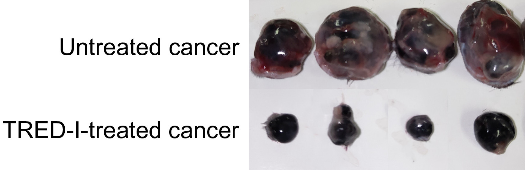
The technique reduced tumor sizes significantly and increased activity of immune cells called cytotoxic CD8+ T cells, the immune system’s primary cancer-fighting cells. , Known as TRED-I (Targeted Reactivation and Demethylation for MHC-I), it was tested in animal cancer models and markedly enhanced treatment efficacy when used in conjunction with existing immunotherapy.
“New modalities for fighting cancer like this are desperately needed because we have few solutions to fight some cancer types,” said Paul de Figueiredo, University of Missouri Bond Life Sciences Center researcher. “This is a radically new approach, and I’ve felt lucky to be part of it.”
MHC class I molecules are a prerequisite for the immune system to recognize and eliminate cancer. When cancer cells are faced with pressure from the immune system, they actively reduce their MHC class I molecules, so cancer cells can hide from drawing the attention of cytotoxic CD8+ T cells. Kobayashi and his team previously identified a gene, called NLRC5, that regulates MHC class I levels. They further found that NLRC5 is suppressed by turning off molecular switches existing on DNA, called DNA methylation, in cancers to reduce MHC class I level. The TRED-I system was able to restore DNA methylation of NLRC5 gene and further activate NLRC5, thus increasing MHC class I levels in cancer without causing severe side effects.
“This work is the result of our team’s research for over 10 years,” Kobayashi said. “It’s great to shed light on moving our findings to potential clinical application. We believe with further refinement, the TRED-I system could contribute significantly to cancer therapy.”
Further research could enable direct delivery of TRED-I system in cancer patients. If successful, such drugs could improve the efficacy for the immune system to eliminate cancer and able to improve the response to existing therapy.
This work was supported by Japan Society for the Promotion of Science (JSPS) KAKEN grant 19K21250, 20K21511, 22H02883, 22KK0112; Japan Agency for Medical Research and Development (AMED) grant JP223fa627005 and 23ym0126801j0002; Japan Science and Technology (JST) START University Ecosystem Promotion Type grant JPMJST2284; Takeda Science Foundation; Bristol Myers Squibb; SENSHIN Medical Research Foundation; Hitachi Global Foundation; Kobayashi Foundation; The Toyo Suisan Foundation; KAKEN grant 20K16433; 19K16681
Contacts:
Professor Koichi S. Kobayashi
Department of Immunology, Graduate School of Medicine
Hokkaido University
Tel: +81-11-706-5056 kskobayashi@med.hokudai.ac.jp
Department of Microbial Pathogenesis and Immunology
Texas A&M Health Science Center
kobayashi@tamu.edu
Professor Shinya Tanaka
Department of Cancer Pathology, Graduate School of Medicine
Hokkaido University
Tel: +81-11-706-7806 tanaka@med.hokudai.ac.jp
Institute for Chemical Reaction Design and Discovery (WPI-ICReDD)
Hokkaido University
Hidemitsu Kitamura
Department of Biomedical Engineering
Faculty of Science and Engineering
Toyo University kitamura012@toyo.jp
Institute for Genetic Medicine
Hokkaido University
Paul de Figueiredo
Bond Life Sciences Center principal investigator
NextGen Precision Health Endowed Professor of Molecular Microbiology & Immunology
University of Missouri School of Medicine
Tel: 573-882-6828 paullifescience@missouri.edu
From the fifth floor of the Bond LSC, one can look down and see the bridges that connect the facility and foster daily interactions and collaboration. |Photo by Beni Adelstein, Bond LSC
By Beni Adelstein
When Julia Rodriguez walked into Bond Life Sciences Center in 2004, she and dozens of others were part of a new campus experiment.
As an administrative staff member for the newly minted center, she had a big task ahead of her, but, as Bond LSC approaches its 20th anniversary, she thinks the trajectory and results largely accomplished their aims.
Julia Rodriguez worked as a grant writer at Bond LSC for nearly 20 years. | photo by Roger Meissen, Bond LSC
“When we started, Bond LSC was a brand-new concept on campus, and the administrative burdens at first were crazy,” Rodriguez recalled.
Bond LSC emerged as a center in pursuit of basic science and collaboration. Its narrative entwines the aspirations and discoveries of its researchers, the evolution of its scientific focus and the crucial growth of its research portfolio.
Rodriguez’s role was to help Bond LSC investigators secure grants, ensuring researchers could primarily focus on their science. She met collaborators, colleagues and policy directors from both the National Institutes of Health (NIH) and the National Science Foundation (NSF) to find out the way they did things and figure out what needed to be done to get an award.
“It’s odd to work in a job where a fail rate of 90% is actually a good thing because the federal funding rates are so low,” explained Rodriguez, shedding light on the harsh realities of funding in scientific research.
But landing more than 10% of submitted grants was a triumph and an integral part of how a center collaborates its way to success and nurtures scientists from divisions across campus. Bond LSC investigators actually have a success rate of about 30%.
Donald Burke, a principal investigator who studies the RNA origins of life and ways to deliver cancer treatment, joined Bond LSC just one year after it opened. The professor works in the departments of Molecular Microbiology & Immunology and Biochemistry with MU’s School of Medicine. He learned about the role collaboration plays in the center.
Donald Burke started at Bond LSC a year after it opened. The scientist studies the RNA origins of life and how to use small molecules to better deliver cancer treatment. | photo by Mariah Cox, Bond LSC
“People who want to be successful on their own, off in the corner, have a harder time doing so in modern science because the expectation is that each new study you bring to bear will have multiple facets to it,” Burke said. “I did not do much collaboration before I moved here — that was not how I was trained as a scientist — but I learned how to work as a team.”
The administrative side had to learn a similar lesson.
“It was incredibly specific how you calculated salaries, gave shared credit and how you filled out forms,” Rodriguez said. “Different departments would argue over the correct way to apply for something. That’s why we needed collaborative administration and collaborative facilitation.”
Bond LSC has grown its research expenditures since then. Rodriguez has seen the center increase grant proposals to make gains in awards, especially in the five years since the University of Missouri set a goal to double research funding. Grant obligations to Bond LSC scientists increased from $11.9 million in 2018 to more than $41 million in 2023.
“It’s part of keeping the machine going,” said Walter Gassmann, Bond LSC’s director. “To the public, it might look like scientists run after money, but our job is to conduct experiments to test ideas and reach the next level of insight. Bond LSC’s focus is excellent science so, in the end, we need to compete for a broad portfolio of external funding.”
That funding comes from a variety of sources, including NIH, NSF, private donors and other grants. While Bond LSC has had a wide array of success with funding agencies, increased success with NIH funding emerged in recent years as new biomedical scientists joined the center.
Henry Wan knows that firsthand. He joined Bond LSC five years ago to grow his lab. Wan’s lab alone has garnered around $32 million and three NIH R01 grants since then.
Henry Wan joined Bond LSC fiver years ago to further his study of vaccines, influenza and other viruses. | photo by Roger Meissen, Bond LSC
Wan studies how viruses like Covid-19 and flu spread, specifically looking at how they infect across human and animal boundaries. His goal is to create better vaccines. A vaccine helps the body defend against a disease by preparing it. Think of it like a mugshot for the body’s immune system to recognize what it’s up against and be prepared.
“We have so many conferences, meetings and seminars here that I really enjoy,” Wan said. “The center has been very supportive, and we see the beauty of the multi-interdisciplinary research in Bond LSC. The center truly makes it easy for collaboration between scientists and different branches of science.”
Some of that teamwork involves big data and artificial intelligence (AI). Wan initially saw a degree of separation between the student programmers and biologists in his lab but now they seamlessly blend.
“The computer science students used to just wait around for me to fit them in; programming they can understand, no problem, but biology is hard, Now I don’t even have to go to the meetings. They talk amongst themselves.”
This teamwork can be seen even before a project gets off the ground. Rodriguez said there is an emphasis on identifying larger groups to apply for more substantial collaborative program and center grants. Sometimes that means being flexible.
Rodriguez said now the attitude in the center is, “Oh, there’s more than one way to do something.” She said seeing collaboration expand into the administrative realm across campus has felt like one of the biggest shifts.
“Suppose department chair A and department chair B want something done in a certain way. Previously they might have been more focused on their own students and research.” she explained. “However, now that getting grant funds benefits everyone, they are more likely to work in tandem with the other department.”
“Collaborative research requires collaborative administration.”
That teamwork translates into passion. Rodriguez recalled when a scientist she was working with on a grant rushed into her office to tell her that he had gotten the award they’d so diligently applied for.
“He came in frantic, saying he needed to talk to me so he could call his wife, and I was a little concerned, but he just wanted me to be the first person he told,” she said. “I have seen incredible dedication and passion for seeing that the science gets done. Watching these projects blossom has been truly rewarding.”
Gassmann agrees. As an inaugural principal investigator in 2004 — long before he became center director — he sees this teamwork through and through.
“It’s amazing seeing how, even though we have different areas of science, all of it connects to form a bigger picture,” Gassmann said.
This spirit fosters an environment where other silos are broken down even beyond faculty in favor of collective success.
Rodriguez and colleague Steve Friedman worked with student researchers in 2022 and 2023 to win NIH F30 predoctoral fellowships, a first for MU. Cynthia Tang — a joint M.D.- Ph.D. graduate student in the Wan lab — focused her application on how Covid-19 spreads and how to track it in rural areas.
“My initial proposal was actually rejected,” Tang said. “I had to take on the challenge of putting together a completely new study design in just under two months.”
Despite the initial setback, Tang prevailed with their help and was able to secure that funding to support her research.
Another example of this teamwork includes Burke’s collaboration with Marc Johnson, another principal investigator studying molecular microbiology and immunology. Johnson happens to be his office neighbor.
“I love having people that you can just go next door or even just shout across the hallway,” Burke said.
“We’re like two vines growing synergistically alongside each other,” Johnson remarked. “Even though we have totally different backgrounds, we’ve gotten the chance to collaborate on publications and mentor students.”
So why does all of this matter? Why is funding for research so important?
“I’m a big fan of basic research because you might not always see the immediate impact, but 20 years later there may suddenly be an application that couldn’t have happened without it,” Gassmann said. “Think about all the things that have made life more pleasant. It’s all based on science and a better understanding of the natural world that keeps us safer and healthier.”
Lead grant writer Julia Rodriguez and director Walter Gassmann share a conversation on Rodriguez’s last day working for Bond LSC. | Photo by Beni Adelstein, Bond LSC.
Gassmann said Bond LSC’s scientific triumphs are both a testament to its past and a promise of a brighter future for scientific innovation.
But Rodriguez said someone else will carry the grant writing torch for that. After 20 years, Julia Rodriguez retired on January 8th this year.
“It’s an emotional time. When I started here, we felt timid as we tested the waters of this new type of environment and now we feel like family,” she said. “There are good people all over campus but, boy, I have loved watching the center mold together and I love these scientists.”