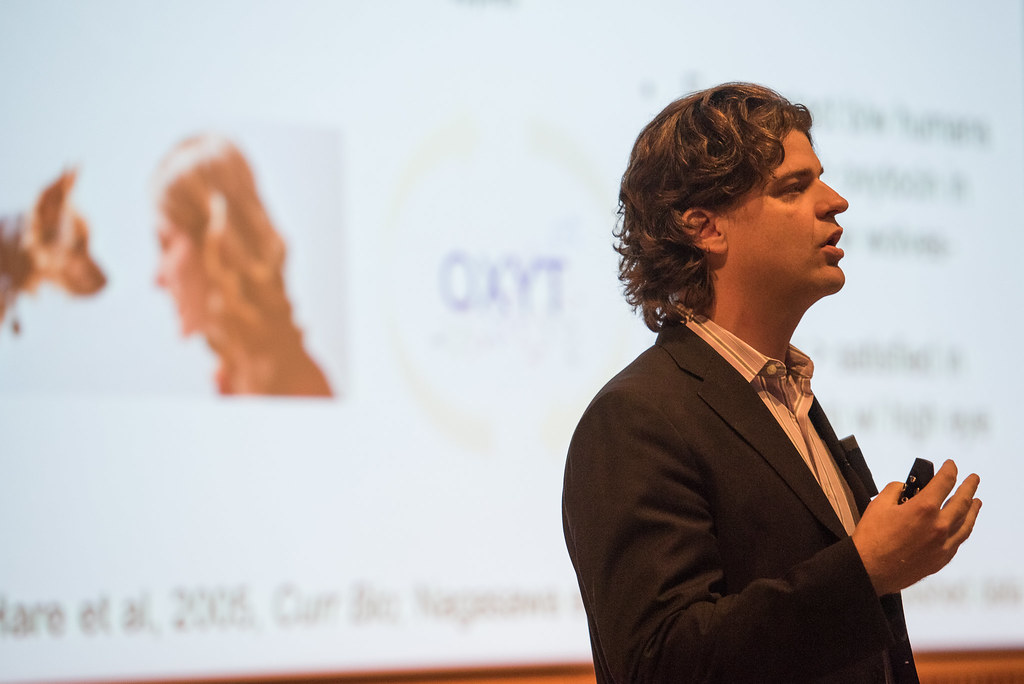
Hare explains survival of the friendliest as component of natural selection
Dr. Brian Hare speaks about how friendliness and natural selection are connected at the 13th annual LSSP symposium, The Science of Love. | photo by Roger Meissen
By Allison Scott | Bond LSC
Dogs really are a man’s best friend if you ask Brian Hare.
Our four-legged friends are a direct result of chance coupled with domestication. And over the course of hundreds of years, that domestication has led to deep bonds between humans and dogs.
“You love your dog, physiologically, the same way as your offspring or partner,” Hare said.
Throughout his career, Hare has analyzed various animals from dogs and foxes to chimpanzees and bonobos to determine how much friendliness actually is a factor in their survival.
Recently, Hare has focused on primates. In his studies, he’s observed distinct differences between chimpanzees and bonobos, both of which are closely related species.
“Chimpanzees are like humans,” Hare said. “Bonobos have a different social system.”
Chimpanzees tend to be the more aggressive, male-dominated of the two. On the other hand, bonobos are the exact opposite and thrive on a more equal approach. Bonobos love to share with anyone and everyone, making them friendlier.
Hare has been able to support this difference through a series of experiments. They show that one bonobo will likely help a stranger in a cage get food that is just out of reach. These experiments showed that bonobos are willing to help others in most situations unless there’s a high cost or risk to self.
The evolutionary elements of friendliness and the traits behind kindness Hare identified as a possible reason homo sapiens won out over other human ancestors like Neanderthals. This is due, in part, to what Hare calls the ‘like me’ trait.
“If we as humans can categorize based on cultural or social characteristics that someone is “like us,” we’re more likely to help them out,” Hare said. “This leads to bonds.”
The opposite is also true, though. This means if we identify someon as “not like us,” we’re less likely to help them. That stark difference between humans and both chimpanzees and bonobos makes for a more complicated communication process, and contributes to humans being both the friendliest and cruelest species alive.
At the end of the day, though, Hare strongly believes the science supports friendliness as a component of natural selection.
“You can win big by being friendly in the evolutionary game,” Hare said.
The 13th annual Life Sciences and Society Symposium, The Science of Love, started Friday, Oct. 6 and Saturday, Oct. 7. It features six experts that research various aspects of love, relationships and connection. The event will conclude on Friday, Oct. 13 with its last speaker, Jim Obergefell, who was the plaintiff in the 2015 Supreme Court case on marriage equality.
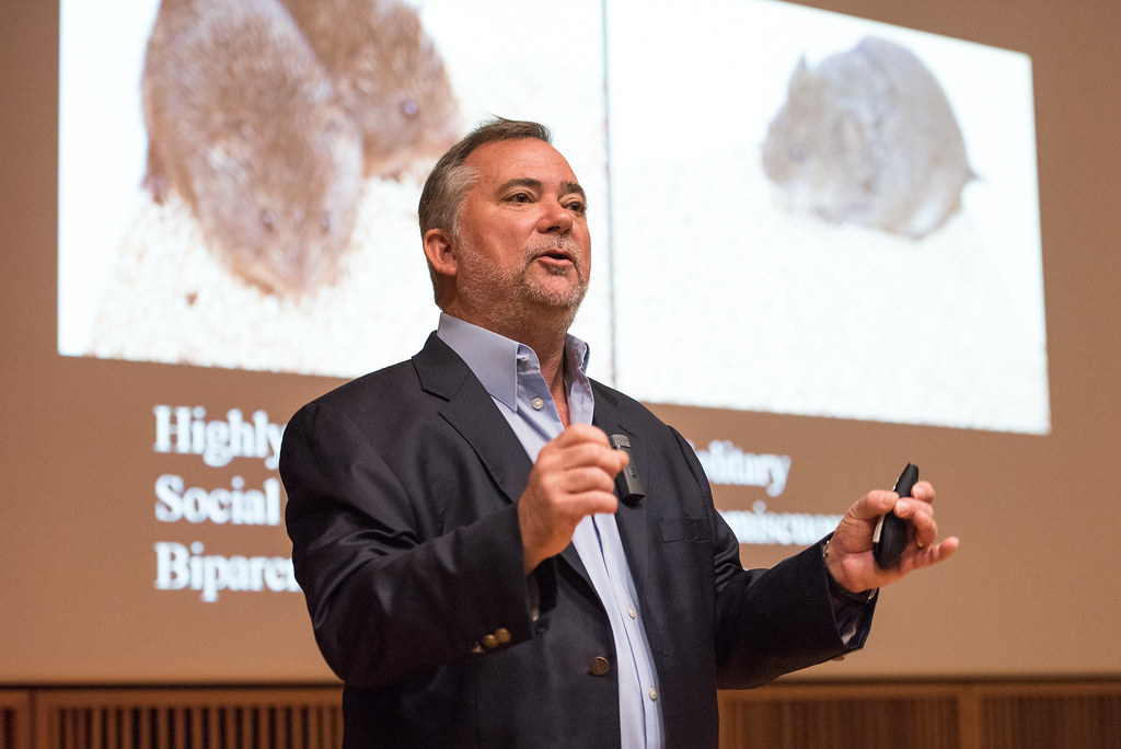
Larry Young explores chemicals behind monogamy in prairie voles, humans
Dr. Larry Young opens the second day of The Science of Love. | photo by Roger Meissen
By Allison Scott | Bond LSC
Upon first glance, it wouldn’t seem that humans and small rodents have that much in common.
However, Larry Young extensively studies the prairie vole because their desire to mate for life.
“Prairie voles mate for life,” Young said. “That’s very unusual, in fact, only three to five percent of mammals do this.”
This commonality between the small mammal and humans allows Young to relate his research on voles to humans. His goal is to understand the neurochemical bond that occurs between two voles after mating.
Neurologically, this “pair bonding” occurs largely because of oxytocin. The brain houses receptors for this hormone that create pleasure from it. The resulting feeling’s mutual, and chemical, leading a bond to form.
“The brain’s reward system houses the receptors that make oxytocin an influential chemical,” Young said. “Prairie voles activate this when they bond.”
Young manipulates the prairie vole’s brain to try an understand why exactly pair bonding exists. He then takes brain scans of them, compares them to mice that aren’t monogamous and sees what’s happening differently in their brains. In doing so, he is able to pinpoint differences in the voles and apply that knowledge to humans.
“We should think of ourselves as part of a continuum,” Young said. “The voles have a similar makeup to humans, but we don’t say that they’re in love – we say they’re bonded.”
That distinction makes a difference in Young’s studies. However, he’s still able to learn a lot about people by observing voles, and it’s those revelations that Young enjoys most.
That insight might one day lead to treatments for autism or other disorders where issues in interaction and bonding affect people.
“The Science of Love isn’t just entertaining,” Young said. “It has the potential to change lives for the better.”
The 13th annual Life Sciences and Society Symposium, The Science of Love, started Friday, Oct. 6 and Saturday, Oct. 7. It features six experts that research various aspects of love, relationships and connection. The event will conclude on Friday, Oct. 13 with its last speaker, Jim Obergefell, who was the plaintiff in the 2015 Supreme Court case on marriage equality.
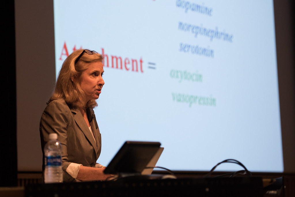
Helen Fisher delves into the relationships we choose and why in our digital age
Dr. Helen Fisher opens the 13th annual LSSP symposium, The Science of Love, on Friday, Oct. 6. | photo by Allison Scott
By Allison Scott | Bond LSC
We might not understand what drives us to establish and maintain romantic relationships, but Helen Fisher has made her living trying to figure it out.
The romantic love expert spoke Friday, October 6, in Bond LSC about the neurological reasons behind why humans behave the way they do.
“Romantic love is located right next to thirst and hunger in the brain – it’s a survival system,” Fisher said. “If we survive another million years, we will continue to fall in love.”
Fisher has worked in tandem with the dating site match.com for more than a decade to tailor sites like chemistry.com to look at how relationships actually work in our brains and in practice. Using data from match.com that represents the U.S. population, Fisher detailed how the digital age impacts the dating scene.
“Fundamentally, love isn’t changing,” Fisher said. “Courtship patterns are.”
He research indicates a tendency toward “slow love,” where partners tend to get sexually involved sooner, but are more cautious about marriage and take much longer than previous generations to pair up in that way.
Technology, meeting online and evolving social norms play huge roles in these changes, but she argued that dating sites aren’t really dating sites at all.
“They’re introducing sites,” Fisher said. “The only real algorithm is your brain.”
These changes in how people meet influence marriage, too. She highlighted how cohabitating influences the perception of marriage and its overall impact, as well as the increased tendency for more casual sexual encounters.
“What we’re seeing now is the expanding of the pre-commitment stage of romance,” Fisher said. “Marriage used to be the beginning of a relationship, but now it’s the end.”
While modern couples often postpone marriage, Fisher notes that most people do marry before they reach the age of 50 and she feels positively toward relationship trends and the direction that romantic love is headed.
“I’m extremely optimistic,” Fisher said. “We’re marrying later and moving toward relative marriage relationship stability.”
The 13th annual Life Sciences and Society Symposium, The Science of Love, started Friday, Oct. 6 and Saturday, Oct. 7. It features six experts that research various aspects of love, relationships and connection. The event will conclude on Friday, Oct. 13 with its last speaker, Jim Obergefell, who was the plaintiff in the 2015 Supreme Court case on marriage equality.
Computer scientists create applications to speed up research in the lab
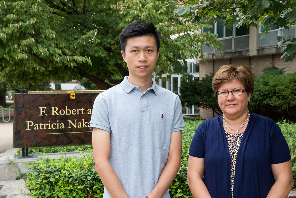
Ph.D. student Ke Gao and computer scientist Filiz Bunyak collaborate with researchers at the Bond Life Sciences Center. The pair helps advance high-throughput phenotyping by developing applications and algorithms for image analysis. | Photo by Samantha Kummerer, Bond LSC
By Samantha Kummerer, Bond LSC
Three years ago, Ke Gao stood uncomfortably beside rows of biomedical students and plant scientists at the Bond Life Sciences research fair. His poster wasn’t discussing the DNA of seeds or how plants transport nutrients but rather a scientific device.
“At the beginning, the visitors didn’t understand what we were presenting, but once I explained how our application can help them accelerate their research and how we can really turn their phones into a research device, they got really excited,” Gao explained.
Gao’s presentation highlighted a mobile app that transforms images of seeds into objective, quantitative data.
It started with a simple problem. Plant scientists were manually comparing hundreds and in some cases thousands, of seed photos. The process was meticulous, slow and subjective.
The solution began with a collaboration with Michele Warmund (Plant Sciences), Tommi White (MU Electron Microscopy Core) and Filiz Bunyak (Computer Science) that led to a MU Interdisciplinary Innovations Fund grant.
Gao was part of this team that developed an algorithm to turn the photos of seeds from the field into data with the touch of the button.
Gao explained the app is very similar to Instagram.
A user takes or uploads photos of seeds. Then the app calculates measurements describing shape, color and size characteristics of the seeds. This data can be emailed or stored in a database.
Some experiments need thousands of seeds analyzed; this would be a massive feat for even a group of students. With this app, hundreds of seeds can be photographed and measured from a single photo. The app analyzes each seed individually and also computes measurement averages for groups of seeds.
There are other apps that analyze seeds, but this is the first mobile application as far as the team knows. Its ability to analyze multiple seeds at once, even if they are touching is also an outstanding ability. Bunyak’s previous experience developing applications to quantify microscopy images and videos of touching and clumping cells helped them design the algorithm to make that function possible.
This isn’t just a problem for researchers in this one lab or even at the University of Missouri.
MU Computer Science professor, Filiz Bunyak, said noninvasive methods to observe and understand biology, imaging equipment and corresponding computing devices have advanced considerably in recent years, leading scientists to produce large amounts of data. The ability for researchers to analyze and quantify this large amount of complex and unstructured data, however, was still missing. Bunyak said this app began as a project to advance scientists’ capabilities to automatically analyze image-based plant phenotyping.
Further collaboration
Bunyak and her students are advancing the field of high-throughput phenotyping beyond this mobile app.
High-throughput phenotyping (HTP) refers to the process of connecting an organism’s DNA makeup to its physical characteristics; it is also a hot topic buzzing through the science community in the last five years.
Two years ago, Bond LSC scientist David Mendoza, who studies how plants collect nutrients, said he never imagined he would be doing HTP.
“The old way of doing this is growing plants on plates and, I’m not kidding, with a ruler you measure how long the roots are,” Mendoza explained of the traditional process that now seems archaic.
Now, the lab is working with computer scientists to design a robot to code the measurements for multiple roots at a single time. For a student, it would take 15 minutes, but now it’s complete in an instant.
Speed isn’t the only reward researchers are reaping.
Bunyak said computational image analysis allows researchers to come up with new ways to quantify and study data that they were not even able to do before, leading to the design of novel experimental methods.
Ruthie Angelovici is another Bond LSC researcher who uses computer scientists to aid in her research.
She said without computer imaging there would be no way for her team to do research that measures plants physical and biochemical traits. Angelovici’s lab uses Bunyak’s mobile app system but on a computer. Eight plants are photographed at once and the application keeps track of features of plants such as shape, color and area as they develop.
What is really revolutionary to Angelovici is the ability for the data of plant growth parameters to be stored and revisited without the need to re-grow. This contrasts with past experiments where researchers would scribble some notes and never be able to return.
“It’s not lost and I think that’s a big step in this field,” Angelovici said.
The collaboration is creating more than advanced tools by fostering a new way to think and approach research.
Rather than buying pre-existing software, the groups from Bond LSC utilizes the resources on campus to build their own devices.
“I would have been in front of a black box that is doing things for me and that would not have given me the tools to teach to my students,” Mendoza reflected. “Now I know what they need to learn to be competitive. Now I know what the gaps are and how they can be filled. I think that was worth it.”
Mendoza’s team publishes all the instruction to its robot online, so the technology can aid other labs in making faster discoveries at a lower price.
Angelovici compared it to buying a cake versus making a cake — at the end of the creation process, she said she would have the knowledge to do a lot of other experiments.
This new way of thinking already began to pay off this summer when her lab expanded computer software to analyze seed size.
“We only approached it because we saw how things worked together. I just pitched a project to engineering about seed collector. Again, this opened my eyes that even undergraduates can do something not so difficult for engineers, but I have no clue how to do it,” Angelovici said.
Mendoza agreed the collaboration is exciting but challenging, “You got a Ph.D. and you got a faculty position and you think you know stuff. When I started this I realized how much I don’t know, but at the same time it reminded me that it is really cool to learn something new.”
Both teams continue to work towards maximizing the functions of their individual machines, but even after the projects reach fruition the collaboration will not be over.
“On the contrary, I think we’re going to keep building more and more and better,” Mendoza said.
Nowadays, Gao no longer feels out of place at the Life Sciences fairs. Researchers from various labs come up to him and ask how they can implement his app in their own lab.
“It seems like I’m doing something that can really help people, so that’s the best part of this process,” he said.
Ruthie Angelovici is an assistant professor in the Division of Biological Sciences and is a researcher at Bond Life Sciences Center. She received her degrees in plant science from institutions in Israel — her B.S. and M.S. from Tel Aviv University, and her Ph.D. from the Weizmann Institute of Science in Rehovot. She was a postdoctoral fellow at the Weizmann Institute and at Michigan State University and has been at MU since fall of 2015.
David Mendoza is an associate professor in Plant Sciences, Life Sciences Center investigator and a member of the Interdisciplinary Plant Group. His research focuses on the mechanisms plants use to resist toxic elements or acquire nutrients. He received his Ph.D. in biochemistry from UNAM in Mexico City and continued on to do post-doc training at UC San Diego.
Filiz Bunyak is an assistant research professor in the Department of Computer Science. She received her bachelors and masters degree from Istanbul Technical University and her Ph.D. from the University of Missouri- Rolla. Her work focuses on computer imaging, image processing, and biomedical image analysis.
Ke Gao is a doctoral student in the University of Missouri’s Department of Electrical Engineering and Computer Science. He earned his bachelor’s of science from the Henan University of Science and Technology in China.
Vinit Shanbhag is a Ph.D. candidate in biochemistry and works in Michael Petris’ lab in Bond LSC. | photo by Allison Scott
By Allison Scott | Bond LSC
“#IAmScience because I like to discover. The excitement of uncovering things that could have an impact on millions of lives is fascinating.”
Vinit Shanbhag isn’t your typical student. His extensive background both overseas in India and at the Florida Institute of Technology serve to prove just that and prepared him for his next adventure at Mizzou.
“When I came here for the on-campus interviews, the department was impressive,” said Shanbhag, who is pursuing a Ph.D. in biochemistry. “The excellent infrastructure, paradigm-shifting research and challenging educational environment influenced my decision to attend MU.”
Shanbhag intentionally joined the lab of Michael Petris at Bond LSC to further his experience.
“I was particularly interested in joining the Petris lab due to my immense interest in cancer research,” Shanbhag said. “That interest has now evolved into an aspiration to pursue a career in the field.”
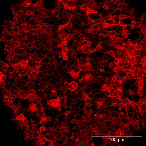
There he studies how an essential dietary nutrient copper is required for the process of tumor formation and metastasis. In a specific study he has deleted a copper-transporting gene (ATP7a) in cancer cells and demonstrated a defect in their ability to grow into larger tumors and spread to other organs in animals.
“By understanding the mechanisms that regulate key processes in cells, one can distinguish between the normal and diseased,” Shanbhag said. “Uncovering these differences at the molecular level is key to the development of novel clinical interventions.”
Shanbhag’s work has been recognized as he was invited to present his research at the Gordon Research Seminar in Vermont earlier this year. While there, he shared the work he’s been doing in his lab and gave a presentation, in addition to showcasing a poster detailing his work.
“People were impressed,” Shanbhag said. “After my talk people came up and asked me questions. Our observations are very interesting and the goal is to develop a drug that could potentially block the function of ATP7A and inhibit cancer progression. The people I spoke with encouraged us to keep going.”
Although he’s presented at departmental seminars, this recognition stands out as a great experience for Shanbhag.
“This was my first invited talk,” Shanbhag said. “I applied for it and got the news of my invite pretty quickly, so I was excited.”
The hope is that Shanbhag’s research will serve as the premise for further development in understanding and eventually eliminating cancer.
“Ultimately, I hope to discover new ways to kill cancer cells and provide cost-effective treatment options for cancer patients,” Shanbhag said.
Sterling Evans is a sophomore plant sciences major conducting research in Gary Stacey’s lab in Bond LSC. | photo by Allison Scott
By Allison Scott | Bond LSC
“#IAmScience because I want to focus my research on problems that exist in agriculture in undeveloped and third world countries.”
Sterling Evans’ mind wasn’t focused on research when he started college, but that would soon change.
The sophomore plant sciences major uncovered his interest thanks to Freshman Research in Plant Sciences (FRIPS) — a program dedicated to introducing research to freshman students from plant-related degree programs.
“I was interested in plant sciences-related fields when I started here, but I had no intention of getting involved in undergraduate research,” Evans said. “Being selected for FRIPS was instrumental in getting me involved with research.”
Along with a handful of students selected for FRIPS each year, Evans got to interact with various professors and mentors around campus on a weekly basis. Because of that exposure, Evans found a place in the lab of Bond Life Sciences Center’s Gary Stacey.
After a year working in Stacey’s lab, Evans just joined a new project that aims to improve the nutritional value of soybeans.
“They’re used as a main source of protein for a lot of countries, so improving their nutritional content would have a huge impact,” Evans said.
The team is applies CRISPR, a gene-editing tool, to model plants called Arabidopsis as a first step.
“We are working on Arabidopsis right now as a proof of concept, because it can be done in a relatively short period of time, before investing as much as a two additional years in soybeans,” Evans said.
While he only spends 15 hours in the lab each week, Evans noticed the lab’s impact on his approach to academics in other ways.
“Research gives me more motivation to think about how to apply information I’ve learned in class to work in the lab,” Evans said. “It has made me more analytical in classes because I have more of a desire to understand things.”
Evans plans to earn a Ph.D. in a plant sciences field and wants to continue research in his career. He’s most interested in helping ensure small communities throughout the world have enough to eat, and he hopes to contribute by studying orphan crops.
“I think they’re cool because they’re really important to small people groups. No one studies them because they aren’t a big deal in the United States or other countries,” Evans said. “If we work on them we won’t have a huge impact on hundreds of millions of people, but we will have a huge impact on small communities.”
That impact all started in a lab. Had he not stepped out of his comfort zone he might never have discovered this path, and he highly encourages other students to give research a chance.
“There are labs for almost everything and there’s an area for everyone,” Evans said. “I didn’t know I wanted to do research until I did it.”
How zebrafish gained its popularity as a model organism
By Samantha Kummerer, Bond LSC
The core of many modern discoveries in developmental biology is swimming in a tank.
These are zebrafish that serve as the lab rats for Anand Chandrasekhar’s research.
Dozens of tanks containing thousands of swimming fish fill the lab in the basement of the Bond Life Sciences Center. There are baby fish, striped fish and clear fish, many genetically modified for experimental reasons, and they are studied from fertilized embryo to adolescence.
Chandrasekhar’s lab uses zebrafish to study migrating motor neurons in the brainstem that control muscle movement in the face and jaw.
Multiple types of neurons positioned precisely throughout the brain connect together to form networks. Those networks give the brain its ability to function.
“For us, it is important to study how these networks form because the brain is what makes us human,” said the Bond Life Sciences Center researcher.
During development, neurons respond to signals that enable them to move to different locations and form networks, Chandrasekhar explained. If the neurons don’t migrate properly to specific locations then the brain can’t function properly.
It is a domino effect. If the brain stem motor neurons don’t migrate correctly then the corresponding neural networks don’t form correctly and then the fish are not able to eat well.
Chandrasekhar’s lab is captivated by this migration. Different neurons move different ways and are set into motion by different signals. What are the signals to tell the neurons to stop or to go? Do these signals vary based on neuron type and species?
The lab also seeks to understand the repercussions of deficient migration using genetically modified fish.
Cell migration doesn’t just occur in the brain or with nerve cells. Cells also need to move in particular ways to form the heart and to fight infections.
A Model Swimmer
The zebrafish has long joined traditional lab rats and mice as scientists’ choice as an ideal test subject.
“When I first started out I wasn’t entirely sure I could do that (work with zebrafish), because how am I expected to handle something in water, moving around and be able to use it to really do experiments?” Chandrasekhar said. “But once you see how you handle fish, how you get them together, it just becomes one more thing that you do and then it makes you wonder how people work with mice.”
Like other model organisms, zebrafish share many genetic similarities with humans. These similarities mean researchers can investigate cancer, heart disease, muscle and tissue disorders in humans by testing and studying fish.
Mice and rats also share a large portion of DNA with humans, but the zebrafish’s unique characteristics enable experiments and observations to be cheap, efficient and fast.
One of those unique traits is a rapid development rate. This allows researchers to study important developmental stages in a single week.
“They’re incredibly efficient in terms of, I can set them up and they’ll be ready tomorrow morning,” said Ph.D. student Devynn Hummel.
The fact that embryo development in mice occurs inside their mother also makes observing early stages of development hard. Zebrafish embryos, on the other hand, grow independently, outside the mother.
Neurons in the hindbrain of a zebrafish embryo are illuminated due to the use of florescent chemicals. The branchiomotor neurons are labeled with green and the commissural axons are labeled with red. | Photo by Suman Gurung
The young zebrafish’s transparency also helps researchers track cells within its body.
Hummel explained that by using fluorescently tagged molecules we are able to zero in on anything from neurons to changes in calcium concentrations, allowing fish to be used in a wide range of different studies.
“We’ve sort of engineered them to allow us to look at different tissues,” Hummel explained. “It really is remarkable the details in imaging zebrafish and what you can see. It’s truly extraordinary.”
An eye of a zebrafish is illuminated 48-hours after fertilization using immunohistochemistry.| Photo by Suman Gurung
Scientists throughout the world are catching on to the advantages of the fish.
Chandrasekhar said there are significantly more zebrafish labs throughout the world than there were 20 years ago.
Despite its advantages, the zebrafish will not completely replace mice as a lab tool.
Chandrasekhar explained there are still certain experiments where mice are more advantageous. The same area of research can be explored using zebrafish, mice or even fruit flies, but the specific questions you ask change based on the tools available.
A snail toxin, MVIIA, is illuminated in a zebrafish embryo. The toxin blocks calcium channels needed for neurotransmitter release. Chandrasekhar’s lab studies motor neurons using zebrafish. | Photo by Devynn Hummel
Beyond Nerves
While Chandrasekhar’s lab concentrates on neurons, understanding how the migration occurs can be applied in a different context because of shared cellular mechanisms.
“Some of these molecules are just tools that different types of cells can use in different ways to accomplish different functions,” he said. “You can study a steering wheel in a car and know that it’s allowing the wheels to turn and the same steering wheel in a truck is allowing the wheels to turn in a truck, but maybe at a different time and a different way.”
Members of Chandrasekhar’ lab already experienced a broader implication of their research when a gene they studied with a role in neuron migration provided insight on Spina bifida, a failure of the fetal spinal cord to close completely.
“We all hope the problem we study has a broader impact on human biology so there’s always a quest to dig deeper and learn something new,” Chandrasekhar said.
The lab is hoping one day soon this fishy tool will help shed even more light on the mechanisms maintaining human health. For now, the researchers and their fish will just keep swimming.
With more than 2,000 fish, the lab has no plans on slowing down.
Anand Chandrasekhar is a Biological Sciences professor at the University of Missouri. He uses mice and zebrafish to study the mechanisms involved with the development of the nervous system. His lab in Bond LSC uses cell biological and genetic methods to understand these mechanisms. He received his Ph.D. in biology from the University of Iowa.
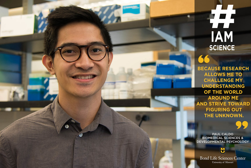
Paul Caldo is studying both biomedical sciences and developmental psychology. He works in Dr. Cheryl Rosenfeld’s biology lab in Bond LSC. | photo by Allison Scott
By Allison Scott | Bond LSC
“#IAmScience because research allows me to challenge my understanding of the world around me and strive toward figuring out the unknown.”
Paul Caldo isn’t your typical undergraduate student. As a junior, Caldo is double majoring in Biology and Psychology, which gives him a unique perspective on science as a whole.
It is in the overlap between his majors, however, that most interests him.
“I am fascinated with development in both psychology and biology because the early stages of life lay the foundation for who and what you will become,” he said. “I have an appreciation for all spheres of academia, and it is becoming more evident to me that an interdisciplinary approach to research will lead to more and more breakthroughs in science.”
As a member of both Dr. Cheryl Rosenfeld’s biology lab in Bond LSC and Dr. Ashley Groh’s psychology lab in Noyes Hall, Caldo gets the best of both worlds while studying the fields he loves. In Rosenfeld’s lab, he’s currently analyzing how endocrine disruptors – which are found in everyday products like sunscreen – impact the development of reproductive organs in female mice.
“By understanding the underlying mechanisms that drive this interaction, our goal is to potentially reverse some of the harmful effects that result from heavy exposure to endocrine disruptors,” Caldo said.
His efforts have not gone unnoticed. Caldo was selected to join an ASH Scholars undergraduate team mentored by Dr. Grohl and collaborator Dr. Amanda Rose. The ASH Scholars program, which provides a $2000 scholarship, is sponsored by the Honors College and the Office of Undergraduate Research. He also received a $200 travel grant that will allow him to present his findings at the Developmental Origins of Health and Disease Conference in Detroit later this month.
“I’m really excited about the travel grant to Detroit,” Caldo said. “It will be my first time attending a national-level conference. I hope to benefit from presenting my work as well as learning from many great scientists from across the country. I think it will be a really enriching experience, and I hope to take away a lot from it.”
After graduation, Caldo hopes to attend graduate school to study developmental psychology using an interdisciplinary bio-behavioral approach to answer research questions. Ultimately, his plan is to earn a PhD in developmental psychology. Until then, though, he’s enjoying his time at Bond LSC learning as much as possible.
“The ambiance is great – working closely with some of the best researchers on campus is an amazing feeling,” Caldo said.
What happens when a chemical engineer, a computer scientist, and an immunologist walk into a lab?
Vaccines are created faster and cheaper.
At least this trio hopes that’s the answer.
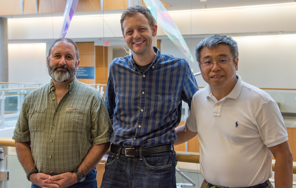
Bond Life Sciences Center computer scientist Dong Xu joined forces with immunologist Jeffery Adamovicz and chemical engineer Bret Ulery for the first time in February 2016. After 18 months and a $100,000 Bond Life Sciences startup grant, the team is closing in on a novel approach to vaccinations. They hope to start collecting initial data in the near future.
“Without this grant, it would be hard to work together,” Xu said. “Bond Life Sciences Center really played an important role for the collaboration research on campus and also for providing this seed money. They really foster these types of collaboration.”
The Problem
Efficiency has plagued the vaccination field for decades.
Traditionally, scientists create new vaccines by continuously guessing which components of the virus or bacteria to test.
“It was a plug-and-play type test system,” explained Adamovicz, who has researched vaccines for years. “We said, ‘well we could try this antigen or that antigen or that antigen and you would mix them together in different combinations and try them and then you would end up selecting one that would work.”
The process is similar to finding a needle in a haystack. It’s like scientists sort through and test every strand of hay until they discovered the needle.
As you can imagine, this method takes a long time to find something usable.
This process becomes a larger issue when viruses emerge that call for immediate measures, such as Zika or Ebola.
Creating vaccines can take a long time because sometimes a variety of different related bacteria cause illnesses. This makes that already inefficient guessing game more complicated by adding, even more, components to test.
Adamovicz explained scientists make a new vaccine each year by predicting which strain of the disease will infect the most people. This is why many people who get a flu vaccination can still get the flu — the strain they caught was not included in the vaccine.
The Solution
Xu, Ulery and Adamovicz decided to combine their strengths from their different fields to approach creating a vaccine in a new way to solve these two problems.
Adamovicz explained this type of collaboration is not routinely attempted when developing new vaccines.
Computer science and engineering enabled the team to more easily sort through thousands of potential options. Xu’s lab and his students created an algorithm to select candidates to test. Without Xu, the team would have to test tens of thousands of different antigen and peptide combinations. But with the algorithm, the team narrowed the combinations to about 10 to test.
Adamovicz said this method has the potential to create vaccines in half the normal time.
“I feel that it is a very meaningful work; that’s why I am interested in doing it because I do see the potential impact,” Xu said.
Rui Zhang, a chemical engineer, works in Bret Ulery’s lab at MU. Zhang is part of the team working with targeted nanoparticles that will induce strong immune responses. | Photo by Samantha Kummerer, Bond LSC
Putting a theory to work
Burkholderia is the first Guinea pig for their new approach.
The potentially deadly bacterial infection occurs in places like Thailand and northern Australia and is caused by a family of related bacteria called Burkholderia pseudomallei. But other strains of Burkholderia also cause disease in patients with Cystic Fibrosis across the globe.
“That was part of the challenge,” Adamovicz said. “It wasn’t that we could take a single bacterium and that’s all we had to worry about. We were worrying about a family of bacteria that had this variation in their genomic sequence and the proteins they produce.”
To eventually create a vaccine that could cure all strains of the bacteria, the team aimed to find a small piece of protein all Burkholderia had within their flagella, the whip-like structure of the bacteria that allows them to propel themselves.
To limit the number of segments within the protein, Adamovicz’s lab, on the biology side, determined some requirements for Xu to write an algorithm.
“It’s not to say this is like Star Trek where you just tell the computer and it does it on its own, it requires a lot of knowledgeable human input to spit out the answers,” said Adamovicz.
Chunhui Xu operates a computer program to examine the protein sequence of a disease called Burkholderia. Xu is a Ph. D. student on the interdisciplinary team working to develop a vaccine against the disease. | Photo by Samantha Kummerer, Bond LSC
Adamovicz explained they wanted a vaccine that would trigger two immune responses. When harmful substances enter the body, like viruses or bacteria, the body responds in two ways. One way is through cellular immunity that responds by activating cells that already exist. The other way the body responds is by other cells making proteins called antibodies, which is called the humoral response.
The segment also had to be conservative, meaning it is less likely to mutate so there is a higher chance a vaccine would continue to be effective against the bacteria and would be effective against more members in the bacterial family. The last criteria Xu’s lab coded for ensured the protein is not similar to others found in humans.
All these limitations lowered more than three thousand possible candidates down to a testable amount and a final list of 10 candidates.
Then the research moves on to Ulery’s Lab. Here a team of chemical engineers take the identified candidates from Xu’s work and engineer targeted vaccine antigen nanoparticles termed micelles. These micelles are comprised of fat-based cores that display vaccine antigens on their surface and have been shown to induce strong immune responses.
Chemical engineering students Caitlin Leeper and Rui Zhang study the physical properties of micelles. The micelles are targeted antigen nanoparticles. Mice will then be injected with the micelles. | Photo by Samantha Kummerer, Bond LSC
“Most of them [subunit vaccines] are somewhat hydrophilic — water-liking peptides, portions of a protein — and we tether a fat to them and what happens is now you have something that likes water and something that doesn’t like water, and you throw it in water and they self-assemble into these small micelles,” Ulery said. “That clustering of all those peptide molecules together actually enhances the immune response to them.”
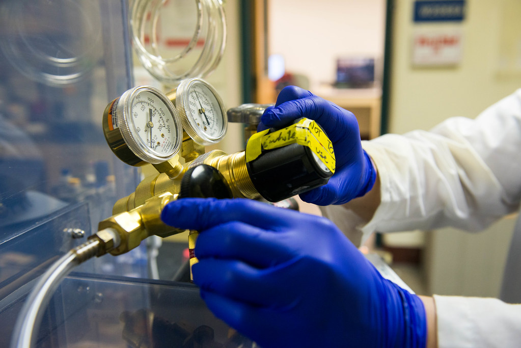
Engineering major Caitlin Leeper operates the reaction vessel that combines peptides and lipids to form micelles. The process is part of the lab’s process to develop a vaccine using chemical and engineering methods. | Photo by Samantha Kummerer, Bond LSC
Then, the work goes back to Adamovicz to evaluate what the portions of proteins are going to make the best vaccine.
This cycle continues as all labs work to enhance and develop the design further.
“It’s a process that takes a village. It doesn’t necessarily take the whole village but you need the right people in the village to work together and that’s kind of what we’re doing,” Adamovicz explained. “We’re recognizing that there are areas in previous work that could have been better optimized.”
After months of back and forth, the team is preparing to feed infected cells some of the selected peptides, the small portions of protein. Those candidates that do well in the cell culture will be tested in mice exposed to the Burkholderia bacteria.
The team hopes this round of data collection will serve as a baseboard for future experiments. If this principle is proven in one antigen, a larger grant may enable the theory to be tested in hundreds of other antigens and applied to other diseases. Over the next four to six months, the team hopes to have a clearer vision for the future of this research.
The team credits the Bond LSC grant for bringing them together and for helping establish initial research to prove an approach like this could be impactful.
But, these types of collaborations don’t stop here.
“Previously, it was small stakeholders working on one protein their entire life, but that’s more basic research. To solve real-world problems requires many, many aspects, so the way that we are working in terms of interdisciplinary collaboration, I think that presents the future and this is already on-going in terms of an overall biology trend,” Xu explained.
Dr. Dong Xu is a professor in the University of Missouri’s Electrical Engineering and Computer Science Department and the Bond Life Sciences Center. His Digital Biology Lab develops and uses computers and software programs to help biological and medical researchers analyze large amounts of data.
Dr. Jeffrey Adamovicz is an associate professor in Veterinary Pathobiology and the director of the Laboratory of Infectious Disease Research. He works on developing vaccines and on animal models for zoonotic diseases.
Dr. Bret Ulery is an assistant professor in MU’s Chemical Engineering Department. He directs the Biomodulatory Materials Engineering Lab that creates new biomaterials for applications in immunology and regenerative medicine. He earned his doctorate degree from Iowa State University.
Anna Gres studies HIV capsid protein using X-ray crystallography. She recently finished her five-year research project at the Bond Life Sciences Center. | photo by Roger Meissen, Bond LSC
By Samantha Kummerer, Bond LSC
Ph.D. candidate Anna Gres frequently described her success at the Bond Life Sciences Center as being lucky.
“It was challenging and stressful, but I think everything worked out well for me and I was lucky in a way. I was fortunate to get to the good labs, interact with great people, attend courses, attend conferences and grow both professionally and personally,” she said.
Her mentors and colleagues use other words to describe her.
Bond LSC scientist and mentor Stefan Sarafianos called her “super spectacular” and her work “nothing short of outstanding” when introducing Gres before her dissertation defense on September 1.
When Gres came to the University of Missouri five years ago as a chemistry student, she knew she wanted to learn about crystallography, a technique that examines the precise arrangement of atoms and the geometry of molecules by looking at the image of the crystal under an X-ray beam. This interest led her to Sarafianos’ lab and into research on HIV.
“I learned a lot of new and exciting things, and I love this about research in general, you have multiple opportunities to evolve if you invest time,” Gres said of her time in Bond LSC. “I’m thrilled I decided to pursue a Ph.D. and do research. It’s definitely something I enjoy a lot.”
She studied a specific HIV protein. Her research involved using X-ray crystallography to determine the structure of the HIV-1 capsid protein. The capsid surrounds the virus’ genetic material, protecting it from a cell’s defenses while transporting the viral DNA to the cell nucleus. Her research produced one of the most complete models of the capsid protein. Previous attempts to understand the protein worked with engineered versions of it rather than the native version.
Gres’ research was the first to reveal the closest representation of the hexameric capsid lattice in intact HIV-1, a discovery that will open the door for further insights.
She explained this knowledge allows scientists to understand the viral life cycle better, study interactions of the viral protein with cellular proteins, further develop capsid-targeting antivirals and improve current ones.
Anna Gres spent her time at the University of Missouri researching the shape of a protein that impacts HIV. She will be leaving HIV research behind when she moves to Sweden for a post doctorate position. | photo by Roger Meissen, Bond LSC
Gres also studied complexes of capsid protein with host cell factors and small molecules that interact with the protein and prevent infection. Other time in the lab was devoted to exploring more than 30 mutations in the protein. Around half of the mutants still need to be completed and analyzed.
Currently, no approved drugs target this protein although it plays a critical role in the virus. Previous clinical trials failed, but recently the protein re-emerged as a promising target. Earlier this year, Gilead, a biopharmaceutical company, announced it would begin capsid inhibitor testing after initial studies revealed its potential as a treatment strategy.
When Gres finished her dissertation defense, she did so with tears in her eyes as she thanked her mentors, family, and friends for their support throughout this five-year journey.
“Work can be stressful; sometimes you need to vent because ‘the project is not working.’ It’s nice when you have people who can listen to you and calm you down and tell you it’s going to work out eventually,” she said.
Her research more than worked out and will continue to provide insights on the disease that affects millions each year.
As for Gres, she is leaving HIV research and the United States behind.
Soon she will be moving to Sweden to start a post doctorate position at the Uppsala University studying chromatin remodeling factors. Gres will bring her skills in structural biology and will transition to cryo-electron microscopy. She explained the topic would be new for her, but that’s why she chose it, so she can continue learning.