This collection of Science Haikus were inspired by the #sciku Twitter campaign by Popular Science, which highlighted a few science haikus from readers. As a response, I rallied some of the scientists at the Bond Life Sciences Center to come up with haikus relating to their discipline of science.
The response was wonderful — Dr. Burke, of the immunology and virology core sent me a whopping 26 haikus — and many more scientists participated than I originally anticipated.
A haiku is a rigid form. It’s also a famously concise rhetoric, forcing the author to condense their meaning into the designated number of syllables (not always an easy task).
The funding environment in sciences is becoming tighter and tighter all of the time, too. There’s less money to go around and scientist are more than ever being asked to condense their grant proposals down to two or three pages (a contrast to the 15 page proposals of the 1990s).
Could it only be a matter of time until the National Institutes of Health will be giving money to research based on haiku proposals?
Jingyou Yu, a graduate student, does cell surface staining in Shan-Lu Liu’s virology lab. The staining illuminates cell marker expressions in experiments that deduce how viruses spread once they are contracted. | Paige Blankenbuehler
News headlines seem to feverishly spread as if they were a pandemic of the brain.
Ebola hemorrhagic fever has been the most talked about disease of the year, appearing in thousands of headlines across the world since May. Through the noise of misinformation and sensationalism, fundamental information about the pandemic becomes harder to distinguish.
In an interview with Decoding Science on Tuesday, Shan-Lu Liu, MD, PhD, a Bond Life Sciences Center investigator who studies Ebola, weighed in on the latest news.
Liu, also an associate professor in the MU School of Medicine’s Department of Molecular Microbiology and Immunology, and his lab are particularly interested in the early behaviors of the virus in transmission and how it can navigate around the host immune response.
Shan-Lu Liu, Bond Life Sciences scientists and associate professor in the MU School of Medicine department of molecular microbiology and immunology.
Q: Talk about the transmission. Ebola doesn’t spread through air, but how easily can it be transmitted through fluids?
A: It’s hard to say. It’s really not like: touch an infected person and you got it. I don’t see that could happen so easily. As an RNA virus, it’s not that stable outside of the body, unlike hepatitis B virus (HBV) where you need to boil the virus for 10 minutes and it becomes not infectious. Because Ebola is not that stable, that should not be the reason why it’s so efficient to transmit.
I think the transmission is one of the biggest things it’s, you know, I don’t think we have a complete understanding. We do know that it spreads by contact through body fluids and many people don’t realize that the handling of the deceased — that’s very dangerous. Touching broken skin or mucous membranes like the nose and mouth is dangerous.
Q: Talk about the incubation period and how that relates to symptoms and spreading of the virus.
A: The incubation time is 2-21 days. At first, the person will have flu-like symptoms, so you know, that’s why it’s hard to notice in the early stages. Some doctors or nurses say ‘just give him antibiotics send him home.’ But in stage two, you get the hemorrhage and it gets serious. The mortality rate is high, from 50 to 90 percent.
I think the fatality is definitely related to the late stages of the disease, especially with the hemorrhaging fever. The early stages are almost unnoticeable but that’s the time transmission might spread easier through contact with an infected person’s fluid. Before symptoms, the virus doesn’t spread.
Q: Last week, an article seemed to contradict with the CDC estimate. The headline: “Some good news about Ebola: It won’t spread nearly as fast as other epidemics.“ What do you make of that?
I don’t know, it’s hard for me to make a comment. Nobody knows. Things can always change. We didn’t expect to see a diagnosis in the United States — like this you know, this patient from Liberia was able to travel on a plane from virus country. Who can expect that? Anything can happen. There seem to have been some mishaps because he came from that area, right? Communication is more important now but it’s hard to predict because anything could happen.
Q: How has the Ebola virus behaved in previous outbreaks?
A: The first outbreak was in 1976 in Sudan and Congo — (Democratic Republic of Congo, known as Zaire at the time). It was from contaminated needles in a hospital and originally came from fruit bats — they are one of those animals that could transmit Ebola from animals to humans. The fruit bats transmitted the virus to primates, primates transmit to humans. It’s hard to notice in the early stages. Editor’s note: The 1976 outbreak was the first occurrence of Ebola in humans. The outbreak affected one village, infecting 318 people that resulted in 280 deaths.
Q: Much of the media has reported a vaccine for ebola was delayed. How could this happen?
A: Drugs and vaccines are a little different. The Ebola vaccine was delayed, that’s for sure. That’s because, the vaccine on trial has to go through tedious steps to get approval and so thats why when this outbreak occurs the NIH (National Institutes of Health) decides to go ahead quickly. One of the things for ebola vaccine is um, the pharmaceutical companies and the industries are not interested in developing vaccines. Do you know why? It is not a big market. Only a hundred — or a thousand or more — people will be infected by ebola, unlike other vaccines like the HPV vaccination where 200 million people need it. The companies are not interested in developing it, because there’s no money in it.
A company needs to spend a lot of money to develop a vaccine, but they don’t see the market — the market can’t do it. But somebody needs to do it. Imagine if, if the virus spread like this, you know, unpredictable, it could be worse. In terms of therapy, the drugs and antibodies, we know they are really effective. And they are specific, so they can reach the market effectively.
Q: Will a drug be enough to prevent wide spreading of Ebola?
I think the companies and governments are speeding up to make those available. To see this prediction (the CDC 1.4 million estimate), they have to be prepared. People have put increasing attention on antibodies because a vaccine is not in the near future. So what’s the approach? A “therapeutic vaccine.” The so-called therapeutic vaccine is an antibody so you engineer, you use you know, molecular engineering technique to generate those antibodies and they can neutralize and block viral infection. It’s more realistic for Ebola and even for HIV. The HIV vaccine has failed so many times. So that’s why I think one of the new approaches is to use a new broad neutralizing antibody.
Q: Does Ebola stay in the body, like chicken pox?
A: Ebola do not cause latent infection. HIV can become latent and become chronic. So influenza virus, ebola viral infection and others normally do not lead to latency. I think for Ebola — for this type of infection — once you block the patient and clear the virus it should be good.
Q: Has the media done a good job in educating the public?
I think in terms of news coverage they are pretty careful. I looked at the news conference by the CDC director and by those doctors in Dallas, and when they make statements they are careful not to exaggerate and also give very cautious measurements. The news media need to be aware of the danger of the virus. In the meantime, you have to be aware of the possibility of being affected.
Again, I think it is a very important problem. It’s important to let the public know the situation. If you see people who have recently traveled from those West African countries, you have to be cautious — air travel is so common. But I think the media have generally done a good job.
Q: Has the government done a good job keeping the pandemic under control?
I don’t know what they do. The air travel is a problem. Intensified screening process, that should definitely be done. It’s very bad for people from the outbreak area, and I just hope that this community won’t be affected.
To control, they should be careful. A person with any sign of the disease — they need to be quickly monitored and treated.
Q: What’s the most important take-away message for the public?
A: I think it’s an important problem and we need to solve it urgently. I hope this outbreak will teach us a lesson in terms of how important emerging infectious viruses are as it comes and goes is to public health. Based on literature and reports, if people do not have obvious symptoms, they do not produce an infectious virus. The incubation time has a big range but again, we are still trying to understand the process better. Infection is a complex process. We need to better understand the viral transmission so I think for now, we need to be very cautious.
Liu and his lab do not work with the contagious Ebola virus on University of Missouri campus. All of the studies involve use of a recombinant or pseudotyped Ebola virus which is not infectious.
A yellow light indicates oxidant production in the tissue of a migrating fly larva. Source: Tobias Dick, German Cancer Research Center | Illustration by Paige Blankenbuehler
University of Missouri research characterizes a novel compound
By Paige Blankenbuehler | Bond LSC
Your body has an invisible enemy.
One that it creates all on it’s own called oxidative stress, long thought of as an underlying cause of some of humanity’s most insidious diseases – cancer, Alzheimer’s, Parkinson’s Disease, cardiovascular disease and diabetes.
Every day, our bodies are exposed to harmful free radicals known as reactive oxygen species as a result of our environment.
But, when something goes wrong with this energy extraction process, cells become inundated with reactive oxygen compounds that cause oxidative stress. The search for drugs to treat the problem have been ongoing, and with a complicated problem like oxidative stress, it’s all about finding the right combination.
Recent research by Bond Life Sciences Center investigator and Biochemistry Department Professor, Mark Hannink, provides a new approach for addressing the problem of oxidative stress and a starting point on developing a drug in pill form.
Mark Hannink and Kimberly Jasmer, a Ph.D. student in his lab, recently helped characterize a new molecule (called HPP-4382) that provides a novel way to treat oxidative stress. Their research was done in a partnership with High Point Pharmaceuticals, LLC, of North Carolina, where this new molecule was developed.
Oxidative stress can cause damage to the building blocks of a cell, resulting in excessive cell proliferation in the case of cancer or cell death in the case of neurodegenerative diseases like Parkinson’s.
Often, the majority of stressors are actually created inside our own cells as a byproduct of how our cells extract energy from the food that we eat and the air that we breathe.
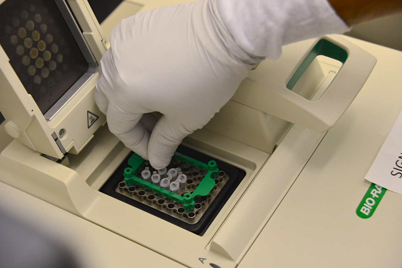
Specimen of protein are prepared for an experiment in a lab at the Bond Life Sciences Center at the University of Missouri.
Understanding oxidative stress
Most simply, oxidative stress is an imbalance that happens when the body uses oxygen to produce energy.
Superoxide, a “promiscuous and nonspecific” compound produced as a byproduct of this process, is a highly reactive molecule that can damage DNA and other cellular components, Hannink said.
The superoxide molecule is a “free radical.” That means it’s especially promiscuous and reacts with many different types of cellular molecules, leaving destruction in its wake, he said.
That damage can lead to a long list of problems, including cancer or neurodegenerative diseases like Parkinson’s disease.
In response to oxidative stress, the cell produces protective “anti-oxidant” proteins, which help remove the harmful reactive oxygen species and minimize damage.
But a heavy anti-oxidant response could be dangerous, too. The bottom line: it’s about maintaining a fine balance between “oxidants” and “anti-oxidants”.
Search for right combination continues
A drug that corrects the imbalance of oxidative stress could one day have wide applicability.
Jasmer developed a test to measure how specific compounds altered gene expression. The genetic response to oxidative stress has both an “ON” switch and an “OFF” switch.
Using this test, Jasmer determined how each compound affected specific genetic switches and, in turn, how the response to oxidative stress is regulated.
This test helped identify which molecules might be promising candidates for treating oxidative stress, leading them to one in particular that seemed to have the desired properties: HPP-4382.
But creating effective drugs is a long process of trial and error. Once molecules have been identified that show efficacy in lab-based assays, scientists try different combinations to increase their potency and drug-like properties, and High Point is currently testing other molecules that behave like HPP-4382.
The compound serves as a good starting point for researchers who are interested in understanding how oxidative stress affects cellular processes, such as cell proliferation or cell death.“Now we have a better understanding of what this compound is doing,” Hannink said. “This compound can be used to test different ideas of how the balance between oxidants and anti-oxidants is achieved in healthy cells and how perturbation of this balance can lead to different diseases.”
Shan-Lu Liu, Bond LSC scientist and associate professor in the MU School of Medicine’s Department of Molecular Microbiology and Immunology. Courtesy Justin Kelley, University of Missouri Health System.
Shan-Lu Liu initially thought it was a mistake when a simple experiment kept failing.
But that serendipitous accident led the Bond Life Sciences Center researcher to discover how a protein prevents mature HIV from leaving a cell.
“It’s a striking phenomena caused by this particular protein,” Liu said. “The HIV is already assembled inside the cell, ready for release, but this protein surprisingly tethers this virus from being released.”
The TIM – T-cell/transmembrane immunoglobulin and mucin – family of proteins hasn’t received much attention from HIV researchers, but recent research shows the protein family plays a critical role in viral infections. From Ebola and Dengue to Hepatitis A and HIV, these proteins aid in the entry of viruses into host cells.
But its ability to stop the virus from leaving cells remained unknown until now. Liu’s lab stumbled onto this finding in November 2011 when trying to create stable cells for a different experiment. After two months of troubleshooting the HIV lentiviral vector – where genes responsible for creating TIM-1 proteins were inserted into a cell to create a stable cell line that expresses the protein – Liu was confident the vector’s failure was not only interesting but also important.
Minghua Li, coauthor of the study and an MU Area of Pathobiology graduate student. Courtesy Justin Kelley, University of Missouri Health System.
The lab spent the next two years trying to figure out what was happening. Minghua Li, an MU Area of Pathobiology graduate student, carried out experiments that confirmed the protein’s power to inhibit HIV-1 release from cells, reducing normal viral infection. His experiments showed TIM proteins prevent normal deployment of HIV, created by an infected cell, into the body to propagate.
TIM proteins stand erect like topiary on the outside and inside surfaces of T-cells, epithelial cells and other cells. When a virus initially approaches a cell, the top of each TIM protein binds with fats – called phosphatidylserine (PS) – covering the virus surface. This allows a virus, such as Ebola virus and Dengue virus, to enter the cell, infect and replicate, building up a population inside.
But as the virus creates new copies of itself, the host cell’s machinery also incorporates TIM proteins into new viruses. That causes problems for HIV as it tries to leave the cell. Now these proteins cause the viruses to bind to each other, clumping together and attaching to the cell surface.
“We see this striking phenotype where the virus just accumulates on the cell surface,” said Liu, who is also an associate professor in the MU School of Medicine’s Department of Molecular Microbiology and Immunology. “We consider this an intrinsic property of cellular response to viral infection that holds the virus from release.”
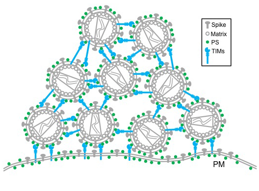
This model shows the interaction between TIMs and PS among the round HIV virions, as well as that between viral producer cells. This collectively leads to accumulation of HIV virions on the plasma membrane on the outside of the cell. Courtesy Minghua Li.
Further research is needed to determine overall benefit or detriment of this curious characteristic, but this discovery provides insight into the cell-virus interaction.
“This study shows that TIM proteins keep viral particles from being released by the infected cell and instead keep them tethered to the cell surface,” said Gordon Freeman, Ph.D., an associate professor of medicine with Harvard Medical School’s Dana-Farber Cancer Institute, who was not affiliated with the study. “This is true for several important enveloped viruses including HIV and Ebola. We may be able to use this insight to slow the production of these viruses.”
The National Institutes of Health and the University of Missouri partially supported this research. Additional collaborators include Eric Freed, PhD, senior investigator with the National Cancer Institute (NCI) HIV Drug Resistance Program; Sherimay Ablan, biologist with the NCI HIV Drug Resistance Program; Marc Johnson, PhD, Bond LSC researcher and associate professor in the MU Department of Molecular Microbiology and Immunology; Chunhui Miao and Matthew Fuller, graduate students in the MU Department of Molecular Microbiology and Immunology; Yi-Min Zheng, MD, MS, senior research specialist with the Christopher S. Bond Life Sciences Center at MU; Paul Rennert, PhD, founder and principal of SugarCone Biotech LLC in Holliston, Massachusetts; and Wendy Maury, PhD, professor of microbiology at the University of Iowa.
Carbon’s next-door neighbor on the periodic table typically receives little attention, but when it comes to corn reproduction boron fills an important role.
According to University of Missouri scientists, tiny amounts of boron play a key part in the development of ears and tassels on every cornstalk. The July 2014 edition of the journal Plant Cell published this research.
“Boron deficiency was already known to cause plants to stop growing, but we showed a lack of boron actually causes a problem in the meristems, the stem cells of the plant,” said Paula McSteen, a Bond Life Sciences Center researcher. “That was completely unknown before, and for plant scientists that’s an important discovery.”
Amanda Durkbak, MU Biological Sciences post doc; Paula McSteen, Bond LSC researcher and associate professor of Biological Sciences; research specialist Sharon Pike; and Walter Gassmann, Bond LSC researcher and professor of Plant Sciences.
Meristems are a big deal to a plant. These pools of stem cells are the growing points for each plant, and every organ comes from them. They are how plants can survive for 500 or 5,000 years, continuously making new organs in the form of leaves, flowers, and seeds throughout its life.
“When you mow your grass, it keeps growing because of the meristems,” said Amanda Durbak, first author on the paper and MU biological sciences post doc. “In corn, there are actually hundreds of meristems at the tips and all sides of ears and tassels.”
But without enough boron, these growing points disintegrate, and, in corn, that means vegetation is stunted, tassels fail to develop properly and kernels don’t set on an ear. This leads to reduced yield. Missouri and the eastern half of the U.S. are typically plagued by boron-deficient soil, an essential micronutrient for crops like corn and soybeans, indicating that farmers need to supplement with boron to maximize yield.
The tassel-less mutant
The team’s discovery started with a stunted, little corn plant that just couldn’t grow tassels, only created a tiny ear and died within a few weeks. These maligned reproductive organs piqued McSteen’s interest and her team of collaborators set out to figure out which gene was affected in this mutant plant. Graduate student Kim Phillips mapped the mutation to a specific gene in the corn genome involved in transporting molecules across the plant membrane.
But, what was this defect preventing the plant from receiving? Two experiments helped find the answer.
They started by looking at similar genes in other plants and animals. Simon Malcomber of California State University-Long Beach compared the gene – named the tassel-less gene after its mutant appearance – to similar genes in other plants and animals. He found that many were known to make a protein that transports boron and a few other elements.
From field to frog
To clinch this hypothesis, McSteen looked to Bond LSC scientist Walter Gassmann and the African clawed frog. Gassmann harvests eggs from these frogs and uses them to “test” the function of genes from both plants and animals.
“What we do is we inject the frog egg with RNA made in a test tube from the corn’s DNA,” Gassmann said. “The egg is a single, living cell that will actually use the message provided by this RNA to make the boron transporting protein and put it in the egg’s membrane.”
Frog eggs don’t naturally have the ability to transport boron, so an uninjected egg in a solution of boron can’t move the element into the cell.
“Corn RNA provides the egg with instructions to make a boron transporter protein, so the boron solution should move from outside to inside the egg,” Durbak said. “The egg should swell, showing this protein moves boron, and, in fact, these eggs swell so much they explode.”
A tank and a bucket guaranteed boron was the culprit. Durbak went back to the cornfield, watering some mutant tassel-less corn with boron fertilizer and other mutant plants with only water.
Only the ones given boron recovered and grew like normal corn plants, showing that the mutant corn has difficulties obtaining enough boron under natural, low boron conditions without this boron transporter. The boron content of the plants were later tested at the MU Research Reactor and the MU Extension Soil and Plant Testing Laboratory, affirming their observations.
A closer look
But, what does boron deficiency look like on a cellular level?
To see, the team collaborated with biochemist Malcolm O’Neill at University of Georgia. He looked at the cell walls in the plant and discovered that the pectin was affected. Pectin stabilizes the plant cell wall, and many home canners know pectin for its help in making jelly and jam solidify. Pectin is strengthened when boron cross-links two carbohydrates together, giving rigidity to the plant cell wall.
“The effect is that it locks in the cellulose, so without it plant cells won’t have nearly the stability,” McSteen said. “What we think is going on is that plant meristems basically disintegrate because they don’t have the support of pectin.”
While McSteen’s team identified the gene that controls the protein for boron transport into a cell, a research team from Rutgers University identified a gene that controls the protein that transports boron out of a cell. See more about both studies in Plant Cell’s “In Brief” section.
The next step in this research is to look more closely at what happens in these boron-deficient cells early on as they develop to understand the mechanism of boron action in stem cells.
A grant from the National Science Foundation supported this research.
In a second travel log from Bond LSC researcher Cheryl Rosenfeld, learn about the wildlife she encountered in Tanzania this summer. Through the North American Veterinary Community (NAVC), Rosenfeld furthered her veterinary education while encountering wildlife in their natural habitat. See more about the first leg of her trip to Rwanda here.
By Cheryl Rosenfeld
In the early morning hours, our group flew from Kigali, Rwanda to the Serengeti in Tanzania. As we began the descent to the dirt runway, we glimpsed our first sight of wildebeest and the awe-inspiring Serengeti plains and I soon boarded “tano,” Swahili for the fifth 4×4 in our convoy. “Tano” soon came to have special meaning, as there are many groupings of five to see in Tanzania: “The Big Five, The Ugly Five, etc.” Emanuel, whose life-long ambition was to attend college to be a guide, led us to see all of these groups of five and then some.
Cheryl Rosenfeld
From the time we pulled away from the meeting spot, I started photographing animals on one side of the vehicle, but it seemed even more intriguing ones would appear on the other side of the road.
When the NAVC and our veterinary guide, Dr. Carol Walton, organized this trip, it was anticipated that the wildebeest would be in the Western corridor of the Serengeti but she was wrong. It’s no longer the case that the location of the wildebeest migrations can be projected with accuracy. Our lecture the first evening in Tanzania discussed how the wildebeest know to migrate and why past modeling of their migratory pattern is no longer accurate. Theories for the migratory nature of these animals include their ability to smell rain and/or changes in calcium concentrations in the soil. Wildebeest rely heavily on calcium to produce sufficient milk to nurse their calves that expend considerable energy keeping up with the herd.
In the past, the rains, like the wildebeest migration, would occur in similar sites and times throughout the year. Climate change has likely contributed to the rainfall in these sites being less reliable and correspondingly, compromised forecasting where the wildebeest will be located throughout the year. This year the wildebeest decided instead to migrate to the central Serengeti region. Thus, the next day we set off on an over-three-hour journey to this region.
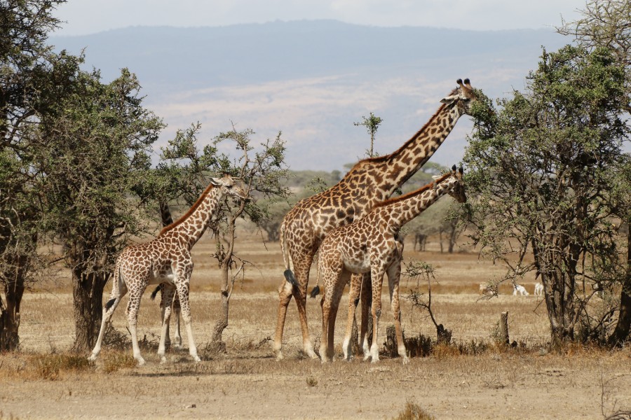
While driving to find wildebeest we came upon new animals and birds including Coke’s Hartbeest, several Masai giraffe, and topis, which seemed to be splattered with oil. Finally, the wildebeest herds starting increasing in size until it reached a crescendo. As far as one could look in all directions there were wildebeest: males, females, and an abundance of baby calves. Eating alongside them were many zebras and their babies, with brown fuzzy fur along their back-end. It was the most amazing spectacle any of us had witnessed.
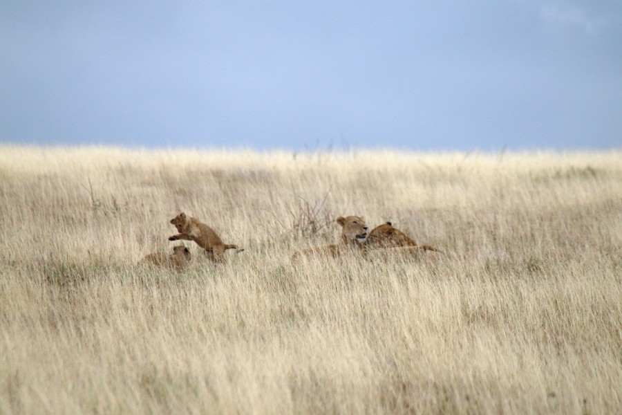
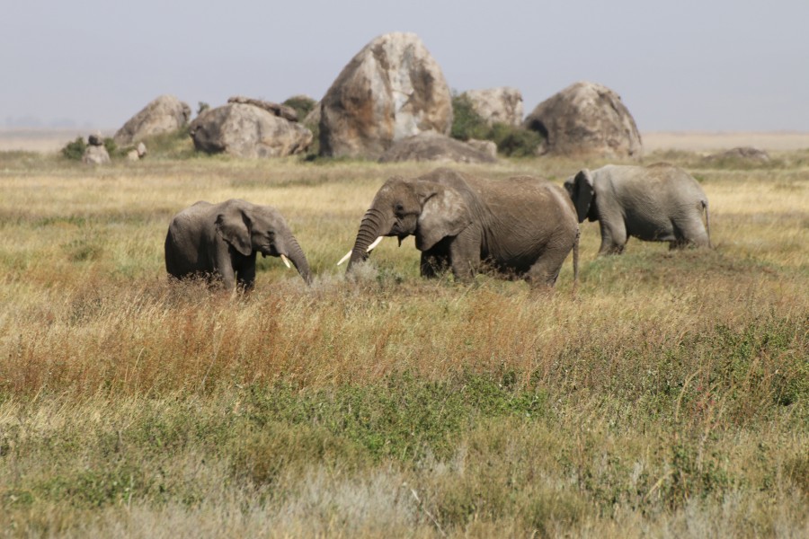
After seeing the vast wildebeest herds, we then came upon the elephants and lions, along with their babies. In the safety of the vehicle, we watched them engage in their natural behaviors for which no zoo experience can replicate. Both species were incredibly affectionate to the offspring and each other. In the case of the lionesses, each time the females, who were likely related, caught up with another that they had not seen in a short while would lead to emotional bouts of jumping on each other, caressing and licking. It was hardly the acts of a deadly predator. Yet, their existence rests on the wildebeest and other prey. With the wildebeest in this area, it seemed all life was flourishing at this time. A true paradise.
However, even paradise is subject to outside threats. One such threat is the massive poaching of elephants in Tanzania and surrounding African countries with 40,000 being killed last year alone. Prior to 2013, Tanzania had a “shoot to kill” policy for those caught poaching elephants or other endangered species. However, this rule has since been relaxed with the elephant numbers now in severe decline. At this unsustainable rate of poaching, the African elephant may go extinct in the wild in the next few decades.
While we did see some groups of elephants, the size of each matriarch-led group seemed less than those depicted in Sir David Attenborough and National Geographic documentaries. Moreover, one elephant that we came across, which I affectionately referred to as “Stumpy,” bore sad evidence of this brutal practice: He lost part of his trunk in a poacher’s snare. Without a full trunk, this elephant exhibited great difficulty grasping various plant items to place in his mouth.
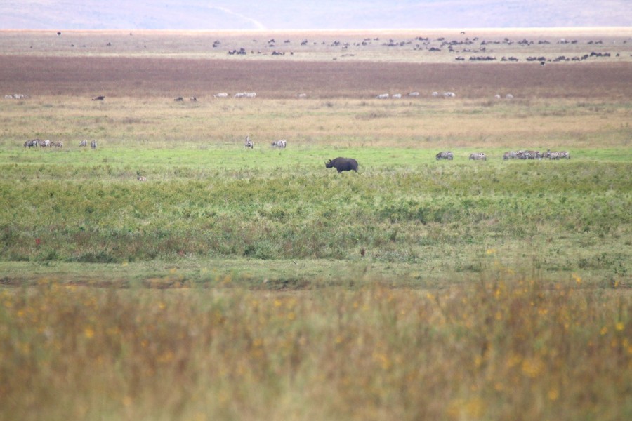
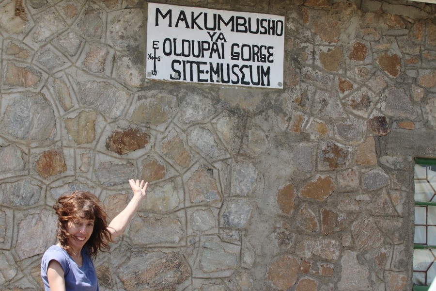
Our subsequent travel took us to the famous “Olduvai (which should actually be Oldupai) Gorge,” one of the most famous paleoanthropological sites, including the Leakeys’ famous discovery. From there we traveled to the Ngorongoro Crater, a gigantic fracture of the earth’s crust that provides habitat for some 30,000 animals, including the most endangered black rhinoceros that only has 20 to 30 left in this area. The difference between the black and white rhino lies not in their coat color but a structural difference in their lips with black rhino possessing pointed and prehensile upper lip to browse on twigs and leaves. In contrast, white rhino possess a square upper lip to graze on the grasses below.
When Dr. Walton convinced us to set off at first light, around 6 a.m., our goal was to find one of these amazing creatures. Once again, with Emmanuel’s assistance and eagle eyes, what started out as a black dot far off in the horizon began to morph into a recognizable rhinoceros. In utter delight, we strained our necks and photographed as this beautiful animal continued to move along and forage in the distance. It would be inhumane to allow this animal to go extinct. In the case of the rhino, many Asian countries believe that its horn increases male libido, which has never been proved to be the case. This mistaken notion possibly originated from the fact that male rhinos copulate with females for several hours duration. This behavior is, however, not transferrable to men who consume any part of a rhino.
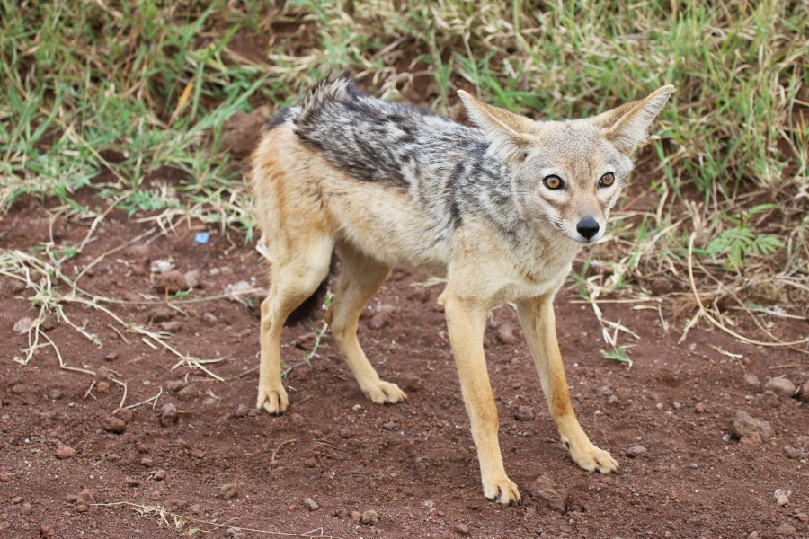
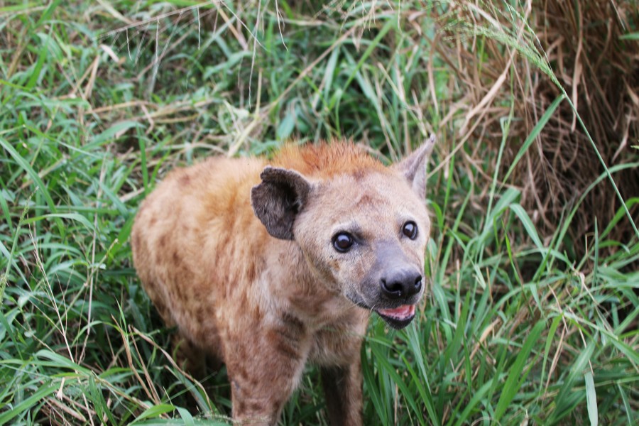
The list of species we saw in Tanzania continued to grow while we drove around the crater with black-backed and common jackals following alongside our vehicle, eland and grants gazelles grazing in the plains, flocks of greater and lesser flamingoes observed in the distance, and more lion prides and spotted hyenas.
The next day, we had our final farewell lunch at the famous Arusha Coffee Lodge, where US presidents have dined. We then set off on our long journey back to the US.
All of us were impacted by the creatures and sites we saw. One male veterinarian, who seemed gruff and quiet for most of the expedition, put it best the night before we left. He unexpectedly stood up after our last lecture and proclaimed that he signed up for the expedition more as an escape from the daily grind of being a veterinarian. However, the trip made such as impression on him to the point that he realized that part of the veterinarian oath on “relieving animal suffering” should include standing up and advocating on their behalf to prevent their brutal murder, as previously occurred in the case of the gorillas and is ongoing for elephants and rhinos. All of us were moved by his emotional comments and the tears welling up in his eyes. Our group was indeed fortunate to see these majestic animals, while they still exist, in their natural environments. I hope there is a wake-up call for nations to come together to prevent their extinction.
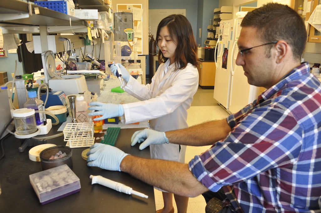
Graduate students Yuleam Song and Dan Salamango inoculate a bacteria culture in Johnson’s lab. The inoculation takes a small portion of a virus and multiplies the sample, allowing researchers to custom-make viruses.
By Madison Knapp | Bond Life Sciences Center summer intern
Modern science has found a way to turn viruses —tiny, dangerous weapons responsible for runny noses, crippling stomach pains and worldwide epidemics such as AIDS— into a tool.
Gene therapy centers on the idea that scientists can hijack viruses and use them as vehicles to deliver DNA to organs in the body that are missing important genes, but the understanding of virus behavior is far from exhaustive.
Marc Johnson, researcher at the Christopher S. Bond Life Sciences Center and associate professor of molecular microbiology and immunology in the MU School of Medicine, has been building an understanding of viral navigation mechanisms which allow a virus to recognize the kind of cell it can infect.
Johnson’s research specifically explores the intricacies of the viral navigation system and could improve future direction of gene therapy, he said.
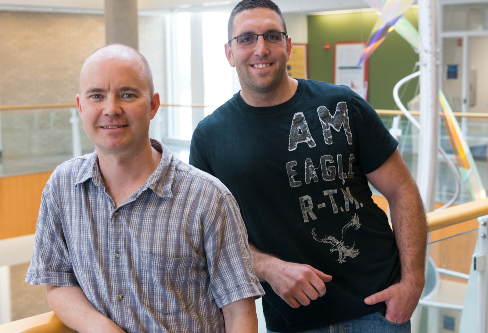
Marc Johnson (left) with Dan Salamango, a graduate student that works in his lab. The lab does important research on the basic function and mechanisms of viral navigation and transport.
Turning a virus into a tool
Conceptualized in the 1970s, gene therapy was developed to treat patients for a variety of diseases, including Parkinson’s, leukemia and hemophilia (a genetic condition that stops blood from clotting).
To treat disease using gene therapy, a customized virus is prepared. A virus can be thought of as a missile with a navigation system and two other basic subunits: A capsule that holds the ammunition and the ammunition itself.
The viral genetic material can be thought of as the missile’s ammunition. When a cell is infected, this genetic material is deployed and incorporated into the cell’s DNA. The host cell then becomes a factory producing parts of the virus. Those parts assemble inside the cell to make a new virus, which then leaves the cell to infect another.
The capsule is made of structural protein that contains the genetic material, and the navigation system is a protein that allows the virus to recognize the kind of cell it can infect.
Viral navigation
Gene therapy uses viruses to solve many problems by utilizing a virus’ ability to integrate itself into a host cell’s DNA; to do this successfully, researchers need to provide a compatible navigation component.
In the body, viruses speed around as if on a busy highway. Each virus has a navigation system telling it which cells to infect. But sometimes if a virus picks up the wrong type of navigation system, it doesn’t know where to go at all.
“What you can do is find a virus that infects the liver already, steal its navigation protein and use that to assemble the virus you want to deliver the gene the liver needs,” Johnson said. “You can basically take the guidance system off of one and stick it onto another to custom design your virus.”
But this doesn’t always work because of incompatibility among certain viruses, he said.
Johnson and his lab are working to understand what makes switching out navigation proteins possible and why some viruses’ navigation systems are incompatible with other viruses.
“I’m trying to understand what makes it compatible so that hopefully down the road we can intelligently make others compatible,” Johnson said.
The right map, the right destination
Johnson creates custom viruses by introducing the three viral components—structural protein, genetic material, and navigation protein—to a cell culture. The structural protein and genetic material match, but the navigation component is the wild card. It could either take to the other parts to produce an infectious virus, or it could be incompatible.
Johnson uses a special fluorescent microscope to identify which viruses assembled correctly and which didn’t.
A successful pairing is like making a match. If a navigation protein is programmed to target liver cells, it’s considered a successful pairing when the virus arrives at the liver cell target location.
The scope of gene therapy continues to widen. Improved mechanisms for gene therapy, and greater knowledge of how a navigation protein drives a virus could help more people benefit from the vehicles viruses can become.
Johnson uses several high-profile model retroviruses, including human immunodeficiency virus (HIV), which affects an estimated 35 million people worldwide each year, according to the World Health Organization.
Understanding nuances of HIV in comparison to other viruses allows Johnson to pick out which behaviors might be common to all retroviruses and others behaviors that might be specific to each virus.
Johnson said his more general approach makes it easier to understand more complex viral features.
“If there are multiple mechanisms at work, it gets a little trickier,” Johnson said. “My angle is more generic, which makes it easier to tease them apart.”
Lauren and Claire Gibbs share contagious laughter, ambition and a charismatic sarcasm.
Both are honor students at Shawnee Mission East High School in a Kansas City suburb.
They also share a neuromuscular disease called spinal muscular atrophy (SMA), designated as an “orphan disease” because it affects fewer than 200,000 people in the U.S.
However, the landscape for individuals with SMA is quickly changing with the development of new drugs.
More than 7 million people in the United States are carriers (approximately 1 in 40) of the so-called “rare” neurodegenerative disease, SMA.
Lauren,17 (left) and Claire, 16 (right), say their shared SMA diagnosis has strengthened their relationship and presented them with opportunities to travel and share their experiences. | Photo provided by the Gibbs family.
Faces of SMA
The success of therapeutics in lab experiments provides a new layer of hope for individuals and families living with the disease.
Lauren, now 17, fit the criteria for SMA Type III, while Claire, now 16, showed symptoms of a more severe manifestation of the disease, SMA Type II.
Lauren and Claire Gibbs were diagnosed on the same day.
Despite their numerous similarities, the biggest disparity between them is mobility.
Claire uses a power wheel chair while Lauren is able to use a manual chair. It’s not unusual to see Lauren being pulled along in her chair, playfully hanging onto the back of Claire’s motorized chair.
Lauren is participating in a clinical trial with ISIS-SMNRx a compound developed by Isis Pharmaceuticals, a leading company in the antisense drug discovery and development based in Carlsbad, Calif. Lauren feels that she has gained stamina and a greater ability to walk — a feat that wasn’t routine just five years ago.
Prior to the trial, Lauren was able to walk only for short distances.
Tim and Natalie Gibbs with their daughters Lauren, 17 (left) and Claire, 16 (right) in Washington D.C. The Gibbs have been visible advocates in the fight for a cure for spinal muscular atrophy. | Photo provided by the Gibbs family.
Bringing New Hope
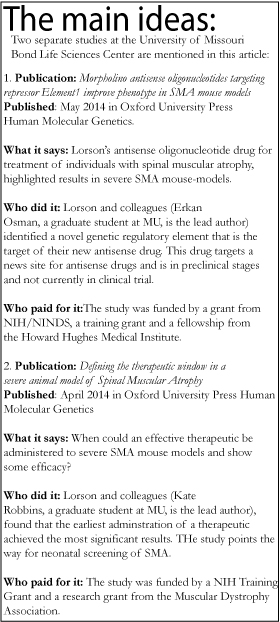
A new experimental drug developed by researchers at the Christopher S. Bond Life Sciences Center, is bringing hope to individuals with the orphan disease affecting one in 6,000 people.
Christian Lorson PhD, investigator in the Bond Life Sciences Center and Professor of Veterinary Pathobiology at the University of Missouri, has been researching SMA for seventeen years and has made a recent breakthrough with the development of a novel compound found to be highly efficacious in animal models of disease. In April, a patent was filed for Lorson’s compound for use in SMA.
Lorson’s therapeutic, an antisense oligonucleotide (a fancy name for a small molecule therapeutic that falls under the umbrella of gene therapy), repairs expression from the gene affected by the disease. The research was published May in in the Oxford University Press, Human Molecular Genetics.
The drug developed by Lorson’s lab is conceptually similar to ISIS-SMNRx already in clinical trial developed by Isis Pharmaceuticals and a team of investigators at Cold Spring Harbor Laboratory headed by Dr. Adrian Krainer.
Antisense drugs are not a new practice, but their wide-spread adoption seems to be on the cusp with recent success stories like the commercialization of an FDA-approved antisense compound produced by Isis in 2013 called Kynamro for the treatment of homozygous familial hypercholesterolemia, a high cholesterol disorder that is passed down through families.
Science behind success
The National Institutes of Health has listed SMA as the neurological disease closest to finding a cure. Discoveries made by the Lorson Lab have contributed significantly to current scientific understanding of the disease mechanisms and to the advances being made in finding an effective treatment for SMA.
These antisense therapies work because of the genetic makeup of SMA —the genetics are incredibly clear: a single, specific gene called Survival Motor Neuron 1 (SMN1) has been pinpointed as the cause of SMA.
SMA is a neurodegenerative disorder, meaning muscles become weaker over time due to sick or dying neurons.
These neurons become less functional because of low levels of the SMN.
Remarkably, the disease can be reversed in animal models of disease if the nearly identical duplicate gene, SMN2, can be “turned on” to compensate for low SMN levels.
Lorson’s antisense oligonucleotide therapeutic provides incredible specificity because it hones in on a specific genetic target sequence within SMN2 RNA and allows proper “editing” of the RNA encoding the SMN protein. The strategy is to “repress the repressor,” Lorson said.
The SMA-specific defect lies at the RNA step – the “cutting and splicing” of important RNA sequences does not happen efficiently in SMN2 RNAs because of a several “repressor” signals.
“The final chapter of the book — or the final exon — is omitted,” Lorson said. “But the exciting part is that the important chapter is still there – and can be tricked into being read correctly: if you know how.”
The new, antisense oligonucleotide seems to know how to get the job done.
The existence of such similar genes as SMN1 and SMN2 in humans creates a rare genetic landscape lending itself especially to a therapeutic development for SMA.
Humans are unique in this duplication — something Lorson calls a “genetic happenstance” that, on an evolutionary scale, may as well have happened yesterday.
Why humans have developed this redundant gene is unknown.
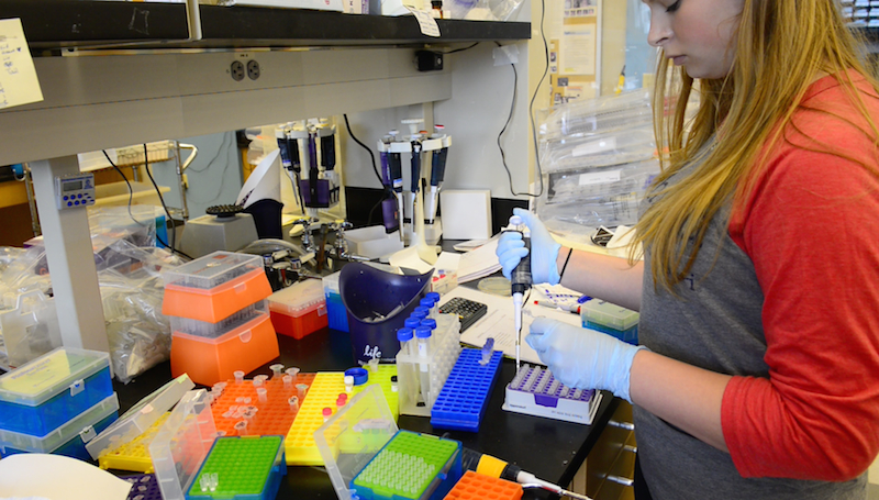
Thalia Sass, an MU biological sciences major, genotypes samples in the Lorson Lab where spinal muscular atrophy is researched.
Timing is everything
In addition to the developments of new SMA therapeutics, Lorson and his lab sought to answer an important biological question concerning the disease: When can a therapeutic be administered and still show some degree of efficacy?
Lorson’s research found that the earliest administration of a treatment provided the best outlook— extending the survival of laboratory mice by 500 to 700 percent, “a profound rescue,” according to his research published in April in the Oxford University Press, Human Molecular Genetics.
A near complete, 90 percent rescue was demonstrated in severe SMA mouse models. But even when the therapeutic was administered after the onset of SMA symptoms, there was still a significant impact on the severity of the disease.
“If you replace SMN early and get (a therapeutic) to cells that are important to the disease, you correct it,” Lorson said. “This provides hope that patients who have been diagnosed will still see some therapeutic benefit even if it is clear that the best results will likely come from early therapeutic administration.”
In Lorson’s study it’s definitive that the earlier a therapeutic can be administered, the better the outcome for individuals with SMA.
“This really points towards a strong push for neonatal screening,” Lorson said. “Infant screening would likely be incredibly beneficial for SMA and that’s something that the SMA community is really excited about.”
A breakthrough for families
On June 2, Lauren and Claire Gibbs attended a routine, annual rehab appointment with Dr. Robert Rinaldi, MD, division of pediatric rehabilitation medicine and attending physician at Children’s Mercy Hospital in Kansas City, Mo Dr. Rinaldi is not associated with the Isis clinical trial.
The appointment was like a reunion among close friends — Rinaldi began seeing Claire and Lauren Gibbs 16 years ago, the first year that he began working at the hospital and when the girls were one- and two-years-old, respectively.
The girls did all of the routine tests —measuring strength of grip and breathing, and assessing range of movement with the occupational and physical therapists.
A little later, Rinaldi sat with Natalie Gibbs, Lauren and Claire’s mother and a relentless advocate for advancement in SMA awareness.
Typically the muscles of individuals with SMA deteriorate over time, but together they inspected the definition of a new calf muscle on Lauren’s left leg.
For a young woman with Type III SMA — this means she can walk for short distances with little discomfort but still uses her wheel chair a majority of the time — Lauren’s new calf muscle is a remarkable achievement.
As Lauren continues to participate in the ISIS antisense therapy clinical trial, her conditions continue to improve dramatically, even with the late administration of the therapy — in her case, 16 years after her diagnosis and onset of effects.
Lauren believes her ability and stamina for walking have increased significantly.
“Quite frankly my jaw almost hit the ground when she stood up — the change was that impressive to me,” Rinaldi said.
Rinaldi, also the co-director of the Nerve and Muscle Clinics at the hospital, had last seen Lauren two years ago. He said the Lauren he saw during a routine rehab appointment in June was like seeing a new person altogether.
“The way she stood up from the wheel chair — how quickly she did that with no support — her posture when she was standing up was more upright, her pelvis was in a much better position, her core was straighter,” Rinaldi said. “It struck me immediately how much better she looked.”
Lauren Gibbs is the first of Rinaldi’s patients to have participated in the ISIS clinical trial.
“It’s moving very fast in this field,” Rinaldi said. “I think the technology that’s evolving in research is opening up more avenues for investigation for us and there’s a big desire to find a cure for these types of diseases.”
The progress has rewarded the Gibbs family’s advocacy in SMA awareness and they’ve been able to set new goals they didn’t imagine were possible when the diagnoses for Lauren and Claire were made. Natalie Gibbs is a long-time member of Families of SMA and is currently on their Board of Directors.
The organization Families of SMA is currently providing funding to Lorson to advance this research area.
“We’re able to see first hand — and our physician who has been watching them for sixteen years has seen — that everything we’re doing in the clinical trials is really making a difference,” Natalie Gibbs said.
Over the course of their daughters’ lives, Natalie and her husband Tim Gibbs say a shift in momentum has accelerated the technology and research toward finding a cure for SMA.
“I am really impressed with the progress Lauren has made with the trial and how well Claire is doing overall,” Natalie Gibbs said. “Even though it’s a progressive and very devastating type of disease, I feel like we’re really conquering it.”
The unusual red color of the Lobelias leaves make them stand out among 200 other species that thrive in the 20-foot plant wall at the Bond Life Sciences Center | Paige Blankenbuehler
By Madison Knapp | Bond LSC
A hidden treasure on the University of Missouri’s campus is a living and breathing work of art.
In the Christopher S. Bond Life Sciences Center, a 20-foot plant wall stands as a towering tribute to the diversity of plant life and coexistence of species — a botanical landscape of more than 200 individual plants from 40 to 50 species including many ferns, vines, perennials, orchids and geraniums.
The display continues to be a visual reminder of the building’s interdisciplinary nature – much like the plants within the wall, the Bond Life Sciences Center facilitates the coexistence and collaboration between an array of researchers within its walls.
One plant cascades down the wall and stands out more than most — it looks more like a large insect than any type of plant typically seen in such displays.
Tillandsia is a genus of succulent plant native to Central America that can undergo long dry spells. The wiry plant thrives on the stone beside the plant wall without the help of a pot of soil, and is seemingly absent an essential, anatomical feature of most flora — roots.
With leaves more like Medusa’s hair, Thallandsia are rootless plants mounted on stone alongside the plant wall. | Paige Blankenbuehler
They still have tiny root protrusions, but the mass is minuscule compared to the thick leaves, which take up and store water and nutrients, rather than the root system which handles that work in most plants.
Approximately 60 Tillandsias pepper the plant wall, including a few mounted directly onto the stone wall with a special kind of non-toxic rubber cement manufactured by Davis Farms based out of San Diego.
The strange plant was added to the green tapestry by Jason Fenton, an office support associate at the Bond Life Sciences Center. He first noticed the rootless plants in Belize while on a trip there in 2003 and had been growing them at his home.
“I think they’re really beautiful and distinctive,” Fenton said. “They help give variety to the wall.”
The rootless plants require special treatment — just a little extra attention from facilities manager, Jim Bixby, who waters the Tillandsias with a spray bottle once a week.
The wall started with a vision from Jack Schultz, director of the Bond Life Sciences Center. The execution and maintenance of the feature is credited to Bixby, who has honed the project since 2010.
After trying several watering systems, Bixby found one that worked. He hung rows of pots with modified flat sides and tailored a simple, drip irrigation system made for easy watering along a grid.
Within the diverse landscape and after additions of a variety of plants over the years, Bixby has seen competition between several of the species.
The wall favors the ferns, which have taken over much of the space, crowding out other plants for the eastern sunlight that flows through the windows, Bixby said.
Several long-stemmed species have found ways to cope, however; venturing out between the ferns’ curtain-like fronds to get their fair share of sunlight.
Supervising editor: Paige Blankenbuehler
The 20-foot tall plant wall outside of Monsanto Auditorium in the Bond Life Sciences Center is a nod to coexistence and diversity. | Paige Blankenbuehler
While summer brings a slower pace for many researchers, others use it as an opportunity to learn for their profession and network with others in their field.
Bond LSC researcher Cheryl Rosenfeld recently traveled to Africa to further her learning as a veterinarian. This continuing education gives her the opportunity to learn the newest techniques in the field and network with others to learn what’s current and on the collective minds in veterinary medicine. Through the North American Veterinary Community (NAVC), Rosenfeld has now gone on three expeditions where participants observe animals in their natural, exotic environments, attend nightly lectures and learn more about the humans near these animals.
Previous expeditions led Rosenfeld to the Galapagos Islands and the Florida Keys, but her June 2014 trip started in Rwanda and ended in Tanzania. Here’s the first of two entries where Rosenfeld shares here experience.
In June 2014 Cheryl Rosenfeld traveled to Rwanda to observe mountain gorillas as part of her veterinary continuing education.
In June 2014 Cheryl Rosenfeld traveled to Rwanda to observe mountain gorillas as part of her veterinary continuing education.
The 1994 Genocide is a omnipresent part of the national psyche of Rwanda.
Sites like this memorialize the millions killed in the 1994 genocide.
Local children from one Rwandan village.
Rosenfeld is pictured in the blue four drums from the left.
By Cheryl Rosenfeld
The fate of animal populations is generally intertwined with the predicament of humans in the area. Nowhere is this truer than in Rwanda. Most people know Rwanda for Dian Fossey’s work with the mountain gorillas and the genocide of more than 1 million Hutus and Tutsis that happened 20 years ago in 1994. In this 100-day period, an average of six individuals were killed per minute. Children that survived were often orphaned and many surviving women suffered being raped and exposed to HIV infection. In all, many still require extensive medical and psychological care. On our flight and checking into our hotel was a medical team from Harvard Medical Center that was there as part of the Clinton Foundation to assist in the medical needs.
We saw the history that continues to shape the country when we first visited a genocide memorial site just outside of Kigali where thousands of individuals were brutally murdered and the Kigali Genocide Museum that was partially funded by an English Jewish Holocaust survivor. The history of the conflict is rooted partially in Western influence that infused a social division. Prior to Europeans colonization, Hutus and Tutsis lived in relative peace and individuals could go back and forth between these two groups. The original difference was that Tutsi individuals owned more than 10 cows. The differential treatment and classification adopted by Europeans began to trigger conflict between the two groups. Prior indicators, including extensive propaganda, were ignored by the United States and United Nations. The museum includes two stained glass windows that depict the evidence that genocide was imminent and failure by other nations to prevent this tragedy. Genocide isn’t unique to Rwanda, though, and the displays describe the commonalities on their sad origins in other countries throughout history. Outside the museum, there are several mass graves where fresh flowers are placed on a routine basis.
I was originally hesitant about traveling to Rwanda because of this history, but am very glad I took the chance. The Rwandan government has worked hard to turn around and instill pride in the country. Their economy is one of the fastest growing in Africa with construction of new businesses and hotels in Kigali. Moreover, the government has placed a ban on plastic bags and hired teams of individuals to keep the country clean. One Saturday a month, all Rwandans, including the President, are expected to participate in clean-up day, which becomes a convivial social event. While there is still sadness in the eyes of many individuals I met, I also saw hope of something better, which was inspiring to witness.
We were soon off to learn about the mountain gorillas that are now the pride of the country. During Dian Fossey’s time, she battled to prevent poaching of these magnificent and intelligent creatures. The country now realizes the worth of preserving and propagating the mountain gorilla populations. In a reasonable and safe way, they developed a tourist industry to view the various troops of gorillas. It currently costs $750 to spend one hour with the mountain gorillas. The government has restricted access to prevent gorilla habituation and stress from too many tourists.
We spent two days with different troops. While waiting in the morning to find out which troop we were responsible for trekking, we were entertained by local dancers. I regrettably made the mistake of indicating I felt fit to track the one of ten groups that was at the furthest distance.
The group that was involved in trekking the first day was called “Snow” in Kinyarwanda. I believe they received this name because they inevitably reside high in the mountains, which used to have snow. As we set off on our hike, many children came out to say “MooRahHoh”- hello in Kinyarwanda and asked for us to take their picture. We were informed that we should easily return before lunch, and therefore were only provided a package of peanuts. Unfortunately, it took us longer to hike through the forests that transitioned from bamboo to masses of stinging nettles and did not return to the hotel until 6:30 p.m. After more than three hours of hiking and our eyes finally fell upon our first mountain gorilla, the silverback of the group. Even knowing that this was the ultimate goal, we were not prepared for this amazing experience of being so close to a creature in the wild that resembled us.
We had the opportunity to meet the rest of the troop, including several 3 to 4 month old babies that were quite entertaining. The enclosed photos and videos only provide a sliver of the spectacle that we were privileged to be part of these two days of gorilla trekking that made our hunger and continued burning sensation on our face and legs from stinging nettles well worth it.