Jessica Whited studies the genetics behind how salamanders grow severed limbs
By Eleanor Hasenbeck | Bond LSC
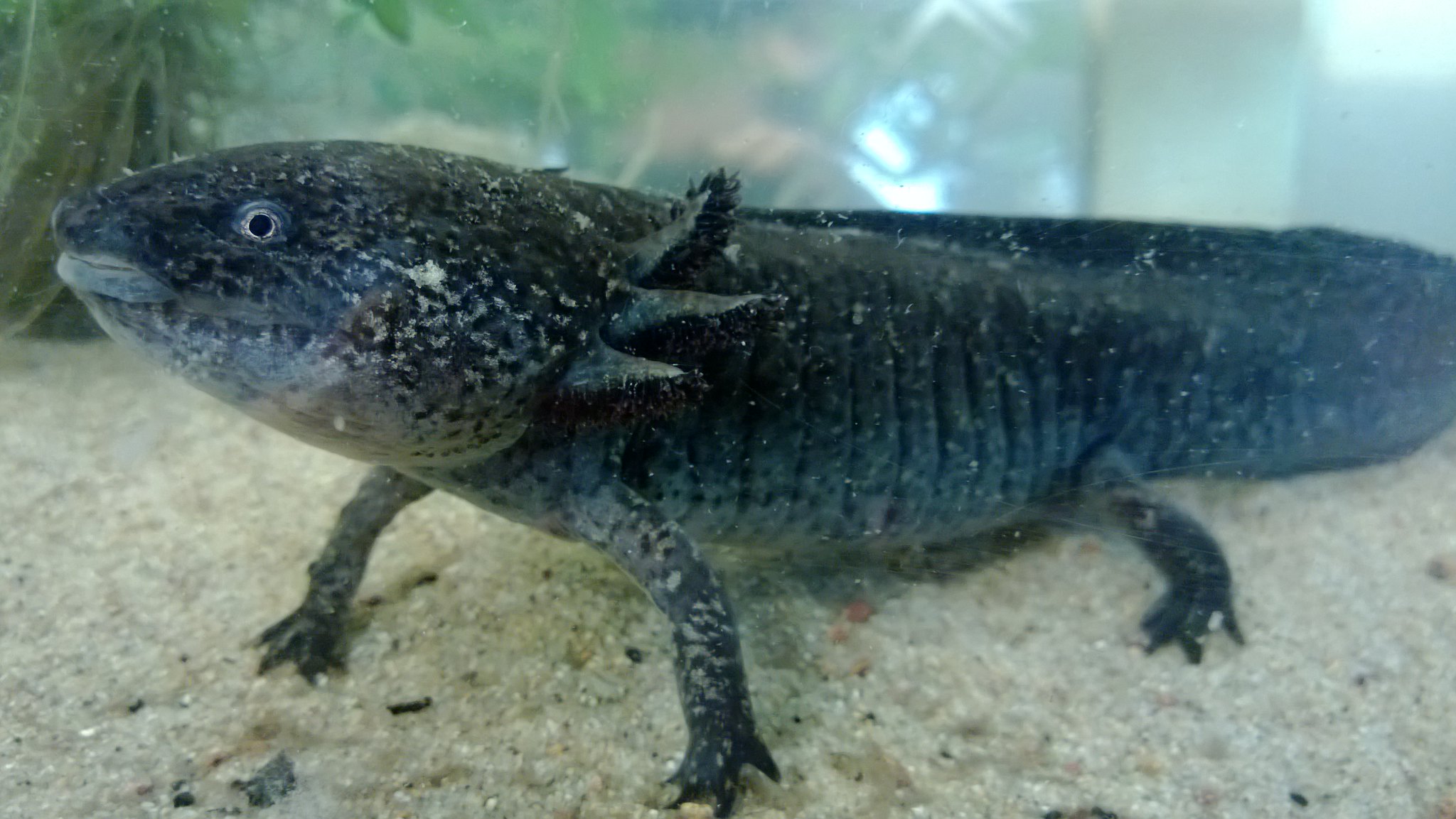
An axolotl rests at the bottom of its tank at Menagerie du Jardin des Plantes in Paris. | photo by Jack Baker, Flickr
It takes about two months for an axolotl to regenerate a lost limb. Humans, as you probably know, don’t regenerate limbs.
But, a basic understanding of how the Mexican salamander regrows limbs advance regenerative medicine in humans according to Jessica Whited, a researcher at Brigham Women’s Hospital and assistant professor at Harvard Medical School.
Whited will speak at 3:30 p.m., Thursday April 13, in Monsanto Auditorium as part of Missouri Life Sciences Week at Bond Life Science Center. Her lecture, “Identifying roadblocks to regeneration in axolotl salamanders” will present the lab’s discoveries and evidence that a specific gene in axolotls can block the animal’s ability to regenerate.
Whited’s lab found axolotls can exhaust their ability to regenerate. When a limb is severed repeatedly, the salamander stops producing blastemas, the mass of cells capable of regeneration that allow the limb to grow back. This could be due to a dysregulated gene blocking the animal’s ability to produce them.
The Whited Lab sequenced the mRNA in axolotls that could regenerate limbs and that could no longer regenerate. They found 912 genes that differed between the two groups. Whited will discuss one of these genes, which her lab considers a potential inhibitor to regeneration.
“It’s much more common for people to think “Oh, what are the things that promote limb regeneration?’ than it is to think about the things that we might have to block to make it happen,” Whited said. “This project has the potential to uncover the roadblocks, which could turn out to be equally critical.”
An MU alumna, Whited received the National Institutes of Health New Innovator Award in 2015 for her work with this unique regenerative salamander. She earned a PhD in biology at the Massachusetts Institute of Technology, and two undergraduate degrees in biological sciences and philosophy at MU.
Whited attended MU as a Bright Flight and Curator’s Scholar. And though it happened nearly 20 years ago, she said receiving those two scholarships were among the most important things that happened in her career. As a high school student, she knew she would go to college, but financially, she didn’t know how it would happen. She also credits her education and undergraduate research experience at MU for preparing her to think at the research bench.
“You have to get an undergraduate education, and it totally prepared me even for graduate school at MIT, which is one of the top programs in the world, in many subjects, but in biology especially,” Whited said. “The idea that you could find a career where you’re using your brain as your primary asset, I figured that out while I was at the University of Missouri, because there were people, our professors, doing that.”
Whited’s lecture is free and open to the public as part of Missouri Life Sciences Week. It occurs at 3:30 on Thursday, April 13 in Bond LSC’s Monsanto Auditorium. See more about events during the week at bondlsc.missouri.edu/life-sciences-week.
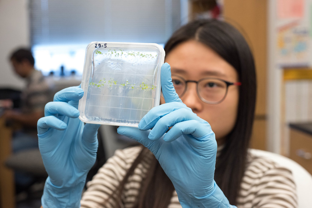
Plants on the left grow with rhizobia bacteria, one type of fixing nitrogen bacteria, in the greenhouse, while the plants on the right grow without the bacteria. | photo by Jinghong Chen, Bond LSC
Jinghong Chen | Bond Life Sciences Center
Since eight years old, Beverly Agtuca knew she wanted to be a scientist.
A trip to Philippines changed Agtuca, an American-born Filipino, and inspired her passion on plants.
“My grandma always told me to work in the field all day so that they can have enough food for us to eat,” Agtuca said. “The life [in Philippines] is so different from here…I want to not just provide food but be that scientist trying to figuring something out, and hopefully saving the world.”
Agtuca is on her way to her dream. She is now a third year doctoral student in Gary Stacey’s lab at Bond Life Sciences Center with a focus on nitrogen-fixing bacteria.
Although she has been involved in research since high school, Agtuca recently faced a new challenge of telling people about her work. The Preparing Tomorrow’s Leaders of Science class tasked her with making a 90-second video to explain her two-year study to the general public.
Her team, “The A Team,” chose to go with the benefits of having nitrogen-fixing bacteria.
For decades, people have been adding nitrogen fertilizers to plants to improve yields, but this can lead to pollution in water systems and ecosystems. Scientists need to enhance plant productivity to meet a huge food demand by the year of 2050.
One little bacteria might make this possible and save the world. Rhizobia, a type of natural bacteria in soil, are able to fix nitrogen via biological nitrogen fixation. These bacteria can convert nitrogen gas into ammonia as a plant nutrient source, while the plants give all the carbon sources back to the bacteria.
“It is like a walky-talky,” Agtuca said. “They are communicating with each other.”
Yet before speaking to the public, Agtuca needs to explain the plant-bacteria interaction to her teammates. Students less well versed in science like Jessica Kaiser, a strategic communication student, thinks of science differently.
“The biggest issue we ran into is jargon, like basic science words that [my teammates] are so comfortable with,” Kaiser said. “We need to focus on what people care about instead of the technical sides, to focus on why it matters to anybody rather than just to a science person.”
Within two weeks, they produced the video “Good Microbes: reducing pollution one farm at a time.” Along with two other teams, their videos will be commented and judged by representatives from Monsanto.
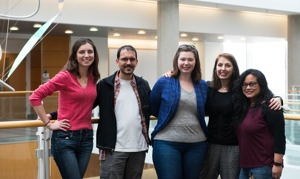
“The A Team” stands together at Bond Life Sciences Center. From left to right: Jessica Kaiser, Sven Nelson, Anna Glowinski, Eleni Galata and Beverly Agtuca. | photo by Jinghong Chen, Bond LSC
The 90-second video is just a glimpse of Agtuca’s study. In the last two years she has been focusing on the use of a new technique — laser ablation electrospray ionization mass spectrometry (LAESI-MS) — that does in situ metabolic profiling of tissues. The lab is using LAESI-MS to investigate the metabolites in a well-characterized model plant-rhizobium system, specifically nitrogen-fixing soybean nodules resulting from root infection by the symbiotic bacterium Bradyrhizobium japonicum.
This work includes a huge collaboration that was developed through a Department of Energy (DOE) grant involving the George Washington University, Washington D.C. and the Environmental Molecular Science Laboratory (EMSL), Pacific Northwest National Laboratory, Richland, WA.
LAESI-MS works like a superhero’s laser-like beams. You first aim the laser on the sample, which then heats it and causes neutral particles to be released into the air. This plume of neutrals is then captured and ionized by the electrospray, and finally analyzed by the spectrometer to figure out the exactly what metabolites in nodules are involved in biological nitrogen fixation.
“It takes about three seconds to analyze one sample using this LAESI-MS technique,” Agtuca said. Other metabolic techniques require extensive pre-treatment of the sample before analysis.
By analyzing the data collected via LAESI-MS, the lab is able to confirm that future plant studies could apply this new approach to understand the interactions between plant and bacteria.
Agtuca’s research is a long way from her first experiences with plants. She still remembers the moment she found her plants in her own garden died. She was less than 10 years old, yet devoted to taking care of her plants with water and fertilizers.
“I was really sad. I could not get my tomatoes, peppers and eggplants to live.…That makes me think that I want to answer why they didn’t grow,” Agtuca said.
More than ever, her future is helping her answer those question for herself.
Gary Stacey is a Bond LSC investigator and MU curators’ professor of plant science and MSMC endowed professor of soybean biotechnology. Read more here about Stacey lab.
Sven Nelson is a USDA/ARS postdoctoral research scientist at the University of Missouri. Anna Glowinski is a Ph.D. student in the USDA/ARS lab. Jessica Kaiser is a graduate student in strategic communication. Eleni Galata works as the team mentor and she is a Ph.D. student in agricultural and applied economics at MU.
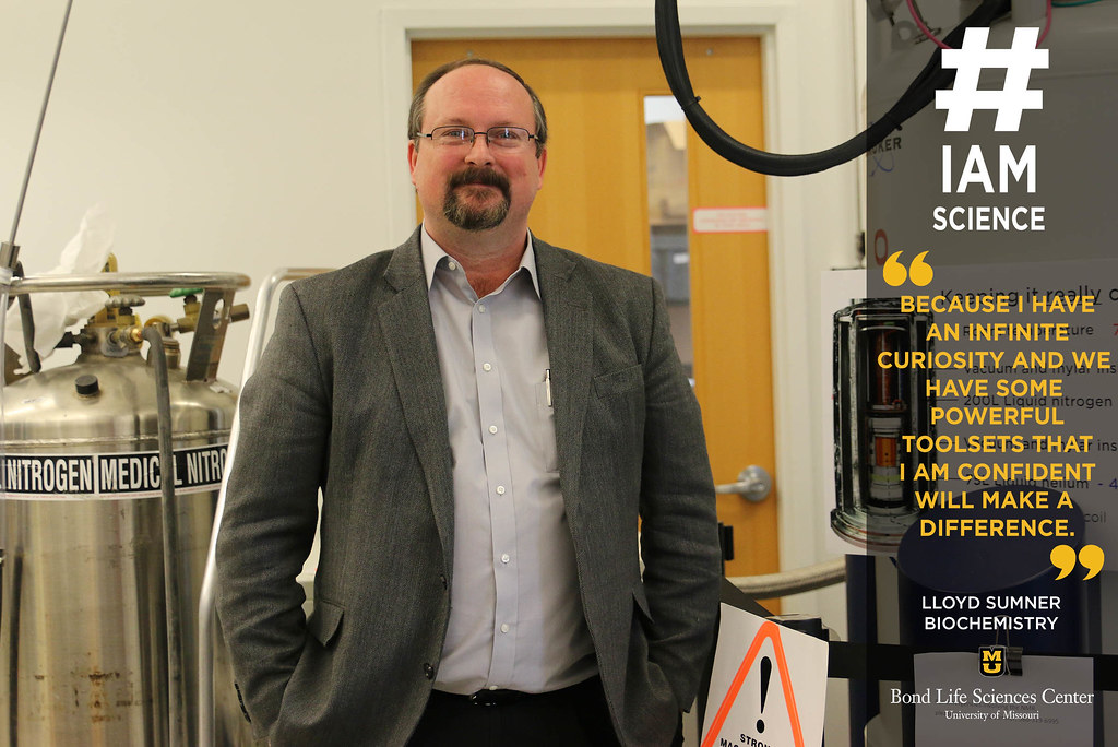
Lloyd Sumner, biochemistry professor and Director of the Metabolomics Center at Bond LSC. | photo by Mary Jane Rogers, Bond LSC
By Mary Jane Rogers | Bond LSC
“#IAmScience because I have an infinite curiosity and we have some powerful toolsets that I am confident will make a difference, not just in plant biochemistry, but in many scientific arenas.”
What change you would like to see in this world because of your research?
“I’m a technology junkie at heart. We are developing tools that can potentially advance many areas, and not just my own personal research program. I want to continue to build upon these tools and also apply them in a meaningful manner. On the plant side, I want to discover and characterize many new biochemical pathways, and use this information to make stronger, healthier and more productive plants. I also want to apply these cutting-edge tools to an ever expanding set of problems; i.e. cancer, veterinary medicine, nutrition, etc. I’m confident that every day when I get up, by the end of that day, week or month that we are making that difference.” -Lloyd Sumner
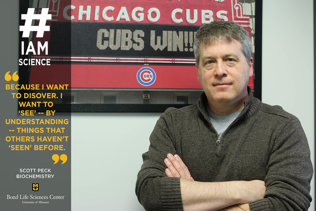
Scott Peck, a biochemistry professor at Bond LSC. | photo by Mary Jane Rogers, Bond LSC
By Mary Jane Rogers | Bond LSC
“#IAmScience because I want to discover. I want to ‘see’ – by understanding – things that others haven’t ‘seen’ before.”
Every day we make decisions based off on what we encounter in the environment. Plants do the same thing. Scott Peck, a Chicago-area native, is a biochemist who studies how plants translate information they receive about the environment (such as changes in light and temperature) into their own chemical “decisions”, also known as signal transduction. For him, it’s about making biology into a puzzle. Put the right pieces together, and you find ways to create more resistant crops or more effective antibiotics. With today’s technology and Peck’s passion for plant communication, anything could be possible.
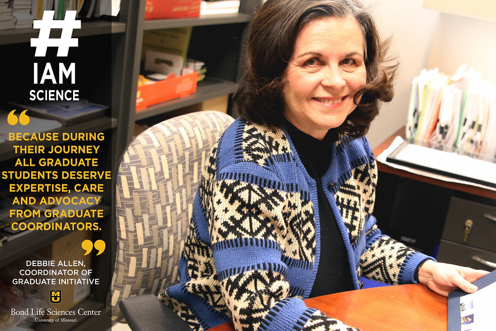
Debbie Allen, the Coordinator of Graduate Initatives at Bond LSC. | photo by Mary Jane Rogers, Bond LSC
By Mary Jane Rogers | Bond LSC
“#IAmScience because during their journey all graduate students deserve expertise, care and advocacy from graduate coordinators.”
As Coordinator of Graduate Life Science Initiatives, Debbie Allen facilitates several activities supporting graduate recruitment, training, mentoring and career services. In other words, she’s been the “mama bear” to many life sciences graduate students over the years, and is passionate about student advocacy. To Debbie, while understanding the hard science her students study is important, supporting those students through their challenges and triumphs, and guiding them closer to their goals motivates her every day.
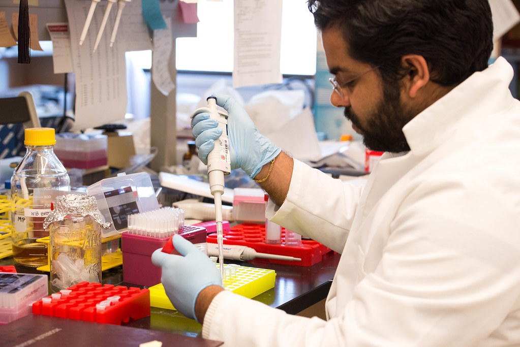
Vinit Shanbhag mixes the CRISPR plasmid DNA with cells. The lab will test whether the gene of interest has been knocked out of the cells later. | photo by Jinghong Chen, Bond LSC
Jinghong Chen | Bond Life Sciences Center
It might be strange to say, but in a way the Australian soil led scientist Michael Petris to where he is now.
In certain areas of Australia, soils suffer from extremely low level of copper bioavailability, resulting in poor growth and neurological problems on sheep.
Petris, a Bond LSC investigator and professor of biochemistry who was born in Australia, now spends his time studying how copper, an essential mineral in human body, works in cells to build and maintain essential functions.
Recently published work from his lab focuses on how the ATP7A protein, one of the major proteins, cycles within the cell.
“Copper is solely acquired from diet. The absorption of copper from the intestine in the blood needs ATP7A,” Vinit Shanbhag, a Ph.D. biochemistry student at Petris’ lab and an author of the study, said. “It transports copper to different copper dependent enzymes and exports free copper from the cell to the outside.”
After exporting copper at the cell membrane, ATP7A needs to come back to its steady-state location within the Golgi apparatus of cells – via a process called retrograde trafficking. But one question baffled scientists: what are the key elements that lead ATP7A coming back?
Back in the late 90s, Petris discovered the importance of one single di-leucine in retrograde trafficking of ATP7A. For those of you wondering, leucine is an amino acid that forms the building blocks of proteins like ATP7A, while di-leucine consists of two of them connected via a peptide bond.
His team wished to identify other signals for retrograde trafficking, but one technical hurdle stood in the way— the ATP7A gene is unstable when grown in bacterial plasmids, the traditional way of amplifying genes in the lab.
Commercial DNA synthesis was the answer. This method could create artificial genes in the laboratory.
“We reasoned that if we introduced enough silent mutations into a DNA sequence, we could avoid or change the region of instability in the native sequence without affecting the encoded protein,” Petris said.
To stabilize the gene, they changed more than 1,000 nucleotides within a 3,000 nucleotides segment, and thus solving the problem of instability of the ATP7A gene. In doing so, they subsequently found that in fact multiple di-leucines that are required for retrograde trafficking of ATP7A. This approach could be used by other laboratories whose gene of interest is also unstable.
An overlooked mineral
“If you ask [people], is it important to understand iron nutrition? Is it important to understand calcium nutrition? Most people would say of course! … But, perhaps you would not get the same answer for copper, despite the fact there is a little dispute that copper is important,” Petris said.
As an essential micronutrient, copper performs central functions to develop and maintain human skin, bones, brains and other organs.
“If you don’t have enough copper in your body, you cannot use oxygen to make energy,” Petris said. “If you don’t have copper, you would not survive.”
Pregnant women who carry a mutated ATP7A gene on their X chromosome can pass it on to their children in the form of Menkes disease.
Menkes disease is a genetic disorder that results in poor uptake and distribution of copper to cells. The incidence of this disease is estimated to be one in 100,000 newborns, according to U.S. National Library of Medicine.
Infants with Menkes disease typically begin to develop symptoms during infancy and rarely live past the first few years of life. Abnormally high accumulation of copper in kidneys and low-level accumulation in the liver and brain, cause visible symptoms like sparse hair, loose skin and failure to grow.
Despite copper’s importance, it also can be a potentially toxic nutrient.
“Copper deficiency can be a problem but too much copper is also a problem. There should be a balance,” Shanbhag explained.
The liver normally stores excessive copper and excretes it into bile to release it out of the body. Yet people with genetic disorders that preventing copper excretion might suffer Wilson’s disease, leading to life-threatening organ damage.
Shanbhag said people with Wilson’s disease accumulate toxic amounts of copper in liver and other organs, causing Kayser–Fleischer rings that encircle the pigmented regions of the eye, a hue caused by copper deposits in the cornea.
Its clinical consequences differ from chornic liver failure to neurological sysmptoms like tremors, dystonia, ataxia and cognitive deteriortation.
About one in 30,000 people have Wilson disease, according to National Institute of Diabetes and Digestive and Kidney Diseases.
Starving tumors
Vinit Shanbhag mixes the CRISPR DNA with mammalian cells to specifically delete a gene in these cells in lab hood. | photo by Jinghong Chen, Bond LSC
In 2013, Petris’ lab published the first direct evidence suggesting ATP7A is essential for the dietary absorption of copper. Since then they have dug deeper into this copper transporter, and his lab now sets their sights on a greater enemy of human health — cancer.
Tumor growth requires access to large amounts of nutrients. Without an adequate supply of oxygen and nutrients, tumors fail to grow and survive. Scientists have identified that by preventing access to nutrients—for example by blocking the growth of new blood vessels—they could starve the tumor of nutrients.
Copper is a key nutrient for tumor growth. With the new-introduced system CRISPR-Cas9 — a genome editing tool to knock out specfic genes — his lab has explored how to exploit understanding of copper metabolic pathways to withhold copper from cancer cells.
“Copper starvation might be a good approach as an anti-cancer strategy,” Petris said.
Weapon of the immune system
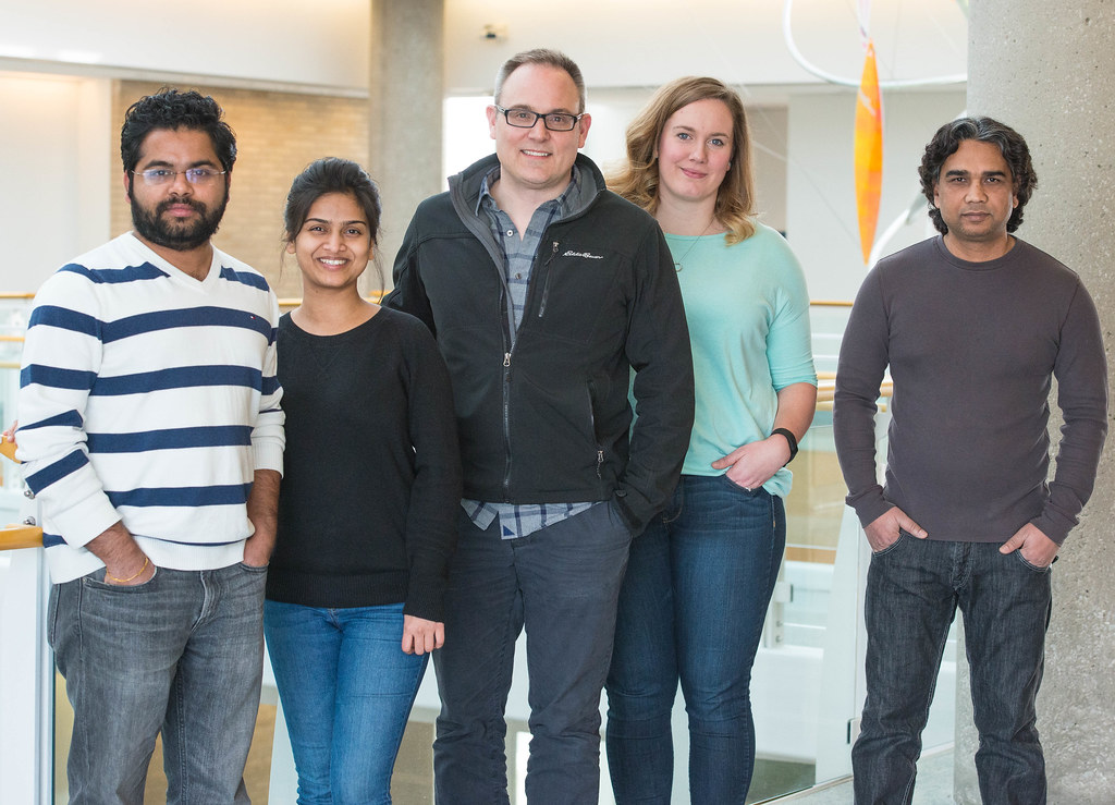
Michael Petris, a professor of biochemistry at MU, stands with his lab. From left to right: Vinit Shanbhag, Nikita Gudekar, Michael Petris, Kimberly Jasmer-McDonald, Aslam Khan. | photo by Jinghong Chen, Bond LSC
Currently, four members study in Petris’ lab to tackle the relationship between copper and various diseases. Petris plans to expand his research to another area: the role of copper in innate immunity against bacterial pathogens.
This is the topic of Petris’ next grant. Nutritional immunity, which describes how the mammalian host withholds nutrients from the invading bacteria during infection, is very well-described for iron and zinc.
Yet copper performs differently.
During infection, the level of copper in blood actually goes up instead of going down. The immune system concentrates copper at sites of infection and within regions where the bacteria are engulfed.
“We speculate that copper is being used as weapon by the host to kill the bacteria,” Petris said. “That is the area we are trying to develop further.”
What if you could have pork without the pig? Nicholas Genovese’s cultured meat could provide a more environmentally friendly meat
This screenshot of a supplemental video included in Genovese’s study shows cells contracting in response to a neurotransmitter. | photo courtesy of the Nicholas Genovese
By Eleanor C. Hasenbeck | MU Bond Life Sciences Center
Scientists are one step closer to that reality. For the first time, researchers in the Roberts’ lab at Bond Life Sciences Center at MU were able to create a framework to make pig skeletal muscle cells from cell cultures.
In vitro meat, also known as cultured meat or cell-cultured meat, is made up of muscle cells created from cultured stem cells.
As a visiting scholar at the University of Missouri, Nicholas Genovese mapped out pathways to successfully create the first batch of in vitro pork. Genovese also said it was the first time it was done without an animal serum, a growth agent made from animal blood.
According to Genovese, his research in the Roberts Lab was also the first time the field of in vitro meats was studied at an American university.
“I feel it’s a very meaningful way to create more environmentally sustainable meats, which is going to use fewer resources, with fewer environmental impacts and reduce need for animal suffering and slaughter while providing meats for everyone who loves meat,” Genovese said.
The research could have environmental impacts. According to the United Nation’s Food and Agriculture Organization, livestock produce 14.5 percent of all human-produced greenhouse gas emissions. Livestock grazing and feed production takes up 59 percent of the earth’s un-iced landscape. Cultured meat takes up only as much land as the laboratory or kitchen (or carnery, the term some members of the industry have coined for their facilities) it is produced in. It uses energy more efficiently. According to Genovese, three calories of energy can produce one calorie of consumable meat. The conversion factor in meat produced by an animal is much higher. According to the FAO, a cow must consume 11 calories to produce one calorie of beef for human consumption.
And while Michael Roberts, the lab’s principal investigator, is skeptical of how successful in vitro meat will be, he said the results could yield other benefits. Researchers might be able to use a similar technique as they used to create skeletal muscle tissue to make cardiac muscle tissue. Pork muscles are anatomically similar to a human’s and can be used to model treatments for regenerative muscle therapies, like replacing tissue damaged by injury or heart attacks.
“I was interested in using these cells to show that we could differentiate them into a tissue. It’d been done with human and mouse, but we’re not going to eat human and mouse,” Roberts said. “The pig is so similar in many respects to humans, that if you’re going to test out technology and regenerative medicine, the pig is really an ideal animal for doing this, particularly for heart muscle,” he added.
While you won’t find in vitro meat in the supermarket just yet, Genovese and others are working toward making cultured meats a reality for the masses. Right now, producing in vitro meat is too costly to make it economically viable. Meat is produced in small batches, and the technology needed to mass-produce it just isn’t there yet. Genovese recently co-founded the company Memphis Meats, where he now serves as Chief Scientific Officer. The company premiered the first in vitro meatball last year, at the hefty price tag of $18,000.
“We are rapidly accelerating our process towards developments of technology that we hope will make cultured meats accessible to everyone within the not-so-distant future,” Genovese said.
Nicholas Genovese was a member of the Roberts lab in Bond LSC from 2012 to 2016. The study “Enhanced Development of Skeletal Myotubes from Porcine Induced Pluripotent Stem Cells” was recently published by the journal Scientific Reports in February 2017.
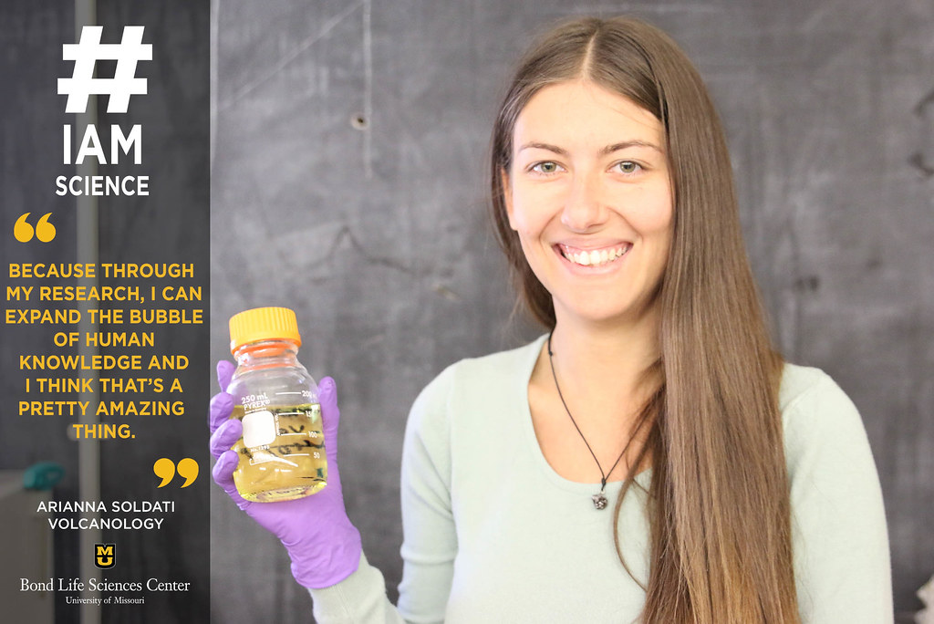
Arianna Soldati, a Ph.D candidate in volcanology at Bond LSC. | photo by Mary Jane Rogers, Bond LSC
By Mary Jane Rogers | Bond LSC
“#IAmScience because through my research, I can expand the bubble of human knowledge and I think that’s a pretty amazing thing. We don’t have a volcano, so we make our own.”
Imagine stirring rock. Sounds impossible? Not to Arianna Soldati, a volcanology expert conducting #MizzouResearch on the viscosity of rock. By heating and then stirring rocky material in a machine that acts like a miniature volcano, she identifies its viscosity, or thickness. As someone who has always been interested in volcanos, she is passionate about saving people from dangerous eruptions. “By knowing the signs, people are more likely to get to safety in time.”
Scientists prove parasite mimics key plant peptide to feed off roots By Roger Meissen | Bond LSC
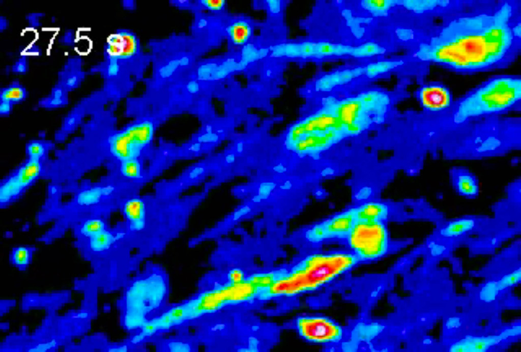
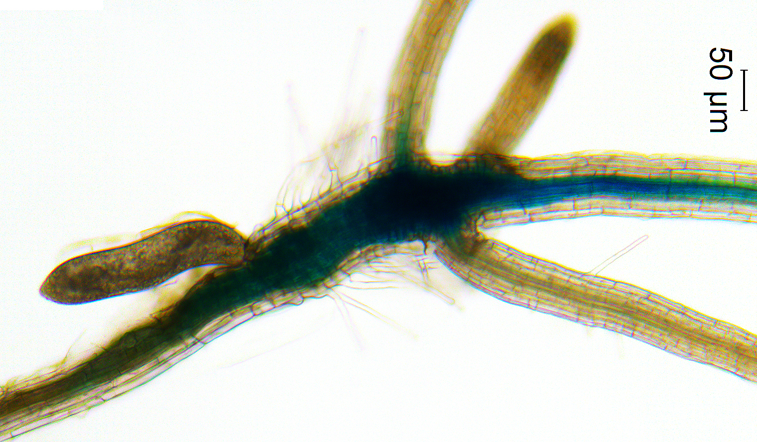
A nematode (the oblong object on the left) activates the vascular stem cell pathway in the developing nematode feeding site (syncytium) on a plant root. | photo by Xiaoli Guo, MU post-doctoral research associate
When it comes to nematodes, unraveling the root of the issue is complicated.
These tiny parasites siphon off the nutrients from the roots of important crops like soybeans, and scientists keep uncovering more about how they accomplish this task.
Research from the lab of Bond LSC’s Melissa Mitchum recently pinpointed a new way nematodes take over root cells.
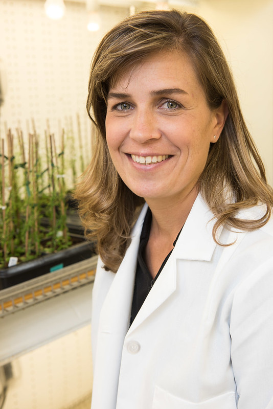
Melissa Mitchum | photo by Roger Meissen, Bond LSC
“In a normal plant, the plant sends different chemical signals to form different types of structures for a plant. One of those structures is the xylem for nutrient flow,” said Mitchum, an associate professor in the Division of Plant Sciences at MU. “Plant researchers discovered a peptide signal for vascular stem cells several years ago, but this is the first time anyone has proven that a nematode is also secreting chemical mimics to keep these stem cells from changing into the plant structures they normally would.”
Stem cells? Xylem? Chemical mimics?
Let’s unpack what’s going on.
First, all plants contain stem cells. These are cells with unbridled potential and are at the growth centers in a plant. Think the tips of shoots and roots. With the right urging, plant stem cells can turn into many different types of cells.
That influence often comes in the form of chemicals. These chemicals are typically made inside the plant and when stem cells are exposed to them at the right time, they turn certain genes either on or off that in turn start a transformation of these cells into more specialized organs.
Want a leaf? Expose a stem cell to a particular combination of chemicals. Need a root? Flood it with a different concoction of peptides. The xylem — the dead cells that pipe water and nutrients up and down the plant — requires a particular type of peptide that connects with just the right receptor to start the process.
But for a nematode, the plan is to hijack the plant’s plan and make plant cells feed it. This microscopic worm attaches itself to a root and uses a needle-like mouthpiece to inject spit into a single root cell. That spit contains chemical signals of its own engineered to look like plant signals. In this case, these chemicals — B-type CLE peptides — and their purpose are just being discovered by Mitchum’s lab.
“Now a nematode doesn’t want to turn its feeding site into xylem because these are dead cells it can’t use, so they may be tapping into part of the pathway required to maintain the stems cells while suppressing xylem differentiation to form a structure that serves as a nutrient sink,” Mitchum said. “To me that’s really cool.”
This means these cells are free to serve the nematode. Many of their cell walls dissolve to create a large nutrient storage container for the nematode and some create finger-like cell wall ingrowths that increase the take up of food being piped through the roots. For a nematode, that’s a lifetime of meals for it while it sits immobile, just eating.
But how did scientists figure out and test that this nematode’s chemical was the cause?
Using next generation sequencing technologies that were previously unavailable, Michael Gardner, a graduate research assistant, and Jianying Wang, a senior research associate in Mitchum’s lab, compared the pieces of the plant and nematode genome and found nearly identical peptides in both — B-type CLE peptides.
“Everything is faster, more sensitive and we can detect things that had gone undetected through these technological advances that didn’t exist 10 years ago,” Mitchum said.
To test their theory, Xiaoli Guo, postdoctoral researcher and first author of the study in Mitchum’s lab synthesized the B-type CLE nematode peptide and applied it to vascular stem cells of the model plant Arabidopsis. They found that the nematode peptides triggered a growth response in much the same way as the plants own peptides affected development.
They used mutant Arabidopsis plants engineered to not be affected as much by this peptide to confirm their findings.
“We knocked out genes in the plant to turn off this pathway, and that caused the nematode’s feeding cell to be compromised. That’s why you see reduced development of the nematode on the plants.”
This all matters because these tiny nematodes cost U.S. farmers billions every year in lost yields from soybeans, and similar nematodes affect sugar beets, potatoes, corn and other crops.
While this discovery is just a piece of a puzzle, these pieces hopefully will come together to build better crops.
“You have to know what is happening before you can intervene,” Mitchum said. “Now our biggest hurdle is to figure out how to not compromise plant growth while blocking only the nematode’s version of this peptide.”
Nga Nguyen hopes to apply her research to increase nutrient contents in crop plants
Nga Nguyen, a doctoral candidate in MU’s Division of Plant Sciences, observes samples of a model plant species, Arabidopsis thaliana, in the Mendoza-Cózatl lab at Bond Life Sciences Center on Feb. 7, 2017. | photo by Eleanor C. Hasenbeck, Bond LSC
By Eleanor C. Hasenbeck | Bond LSC
Plants smell better than animals, at least to Nga Nguyen. That’s one reason why she decided to study them.
“In my undergrad, I studied horticulture,” Nguyen said. “For that you don’t really learn the inside mechanisms of plants, so I decided besides knowing the cultivation techniques, I’d like to also learn about the molecular biology.”
As a fifth year doctoral candidate in the Mendoza-Cózatl lab at Bond Life Sciences Center, she hopes to combine her undergraduate background with her present research in the microbiology of plants to improve the crops of the future.
Nguyen studies how transporter proteins move micronutrients like iron through plants. By understanding how plants move these nutrients in model plants, researchers hope to apply the same understanding and techniques to crops like soy and common beans. Increasing the micronutrient content of these crops could be a useful tool in combatting nutrient deficiencies in areas where people don’t have access to meat and dairy.
But Nguyen says the benefits of studying plants don’t end there. “I hope people pay attention to plant research and study,” Nguyen said. “If you think about it, it’s not just our food, but our clothing and the materials we use.”