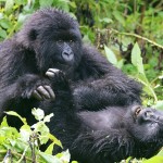
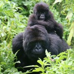
In a second travel log from Bond LSC researcher Cheryl Rosenfeld, learn about the wildlife she encountered in Tanzania this summer. Through the North American Veterinary Community (NAVC), Rosenfeld furthered her veterinary education while encountering wildlife in their natural habitat. See more about the first leg of her trip to Rwanda here.
By Cheryl Rosenfeld

In the early morning hours, our group flew from Kigali, Rwanda to the Serengeti in Tanzania. As we began the descent to the dirt runway, we glimpsed our first sight of wildebeest and the awe-inspiring Serengeti plains and I soon boarded “tano,” Swahili for the fifth 4×4 in our convoy. “Tano” soon came to have special meaning, as there are many groupings of five to see in Tanzania: “The Big Five, The Ugly Five, etc.” Emanuel, whose life-long ambition was to attend college to be a guide, led us to see all of these groups of five and then some.

From the time we pulled away from the meeting spot, I started photographing animals on one side of the vehicle, but it seemed even more intriguing ones would appear on the other side of the road.
When the NAVC and our veterinary guide, Dr. Carol Walton, organized this trip, it was anticipated that the wildebeest would be in the Western corridor of the Serengeti but she was wrong. It’s no longer the case that the location of the wildebeest migrations can be projected with accuracy. Our lecture the first evening in Tanzania discussed how the wildebeest know to migrate and why past modeling of their migratory pattern is no longer accurate. Theories for the migratory nature of these animals include their ability to smell rain and/or changes in calcium concentrations in the soil. Wildebeest rely heavily on calcium to produce sufficient milk to nurse their calves that expend considerable energy keeping up with the herd.
In the past, the rains, like the wildebeest migration, would occur in similar sites and times throughout the year. Climate change has likely contributed to the rainfall in these sites being less reliable and correspondingly, compromised forecasting where the wildebeest will be located throughout the year. This year the wildebeest decided instead to migrate to the central Serengeti region. Thus, the next day we set off on an over-three-hour journey to this region.
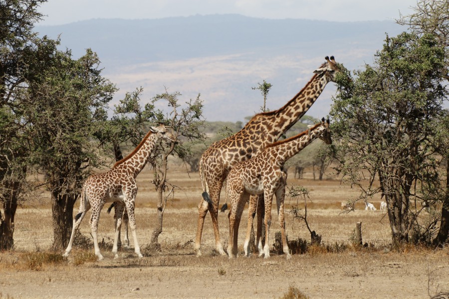
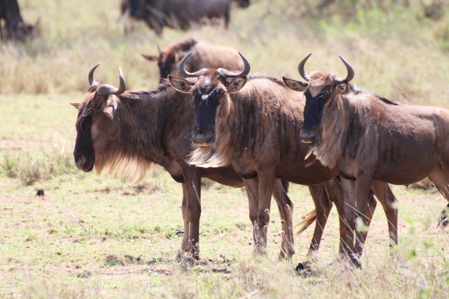
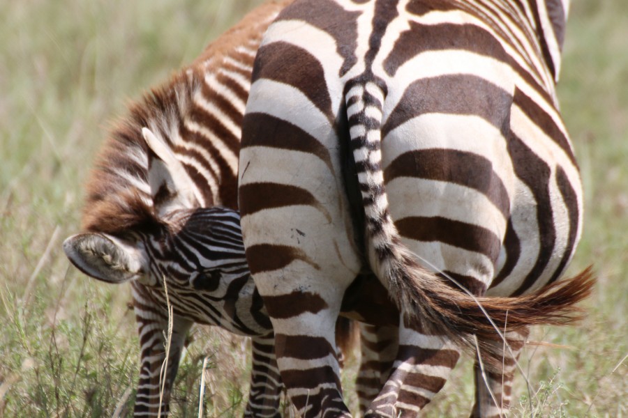
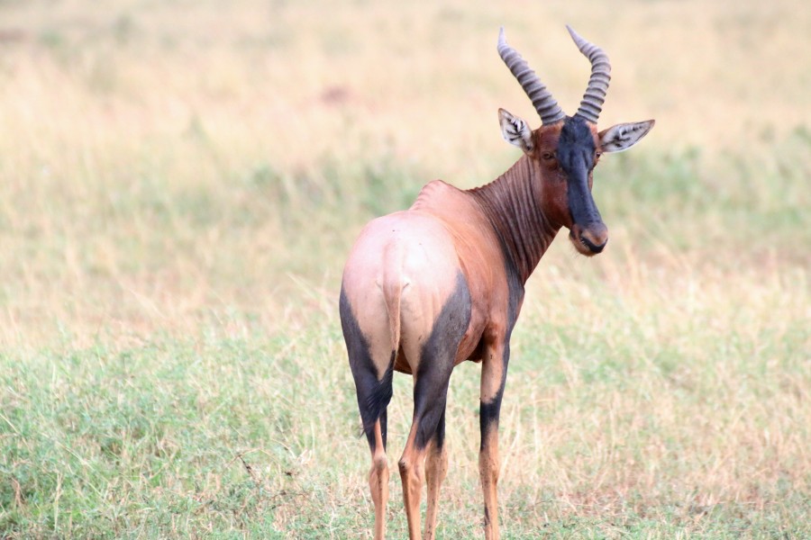
While driving to find wildebeest we came upon new animals and birds including Coke’s Hartbeest, several Masai giraffe, and topis, which seemed to be splattered with oil. Finally, the wildebeest herds starting increasing in size until it reached a crescendo. As far as one could look in all directions there were wildebeest: males, females, and an abundance of baby calves. Eating alongside them were many zebras and their babies, with brown fuzzy fur along their back-end. It was the most amazing spectacle any of us had witnessed.
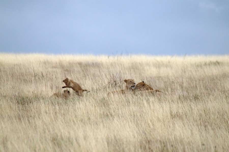
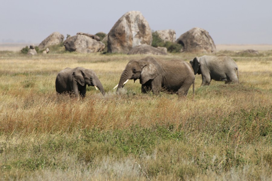
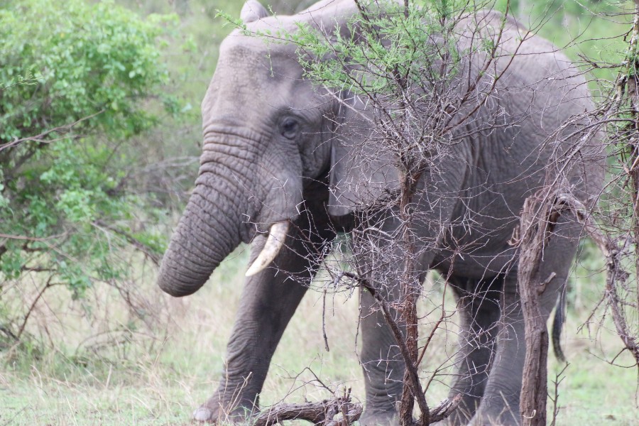
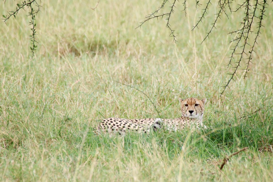
After seeing the vast wildebeest herds, we then came upon the elephants and lions, along with their babies. In the safety of the vehicle, we watched them engage in their natural behaviors for which no zoo experience can replicate. Both species were incredibly affectionate to the offspring and each other. In the case of the lionesses, each time the females, who were likely related, caught up with another that they had not seen in a short while would lead to emotional bouts of jumping on each other, caressing and licking. It was hardly the acts of a deadly predator. Yet, their existence rests on the wildebeest and other prey. With the wildebeest in this area, it seemed all life was flourishing at this time. A true paradise.
However, even paradise is subject to outside threats. One such threat is the massive poaching of elephants in Tanzania and surrounding African countries with 40,000 being killed last year alone. Prior to 2013, Tanzania had a “shoot to kill” policy for those caught poaching elephants or other endangered species. However, this rule has since been relaxed with the elephant numbers now in severe decline. At this unsustainable rate of poaching, the African elephant may go extinct in the wild in the next few decades.
While we did see some groups of elephants, the size of each matriarch-led group seemed less than those depicted in Sir David Attenborough and National Geographic documentaries. Moreover, one elephant that we came across, which I affectionately referred to as “Stumpy,” bore sad evidence of this brutal practice: He lost part of his trunk in a poacher’s snare. Without a full trunk, this elephant exhibited great difficulty grasping various plant items to place in his mouth.
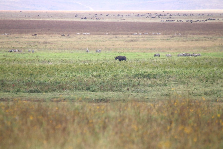
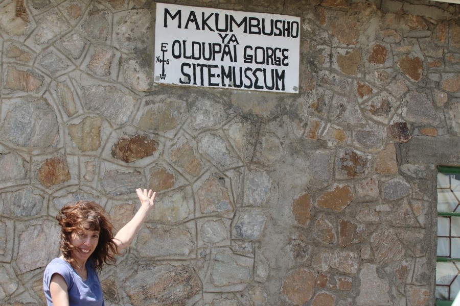
Our subsequent travel took us to the famous “Olduvai (which should actually be Oldupai) Gorge,” one of the most famous paleoanthropological sites, including the Leakeys’ famous discovery. From there we traveled to the Ngorongoro Crater, a gigantic fracture of the earth’s crust that provides habitat for some 30,000 animals, including the most endangered black rhinoceros that only has 20 to 30 left in this area. The difference between the black and white rhino lies not in their coat color but a structural difference in their lips with black rhino possessing pointed and prehensile upper lip to browse on twigs and leaves. In contrast, white rhino possess a square upper lip to graze on the grasses below.
When Dr. Walton convinced us to set off at first light, around 6 a.m., our goal was to find one of these amazing creatures. Once again, with Emmanuel’s assistance and eagle eyes, what started out as a black dot far off in the horizon began to morph into a recognizable rhinoceros. In utter delight, we strained our necks and photographed as this beautiful animal continued to move along and forage in the distance. It would be inhumane to allow this animal to go extinct. In the case of the rhino, many Asian countries believe that its horn increases male libido, which has never been proved to be the case. This mistaken notion possibly originated from the fact that male rhinos copulate with females for several hours duration. This behavior is, however, not transferrable to men who consume any part of a rhino.
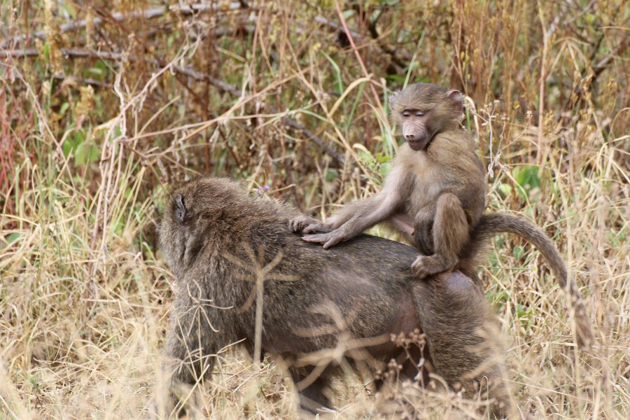
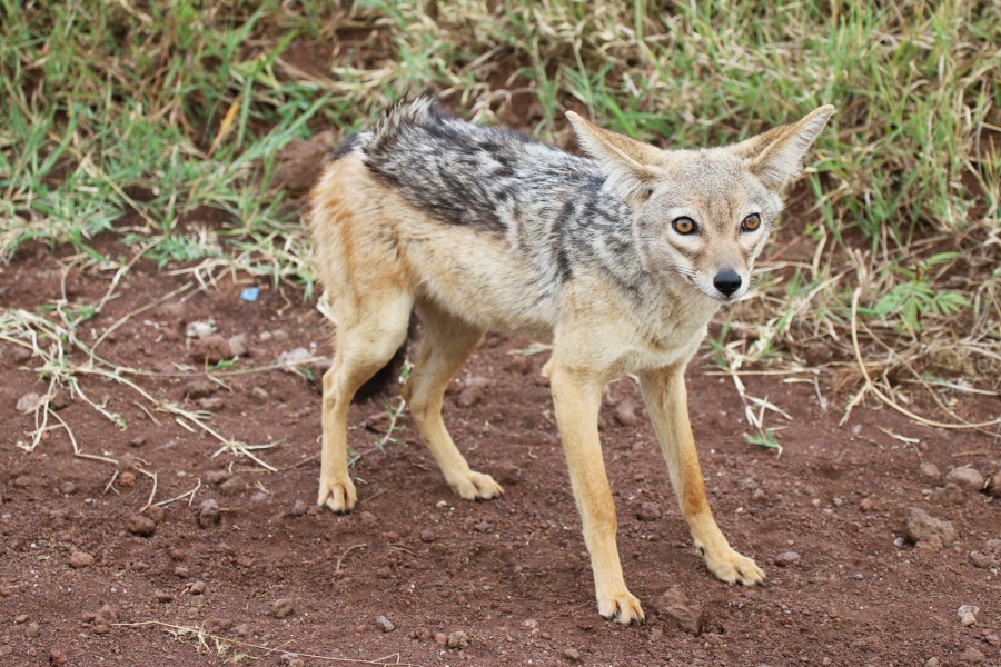
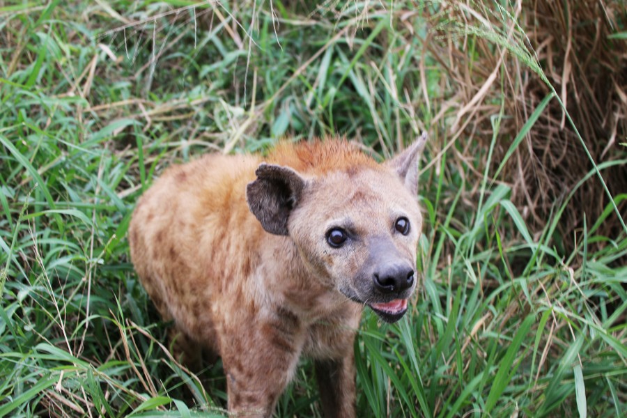
The list of species we saw in Tanzania continued to grow while we drove around the crater with black-backed and common jackals following alongside our vehicle, eland and grants gazelles grazing in the plains, flocks of greater and lesser flamingoes observed in the distance, and more lion prides and spotted hyenas.
The next day, we had our final farewell lunch at the famous Arusha Coffee Lodge, where US presidents have dined. We then set off on our long journey back to the US.
All of us were impacted by the creatures and sites we saw. One male veterinarian, who seemed gruff and quiet for most of the expedition, put it best the night before we left. He unexpectedly stood up after our last lecture and proclaimed that he signed up for the expedition more as an escape from the daily grind of being a veterinarian. However, the trip made such as impression on him to the point that he realized that part of the veterinarian oath on “relieving animal suffering” should include standing up and advocating on their behalf to prevent their brutal murder, as previously occurred in the case of the gorillas and is ongoing for elephants and rhinos. All of us were moved by his emotional comments and the tears welling up in his eyes. Our group was indeed fortunate to see these majestic animals, while they still exist, in their natural environments. I hope there is a wake-up call for nations to come together to prevent their extinction.