Bond LSC connects scientists in “hot topic” research
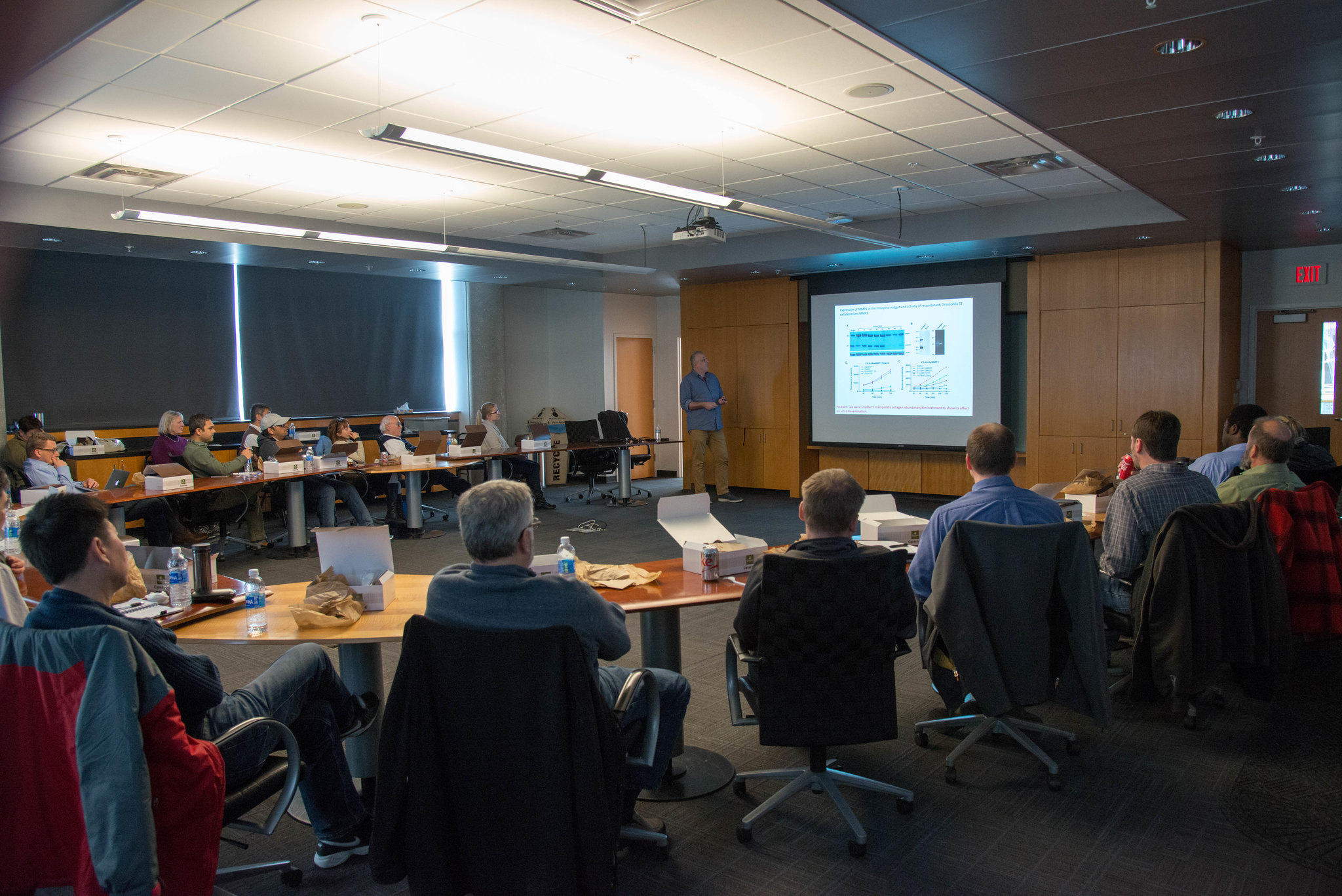
Alexander Franz presents his research on arboviruses and mosquitoes to the Host/Pathogens Research Network. The network brings researchers from across campus together to foster cross-discipline research. | Photo by Samantha Kummerer, Bond LSC
By Samantha Kummerer | Bond LSC
An immunologist, a plant biologist and a biochemist enter a room.
No, that’s not the start of a geeky science joke, but rather is the start of a conversation meant to spur ideas.
As a group of scientists crowd a conference room in the Bond Life Sciences Center in December, they aim to share ideas about their diverse research projects and disciplines.
Today they set about to learn about viruses in mosquitoes from Alexander Franz of MU’s Department of Veterinary Pathobiology.
“The take-home message is that mosquitoes are not just flying syringes or something,” said Franz, explaining the basic science behind his studies. “That is the very wrong idea. This is a very intricate relationship between the virus and the mosquito.”
He spoke to the scientists in attendance about the Chikungunya virus that is spread by mosquitoes to humans throughout the world and currently has no vaccine. Franz’ work examines the genome of the virus and the virus’ expansion into secondary tissues.
The scientists are part of a research network focused on Host-Pathogen relationships. This overarching topic unifies researchers from across campus who share this commonality. The hope is to spark shared projects between scientists that often find it difficult to make connections outside of their discipline.
This group is one of three hot topic areas Bond LSC is currently targeting to get the conversation started. Researchers across campus joined this network two months ago along with groups interested in metabolomics and cancer biology.
During each meeting, one researcher volunteers to present their work to the group and other members are able to jump in, ask questions or offer advice to bounce ideas off one another.
Bond LSC interim director Walter Gassmann kick started the meetings, recognizing both the need for collaborative research and the central location of the Bond LSC building.
“The increasing complexity and sophistication of basic research leads to increased specialization. Yet, fundamental questions can only be tackled by getting at them from different directions,” Gassmann said.
The LSC has always encouraged collaborative discussions for scientists within the center, but Gassmann decided to expand these to include faculty from all buildings on campus as well as include students in the conversation.
“When I became the interim director, I felt Bond LSC could really function as a catalyst for a wider research community on campus. This meant advertising the “hot topics” meetings across campus,” Gassmann said.
Bond LSC investigator Michael Petris said previously multiple small cancer research groups met, but these meetings expand the group and centralize everyone.
Petris is one of the leaders of the cancer biology group and said the effect of these talks is already showing.
After Petris gave the first talk for the cancer biology group, he was invited to exchange techniques, ideas and cell lines with another researcher who was in attendance.
“The discussions that go on in these sort of groups are, for me at least, opening my eyes to the broader spectrum of cancer biology that goes beyond my wheelhouse, my sort of understanding from a narrow perspective,” Petris said.
One way this is occurring is due to the inclusion of clinicians who deal with patients regularly. Petris explained their perspective on problems in cancer and biology may be something the cancer biologist never even thought of before.
The center supports these talks with the aim of sparking collaborative research, publications, and grants in the future.
A full schedule of spring 2018 hot topic meetings will be released in January.
Bond LSC’s Jay Thelen was recently part of a team that looked at how short laser pulses might be used to modify peptides and proteins to make foods edible for those with specific allergies.
Thelen, a biochemistry professor, joined scientists from his department, engineering and Denmark to explore this possibility. What they found was a way to modify molecules quicker and more cheaply than current chemical methods. This could potentially lower costs for specific applications in medicine, pharmacology, biotechnology and more.
We don’t want to give everything away, so read the whole story from MU’s College of Engineering.
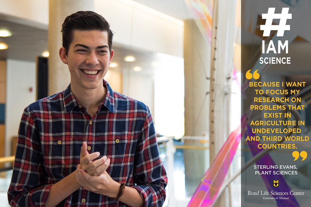
Sterling Evans is a sophomore plant sciences major conducting research in Gary Stacey’s lab in Bond LSC. | photo by Allison Scott
By Allison Scott | Bond LSC
“#IAmScience because I want to focus my research on problems that exist in agriculture in undeveloped and third world countries.”
Sterling Evans’ mind wasn’t focused on research when he started college, but that would soon change.
The sophomore plant sciences major uncovered his interest thanks to Freshman Research in Plant Sciences (FRIPS) — a program dedicated to introducing research to freshman students from plant-related degree programs.
“I was interested in plant sciences-related fields when I started here, but I had no intention of getting involved in undergraduate research,” Evans said. “Being selected for FRIPS was instrumental in getting me involved with research.”
Along with a handful of students selected for FRIPS each year, Evans got to interact with various professors and mentors around campus on a weekly basis. Because of that exposure, Evans found a place in the lab of Bond Life Sciences Center’s Gary Stacey.
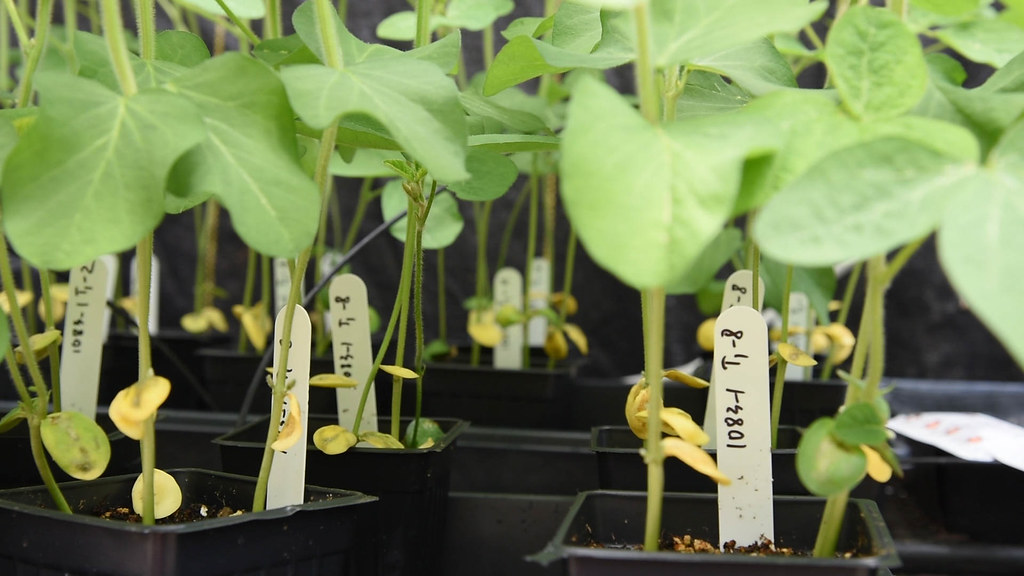
After a year working in Stacey’s lab, Evans just joined a new project that aims to improve the nutritional value of soybeans.
“They’re used as a main source of protein for a lot of countries, so improving their nutritional content would have a huge impact,” Evans said.
The team is applies CRISPR, a gene-editing tool, to model plants called Arabidopsis as a first step.
“We are working on Arabidopsis right now as a proof of concept, because it can be done in a relatively short period of time, before investing as much as a two additional years in soybeans,” Evans said.
While he only spends 15 hours in the lab each week, Evans noticed the lab’s impact on his approach to academics in other ways.
“Research gives me more motivation to think about how to apply information I’ve learned in class to work in the lab,” Evans said. “It has made me more analytical in classes because I have more of a desire to understand things.”
Evans plans to earn a Ph.D. in a plant sciences field and wants to continue research in his career. He’s most interested in helping ensure small communities throughout the world have enough to eat, and he hopes to contribute by studying orphan crops.
“I think they’re cool because they’re really important to small people groups. No one studies them because they aren’t a big deal in the United States or other countries,” Evans said. “If we work on them we won’t have a huge impact on hundreds of millions of people, but we will have a huge impact on small communities.”
That impact all started in a lab. Had he not stepped out of his comfort zone he might never have discovered this path, and he highly encourages other students to give research a chance.
“There are labs for almost everything and there’s an area for everyone,” Evans said. “I didn’t know I wanted to do research until I did it.”
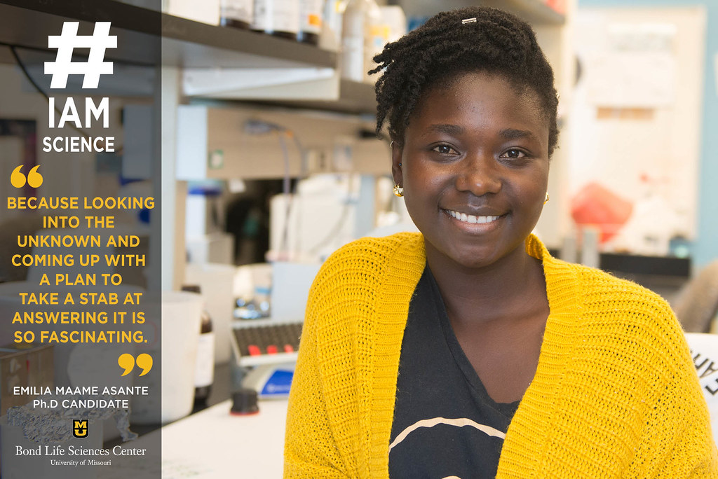
“#IAmScience because looking into the unknown and coming up with a plan to take a stab at answering it is so fascinating.”
No one in Emilia Asante’s family works in a science field or attended graduate school.
“As an immigrant from Ghana, my family was unaware of the American educational system,” she said. “So many of my academic journeys were unknown and I had to navigate it by myself. I was always interested in science and with the help of programs such as the Lang Youth Medical program when I was in grades six through 12 and the McNair Program at Earlham College, I was put on the right path. These programs provided the help I need and allowed me to explore science in a way I never knew existed.”
Asante graduated in 2014 from Earlham College in Richmond, Indiana with a degree in biology. She is currently pursuing a Ph. D in biological sciences at MU, and works in Anand Chandrasekhar’s lab at Bond LSC.
“I love being able to walk down the hall and talk to other labs about my project and the getting feedback in real time,” she said. “I love the community that I have built since being here.”
For her dissertation topic, Asante is using the zebrafish model to understand the consequences of abnormal neuronal migration of the facial branchiomotor (FBM) neurons on circuit formation and behavioral function. Her research aims to explore the relationship between behavior and circuit organization due to neuronal migration abnormalities of the FBM neurons.
“Essentially, I’m trying to figure out what happens when the proper connections are not made,” she said. “For example, autism is a neurological disease. How does the defect in the brain connections lead to the complex behaviors such communication and social impairments? I want to understand how the brain rewire itself when there is a misconnection.”
Asante has always been interested in behavioral science and how things work. After graduation, she hopes to take the training from her research and lab experience at Bond LSC to a translational research position. Translational research means she would help people in a more direct way, compared to research that is more removed or abstract. Labs that deal with translational research might discover how certain genes cause diseases or work with translating ALS models to drug therapy.
“The possibility that I might discover something that will add to the knowledge in my field – no matter how small the contribution – that’s very exciting,” Asante said.
New web-based framework helps scientists analyze and integrate data
By Emily Kummerfeld | Bond LSC
Large-scale data analysis on computers is not exactly what comes to mind when thinking about biological research.
But these days, the potential benefit of work done in the lab or the field depends on them. That’s because often research doesn’t focus on a single biological process, but must be viewed within the context of other processes.
Known as multi-omics, this particular field of study seeks to draw a clearer picture of dynamic biological interactions from gigantic amounts of data. But, how exactly can scientists suitably weave multiple streams of information together, especially considering technology limits and other biological variables?
Trupti Joshi and her team are seeking to find a solution to that problem.
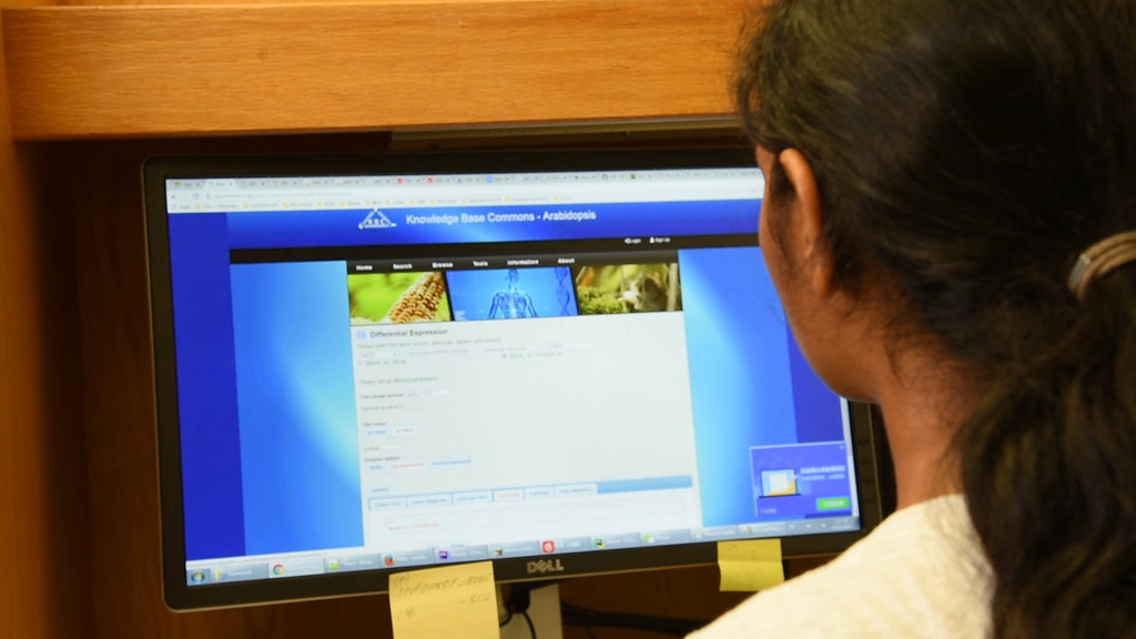
Joshi, as part of the Interdisciplinary Plant Group faculty, works on translational bioinformatics to develop a web-based framework that can analyze large multi-omics data sets, appropriately entitled “Knowledge Base Commons” or KBCommons for short. She describes KBCommons as “a universal, comprehensive web resource for studying everything from genomics data including gene and protein expression, all the way to metabolites and phenotypes.”
Her work began about eight years ago with soybeans. Dubbed the Soybean Knowledge Base (SoyKB), her team had developed a lot of their own data analysis tools for soybean research, but they realized the same tools could help research of other organisms. From there sprouted the Knowledge Base Commons, intended for looking at plants, animals, crops or disease datasets without the need to “reinvent the wheel” each time.
Soybean plants used in research that utilizes Soy KB web-based network. | Emily Kummerfeld, Bond LSC
“Our main focus has been in enabling translational genomics research and applications from a biological user’s perspective, and so our development has been providing graphic visualization tools,” Joshi said.
Those tools provide an array of colorful graphics from basic bar graphs to assorted colored pie charts to help the researcher better analyze the data once data has been added to the KBCommons.
Colorful graphs and comparisons lets many researchers look past the lines of text and tables full of numbers that represent genes, plant traits or other experimental results, and making the interpretation of data much more easier and efficient.
One particular tool allows the researcher to look at the differential genes of four different comparisons or samples at the same time. Differential genes are the genes in a cell responding differently between different experimental conditions. For example, a blood cell and a skin cell both have the same DNA, however, some genes are not expressed in the blood cell that are expressed in the skin cell. With this KBCommons tool, a researcher can examine genes to see “what are the common ones, what are the unique ones to that, and at the same time look at the list of the genes and their functions directly on the website, without having to really go and pull these from different websites or be working with Excel sheets,” Joshi explained.
She envisions KBCommons as a tool to enable translational research as well. Users will be able to compare crops, such as legumes and maize for food security studies, or link research between veterinary medicine and human clinical studies for better therapies.
Intended for a wide range of users, Joshi is keenly aware of its potential users right here at MU.
One current user of the Soybean Knowledge Base (SoyKB) system is Gary Stacey, whose lab at Bond Life Sciences Center studies soybean genomics and to date has been the longest user of the SoyKB resource. Like many researchers, Stacey explained the need for a program like SoyKB that can process enormous amounts of data.
“The reason it’s called “Knowledge Base” is the idea that we’re putting information in, and what we hope to get out is knowledge. Because information is different than knowledge,” he said, “we don’t just want to collect stamps, we want to be able to actually make some sense out of it…By having a place to store the data, and then more importantly have a place to analyze it and integrate it, it allows us to ask better questions.”
This is essential, given that one soybean genome is 1.15 GB in size, and one thousand soybean genome sequences could generate 30 to 50 TB of raw sequencing data and tens of millions of genomic variations (SNPs).
But such numbers are modest compared to the program’s true capabilities.
“The KBCommons system is so powerful that it can allow you to run thousands of genomes at the same time using our XSEDE gateway allocations,” Joshi said. “This whole scalability is a unique feature of KBCommons, which a lot of databases do not provide, and we are happy we have been able to bring that to our MU Faculty collaborators on these projects, so that they can really utilize the remote high performance computing (HPC), cloud storage and new evolving techniques in the field.”
KB Commons is a new web-based network for biological data analysis and integration developed by students. | Emily Kummerfeld, Bond LSC
Mass data capability and colorful graphs aside, her favorite part is who exactly is designing the program.
“What I like most about KBCommons is that it serves as a training and development ground and is developed by students, undergraduate and graduate students from computer science and our MUII informatics program.”
KBCommons is still under development, but publication and access for all users is planned for the end of this year or early 2018. Users will not only be able to view public data sets, but add their own private data sets and establish collaborative groups to share data.
Dr. Trupti Joshi is an Assistant Professor and faculty in the Department of Health Management Informatics, the Director for Translational Bioinformatics with the School of Medicine, and Core Faculty of the MU Informatics Institute and Department of Computer Science and the Interdisciplinary Plant Group.
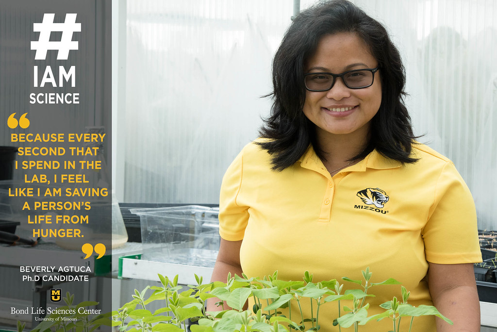
Beverly Agtuca, a Ph.D. candidate that works in Dr. Gary Stacey’s lab in Bond LSC. | photo by MJ Rogers
Beverly Agtuca was born in New York, but has family in the Philippines, a country that struggles with malnutrition and undernourishment. Her overall goal for her research is to help countries that struggle with undernourishment by increasing the agricultural productivity in those countries.
“When I was little, I went on summer vacation to visit my family, which included my grandmother in the Philippines,” she said. “Everyday my grandmother wanted me to go out to the rice fields from 5 a.m. to 10 p.m. with the other children to get rice for our meals. That was not an easy task and that moment changed my life. That’s when I decided that I wanted to be a plant scientist.”
Agtuca graduated in 2014 with honors in Biotechnology and a minor of Microscopy from the State University of New York College of Environmental Science and Forestry (SUNY ESF) in Syracuse, NY. She’s currently a Ph.D. candidate in plant breeding, genetics, and genomics at MU. She chose to come to Bond LSC because of the community and Dr. Stacey, her supervisor and mentor.
“If you ever need help, there’s always help here,” she said. “Everyone at Bond LSC is so kind, including the staff. I love to make small talk with the custodians and they are always supporting me and say I should never give up when I have a bad day.”
Ever since coming to MU in 2014, Agtuca has been keeping busy. In June, she received a travel award to go to the American Society of Plant Biologists (ASPB) in Hawaii. The International Society for Molecular Plant-Microbe Interactions (IS-MPMI) also awarded her a travel award to attend the 2016 meeting in Portland, Oregon, where she gave an oral and poster presentation. She also has two original research publications under her belt and is currently working in Dr. Gary Stacey’s lab at Bond LSC.
The research for her dissertation is focused on the relationship between rhizobia and soybeans. She collaborates with scientists at George Washington University (GWU) in Washington, D.C. and the Pacific Northwest National Laboratory (PNNL) in Richland, Washington to enhance the capabilities of the 21 Tesla Fourier transform ion cyclotron resonance mass spectrometer (21T FTICR) through application of laser ablation – electrospray ionization mass spectrometry (LAESI-MS) technology that can analyze the contents of single plant cells. This 21T FTICR machine was recently installed at PNNL and represents one of only two such machines in the world.
This is revolutionary because few people do single cell analysis. Usually, scientists deal with the law of averages, which dilutes the final measurements. But this technology gives an in-depth glimpse into a single cell so scientists can obtain a more comprehensive bigger picture.
“After we finish building this technology, we want to spread the technique to different research groups so they can answer these research questions on their own,” said Agtuca. “It can help people outside of plant sciences too, and hopefully will help with cancer treatment and disease prevention.”
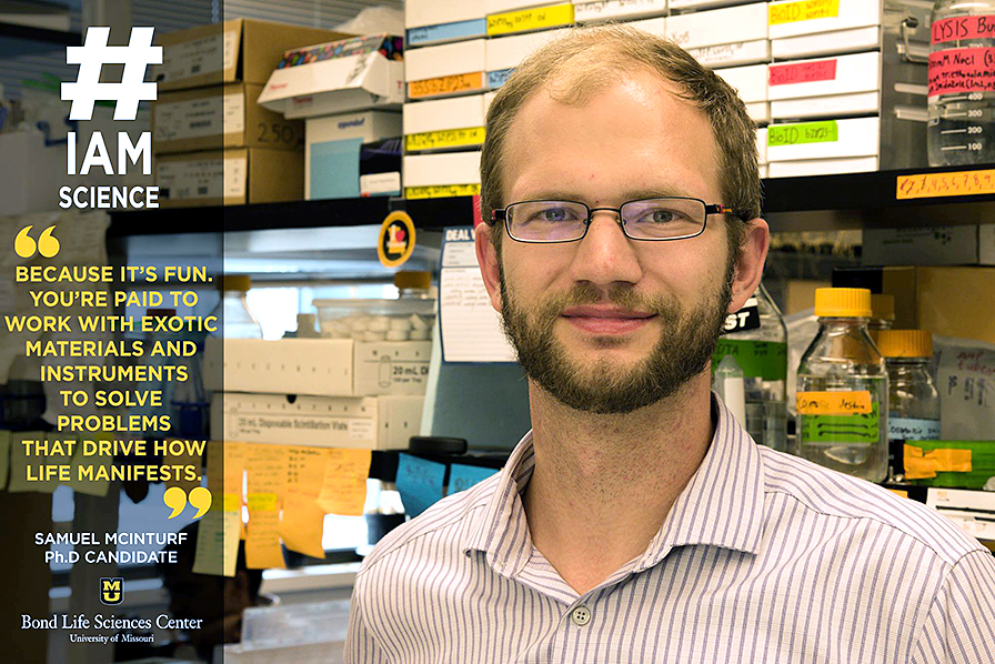
“#IAmScience because it’s fun. You’re paid to work with exotic materials and instruments to solve problems that drive at how life manifests.”
Samuel McInturf’s father is an accountant and his mother is an HR director, but somehow he ended up falling in love with science. By the 4th grade he had already asked his parents to buy him a compound microscope. He completed his undergraduate degree in plant biology at University of Nebraska, Lincoln with a minor in biochemistry. Now, he’s finishing up his fifth year pursuing a Ph.D in plant stress biology and works in Dr. David Mendoza-Cózatl’s lab at Bond LSC.
“I mainly came to Bond LSC to work with Dr. Mendoza,” said McInturf. “The work in his lab was right in line with what I wanted to do and I knew the faculty at Bond LSC was great.”
And he’s enjoyed the last five years he’s spent here.
“Bond LSC has vast resources of knowledge and labs are very friendly towards one another,” he said. “So if you are short up on a reagent, or you need to learn to do an assay, someone is always available to lend a hand.”
McInturf’s thesis deals with understanding the genetic factors that balance the uptake and demand for micronutrients – heavy metals – against their toxicity. He specifically looks are regulators of iron and zinc homeostasis.
In addition to his interest in plant biology, he’s also an engineer of sorts. McInturf helps teach a bioengineering class at Bond LSC with undergraduates. The goal of the class is to build robotics that aid laboratory research, and he has taught three of these classes so far.
“I found the change in scale between building widgets in my bedroom to building full scale devices challenging, but ultimately rewarding,” he said.
For undergraduates interested in continuing a career in science, McInturf advises them not to give up, even when things get tough. He admits that he was intent on dropping out of school up until he was 18, but now he’s almost finished with his Ph.D.
“Ten years ago I was very intimidated by what I saw as the difficulty of science and was wavering on whether I wanted to take the dive into a research-heavy field,” he said. “It took a few years to figure that out, so I guess I would have told myself to get a move on and not be so faint hearted about it.”
McInturf isn’t positive where he’s like to be in 10 years, but he’d enjoy continuing to teach and conduct research at a university like MU.
“I’d love to have Dr. Mendoza’s job one day,” he laughed.
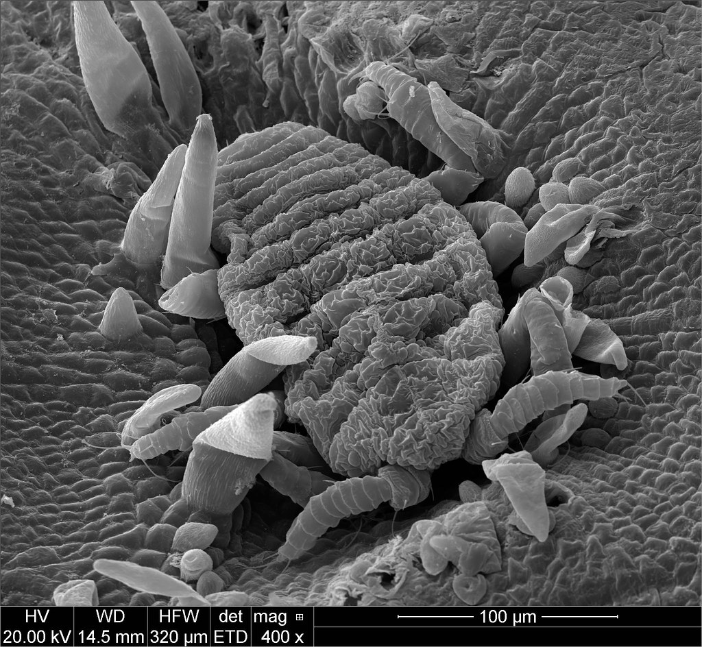
How bossy insects make submissive plants create curious growths
By Samantha Kummerer | Bond LSC
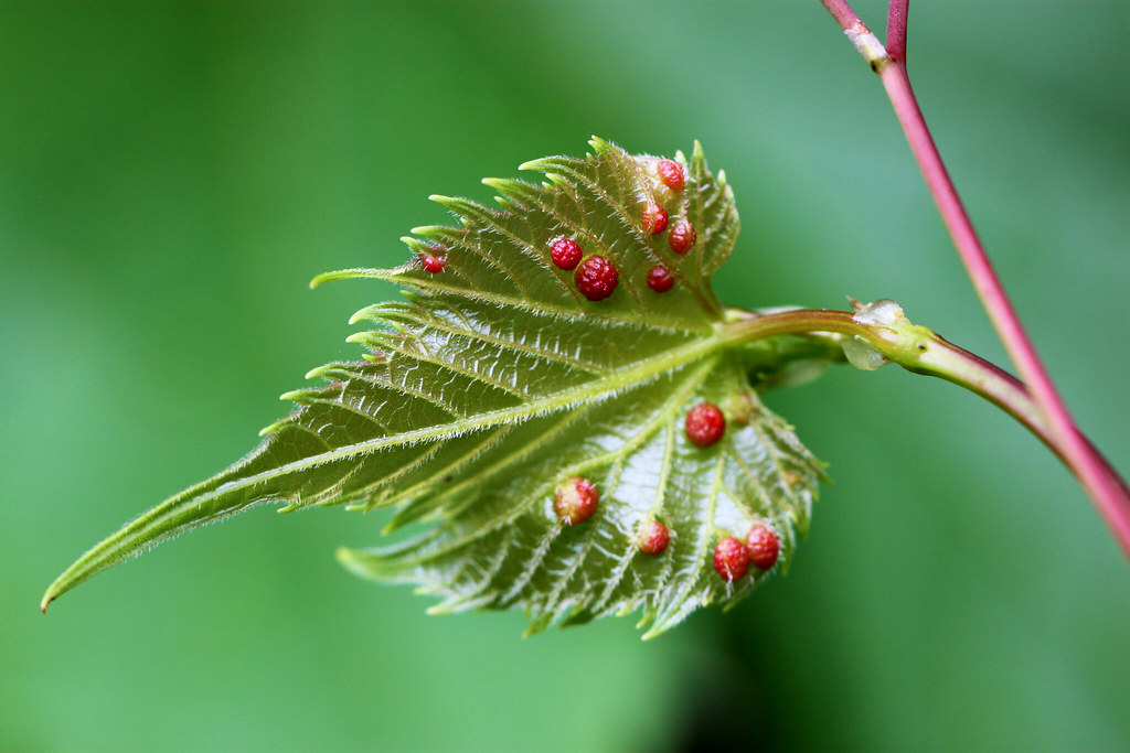
They are bumps on leaves, bulges in stems and almost flower-like growths from plant tissue with a striking amount of variety. They are galls.
These unnatural growths garnered the curiosity of Jack Schulz for years. While he’s spent 40 years studying topics from Insect elicitors to habitat specialization by plants in Amazonian forests, what he’s really wanted to study was galls.
“It’s so weird,” said Schultz, director of the Bond Life Sciences Center. “I’ve always been really curious about how these strange structures form on plants.”
Schultz has spent the last two years trying to answer that question, looking at their development and the underlying genetic changes that make galls possible.
He’s not alone in his fascination.
“I found my very first gall when I was a masters student,” said Melanie Body, a postdoctoral researcher in Schultz’s lab. “I was really excited because it was here the whole time, I just didn’t see it. One of my teachers showed me and it was like a revelation, basically, what I wanted to work on.”
These “strange structures” are often mistaken for fruit or flower buds on a variety of plants from oak trees to grapevines and there’s a good reason why…
“A gall on a plant is actually, at least partly, a flower or a fruit in the wrong place,” Schultz said.
Galls on a leaf. The leaf’s red bumps are not natural, but caused by tiny insects. Inside each gall is many tiny insects. | photo by Melanie Body
These galls can be the size of a baseball or the size of a small bump depending on the plant. They can also range from just small green bumps on the undersides of leaves to vivid complex growths of color.
Despite the variety, the one thing consist across plants is that the gall is not there by the plant’s choice.
“The insect has a pretty good strategy because it starts feeding on the plant and it will create a kind of huge structure, huge organ, where it can live in, so it’s making it’s own house,” Body said.
The reasoning behind the formation is relatively unknown, however, it is hypothesized that the insect flips a switch within the plant. The insect is not injecting anything new, but rather turning off and on certain genes within the plant.
Schultz explained the galling insect has the power to changes the expression of genes and in some instance disorient the plant’s determination of what is up from down.
The Problem
So how does this affect the plant?
Not only is the insect creating the gall against the plant’s nature, it is also using the plant’s energy and materials for the job.
“I think it’s very cool to imagine an insect can hijack the plant pathway to use it for its own advantage,” Body said.
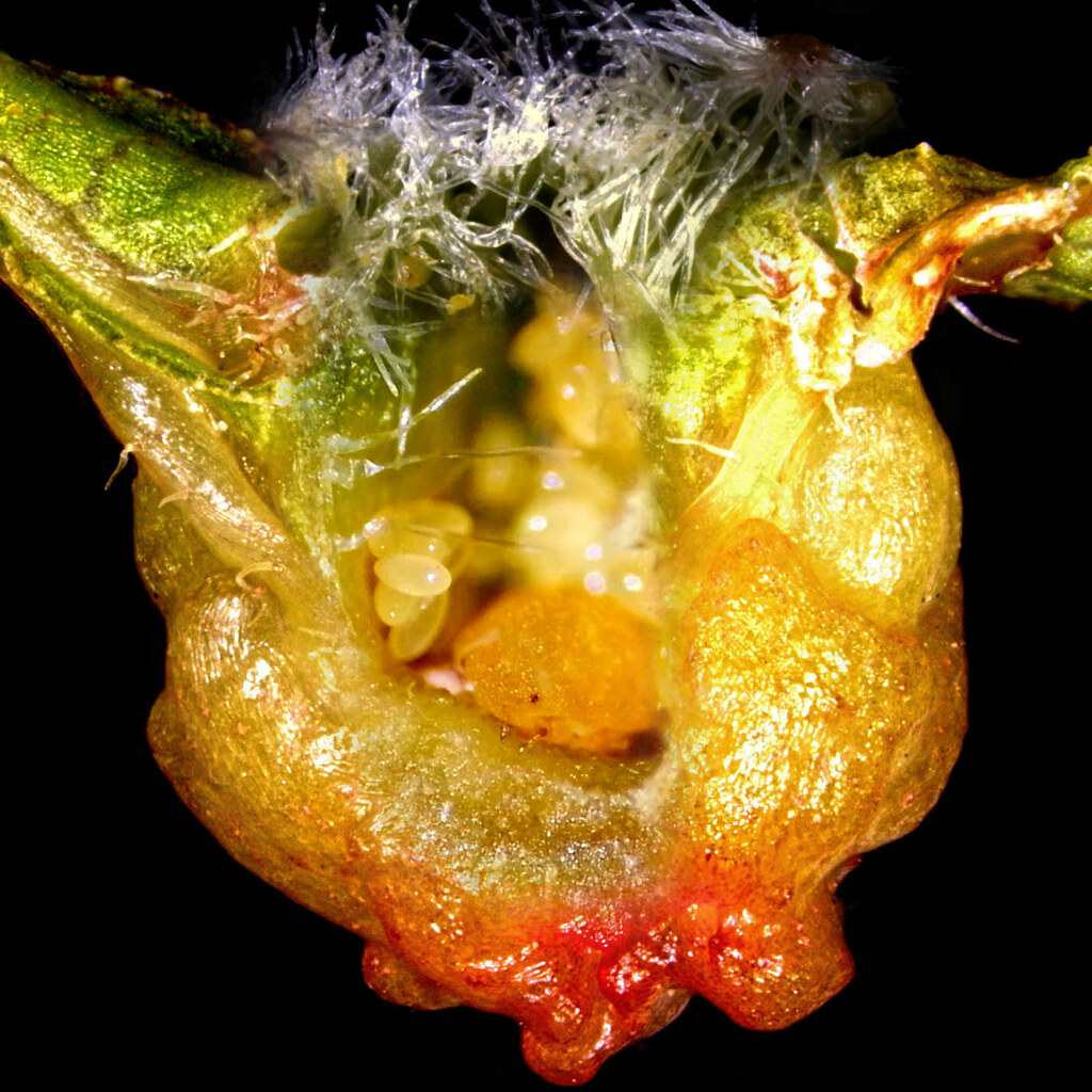
The view of a gall from under a microscope. The mother insect is the orange figure in the foreground and is surrounded by her eggs. | Photo by Melanie Body
The insect receives protection and a unique food source and in turn, the plant is left with fewer resources.
“From the plant’s point of view that’s all materials that could have gone into growth and reproduction, so you can think of these galling insects as competing with the plant they’re on for the goodies the plant needs to grow and reproduce,” Schultz explained. “That’s not so good for producing grapes.”
Grapevines are just one of the many plants that galls can form on, but also the plant Schultz’s research uses.
“In our case we work on grapes, so it can be a big issue if the fruits are not sweet enough anymore, because if you don’t have sugar in the fruit then it’s not good enough for the wine production, so it’s pretty important,” Body explained.
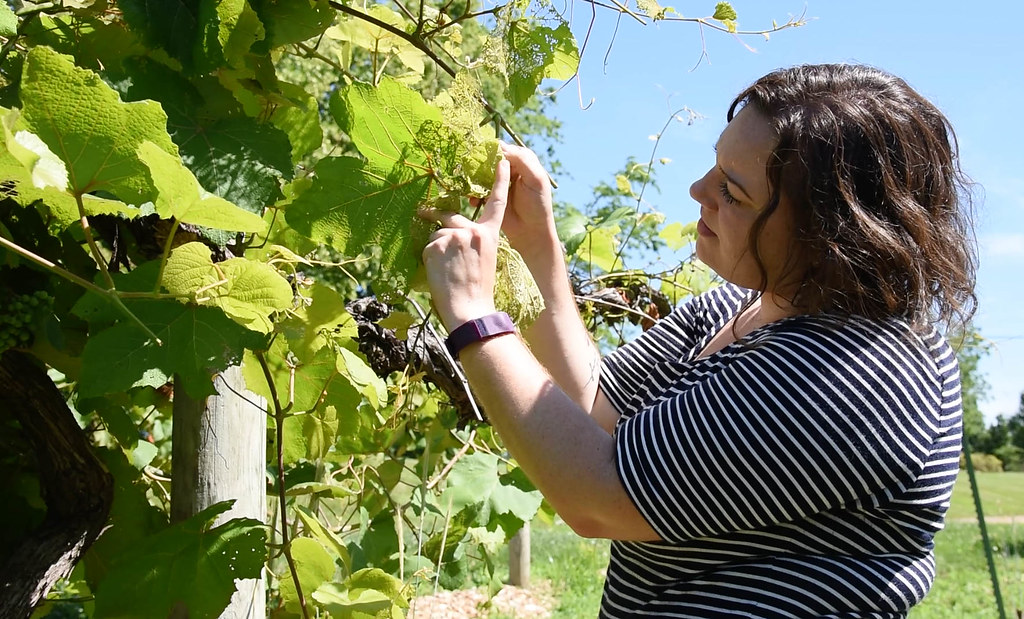
Researcher Melanie Body searches the grapevines for galls at Les Bourgeois Vineyards. | Photo by Samantha Kummerer, Bond LSC
In Missouri, the story of grapevines and galls goes back to the 1800’s.
The story goes, the phylloxera insect found its way over to France. Soon it spread throughout the country; wiping out vineyard after vineyard.
“The great wine blight and the world was going to lose all wine production because of this pest,” said Schultz.
Luckily, a small discovery in native Missouri grapevines led to a solution that allowed wine drinkers to rejoice and scientists to puzzle.
“There’s something about the genes in Missouri grapevines that protects them against this insect,” Schultz explained.
While the European wine industry faced extinction, the phylloxera insect, coexisted with native Missouri grapevines. So, now every grapevine in a vineyard is grafted with insect-resistant roots from Missouri grapes.
But, no one really understands what’s so special about grapevine roots in Missouri.
The research
These galls aren’t new. They’ve actually existed for up to 120 million years. But, here’s what is:
“When we started this research, we thought this is a really well-studied insect,” Schultz said. “It turns out there is an awful lot we don’t know about them.”
The team collects samples of galls from grapevines at Les Bourgeois. Back in the lab the galls are dissected using very small tools and then examined with a microscope. Under the microscope, a colony of the insects emerges. The otherwise miniscule mother insect and her 200 eggs can be seen alongside other insects just moving around the gall.
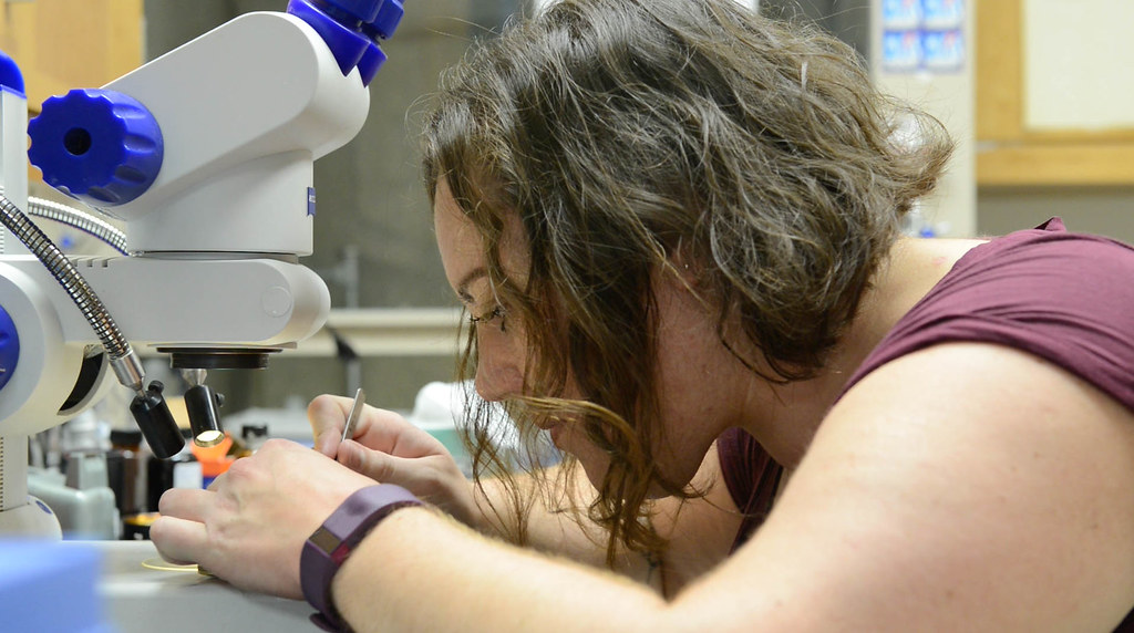
Researcher Melanie Body examines a grapevine gall under a microscope. Body is a member of Jack Schultz’s lab that studies galls and the insects that create them. | Photo by Samantha Kummerer, Bond LSC
Body compares the insects’ round textured bodies to oranges but with two black eyes.
Schultz’s team hypothesized that there are specific flower or fruit forming genes that are necessary for the insect to create a gall.
To answer this, the team looks at which genes are turned on when the insect creates the formation of a gall. Those observations by themselves don’t prove which genes are essential. So, next, the researchers manipulate the genes by changing the gene’s expression.
“If we find that Gene A is always on when the insect causes a gall to form, we can stop the expression of Gene A to test the hypothesis that it needs gene A to get the gall to form,” Schultz explained. “We can ask a plant. If you lack Gene A, can our insects still form galls?”
The researchers are still analyzing the results, but the current findings suggest that some genes do reduce the insect’s ability to make a gall.
A microscopic view of a galling insect in the process of creating a gall. The gall is forming around the insect. | Photo by Melanie Body and David Stalla
Since beginning the research two years ago, Schultz said he has discovered, “all kinds of crazy things”.
Schultz said it was previously believed that the insect was staying in one place when making the circular gall, but actually, the little insect is moving around; something no one realized before.
And that’s not the only myth this research is debunking. For example, many believe there is one insect per gall, but this turns out to be incorrect. The gall can actually become a hospital of sorts where many mother insects flock to move in and lay their eggs.
And although galls sounds like an odd area of study, the research actually falls under basic developmental biology.
Schultz said research on galls could lead to discoveries about flowering and fruiting.
“Finding a situation in which flower or fruit structures are forming in odd places is actually suggesting to us, pathways and signals that are probably not as well studied in developmental – normal flowers and fruits,” Schultz said.
Beyond curiosity, one of the reasons to study the galls is to find a way to reduce the number of pesticides used on grapevines. The small size of the galling-insect causes grape growers to spray a lot of chemicals.
There’s a lot of discoveries, a lot of implications, but also still a lot of unknowns. Schultz doesn’t let that discourage him.
“If we knew everything about all kinds of things in nature, I’d be out of business and we’d have nothing to do,” Schultz reassured.
The curiosity behind the research continues to hold true for both Schultz and Body outside the lab. From collecting galls for each other to photographing the mysterious spheres, the two are always on the look out for the hidden work of the tiny insect.
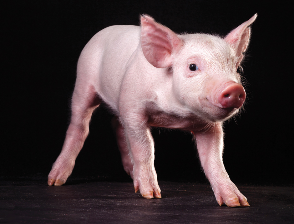
Research quadruples speed and efficiency to develop embryos
New research makes IVF four times more efficient to create pigs like this for genetics research and breeding in labs like that of Randy Prather at MU. | Photo by Nicholas Benner.
By Samantha Kummerer | Bond LSC
What started as a serendipitous discovery is now opening the door for decreasing the costs and risks involved with in vitro fertilization (IVF).
And it all started with cultured pig cells.
Dr. Michael Roberts’ and Dr. Randall Prather’s laboratories in the University of Missouri work with pigs to research stem cells. During an attempt to improve how they grew these cells, researchers stumbled across a method to improve the success of IVF in pigs.
“Sometimes you start an experiment and come up with up with a side project and it turns out to be really good,” Researcher Ye Yuan said.
The Prather lab in the MU Animal Sciences Research Center uses genetically modified pig embryos to improve pig production for agriculture and also to mimic human disease states, such as cystic fibrosis. Roberts’ team in the Bond Life Sciences Center occasionally collaborates with Prather’s lab to produce genetically modified pigs for this valuable research. However, the efficiency of producing these pigs is very low because it depends on multiple steps.
First, scientists remove oocytes (“eggs”) and the “nurse” cells that surround them from immature female pig ovaries and place the eggs in a chemical environment designed to mature the eggs, allowing them to be fertilized in vitro with sperm from a boar. This process creates zygotes, which are single-celled embryos, that are allowed to develop further until they become hollow balls of cells called blastocysts about six-days later. These tiny embryos are then transferred back into a female pig with the hopes of achieving a successful pregnancy and healthy piglets.
However, Roberts said the chance of generating a successful piglet after all those steps is very low; only 1-2 percent of the original eggs make it that far.
The quality of the premature eggs and the process of maturing them significantly reduces the rate of success.
“In other words, all this depends on having oocytes that are competent, that is they can be fertilized, form blastocysts and initiate a successful pregnancy,” Roberts explained.
Normally, researchers overcome the low success rate by starting out with a very large number of eggs, but this takes lots of time and money.
So, lab researchers, Ye Yuan and Lee Spate, began tinkering with the way the eggs were cultured before they were fertilized, making use of special growth factors they used when culturing pig embryonic stem cells.
Yuan and Spate added two factors called fibroblast growth factor 2 (FGF2) and leukemia inhibitory factor (LIF).
This combination helped more than the use of just a single factor and so they decided to add a third factor, insulin-like growth factor 1 (IGF1).
Together the three compounds create the chemical medium termed “FLI”.
“It improved every aspect of the whole process,” Roberts said. “It almost doubled the efficiency of oocyte maturation in terms of going through meiosis. It appeared to improve fertilization and it improved the production of blastocysts.”
In all, the use of FLI medium doubles the number of piglets born and quadruples the efficiency of the entire process from egg to piglet.
While the researchers are still figuring out why the three factors work together so well, Roberts believes it has to do with the fluid that surrounds the immature eggs while they are still in the ovary.
Roberts explained that unusual metabolic changes happen in the eggs and their nurse cells when the three components are used in combination but not when they are used on their own. These components are also found in the follicular fluid surrounding the egg when it is in the ovary.
However, follicular fluid actually contains factors that hinder egg maturation until the time is right, so it would seem counterintuitive to add the fluid to a chemical environment aimed at maturing the eggs. However, when freed from the other components of follicular fluid, the three growth factors act efficiently to promote maturation.
“It just creates this whole nurse environment for that egg. Once you’ve done that you’ve sort of patterned them to do everything else after that properly — fertilization, development of that fertilized egg to form a blastocyst, and the capability of those blastocysts to give rise to a piglet,” Roberts said.
Researchers hope the FLI medium can be translated beyond genetically modified pigs.
“If we could translate this to other species it could be more meaningful,” Yuan explained.
For the cattle industry, FLI has the potential to decrease the time between generations in highly prized animals.
Currently, if an immature dairy cow has desirable traits, the industry has to wait a year or so for that cow to mature and for its eggs to be collected. Using FLI medium immature eggs could be retrieved when the prized female is still a calf. After fertilizing them with semen from a prized bull, production of more cows with desirable traits could be achieved in a shorter amount of time.
The potential implications of this discovery aren’t just for farm animals.
Yuan said if this treatment could be applied to humans it would be a big help for both the patient and the whole field of human IVF.
Currently, in vitro fertilization for humans comes with high costs and risks.
“You try to generate a lot of eggs from the patients by using super-high doses of expensive hormones, which is not necessarily good for the patient and can, in fact, be risky. ” Roberts explained.
These eggs are then collected, fertilized, and the best-looking embryo transferred back to the patient. As in pigs, this overall process isn’t all that efficient. The hope is that the treatment of the patient with hormones can be minimized if immature eggs are collected directly from the ovary by using an endoscope and matured in FLI medium, allowing them to be just as competent as those retrieved after high hormone treatment.
“The idea is it would be safer for the woman, it would be cheaper, and it might even achieve a better success rate,” Roberts said.
The team still has some time before knowing for sure if FLI medium is applicable in other mammals.
Yuan said the focus is now on understanding the mechanism behind how the three compounds work so well together.
For now, preliminary data are being collected with mice and a patent is awaiting approval. Still, the team has high hopes for this almost accidental finding.
“Whenever you’re doing science, you’d like to think you’re doing something that could be useful,” Roberts said. “I mean when we started this out it wasn’t to improve fertility IVF in women, it was to just get better oocytes in pigs. Now it’s possible that FLI medium could become important in bovine embryo work and possibly even help with human IVF.”
Michael Roberts is a Bond LSC scientist and a Curators’ Distinguished Professor of Animal Science, Biochemistry and Veterinary Pathobiology in the College of Agriculture, Food and Natural Resources (CAFNR) and the College of Veterinary Medicine. He is also a member of the National Academy of Sciences.
Randall Prather is a Curators’ Distinguished Professor of Animal Science in the College of Agriculture, Food and Natural Resources (CAFNR) and Director of the National Institutes of Health funded National Swine Resource and Research Center.
How an MU student helped start a Twitter trend and how social media is advancing science.
By Mary Jane Rogers | Bond LSC
In the modern age, science isn’t a solitary endeavor.
You might be a tweet away from connecting with scientists about their work, as one MU student recently proved.
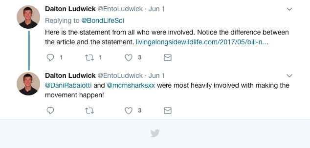
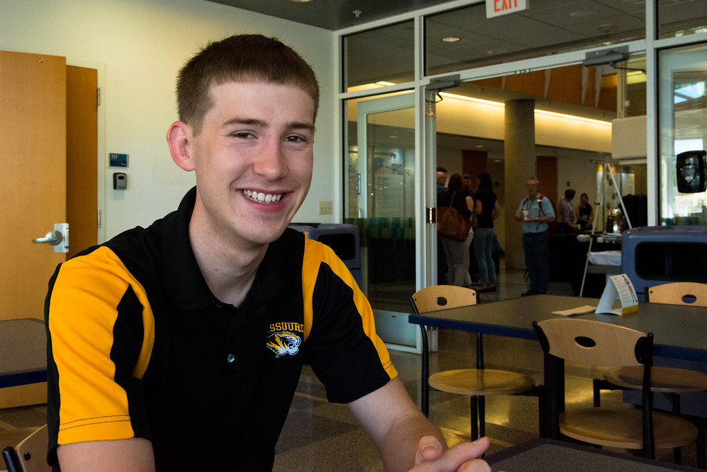
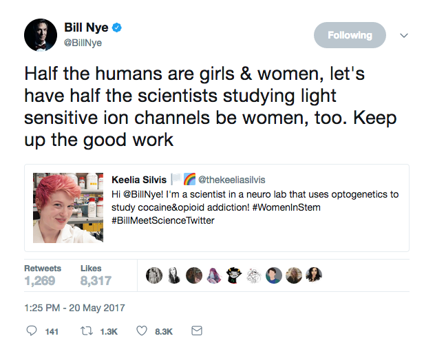
Dalton Ludwick, an MU doctoral student in entomology, helped spur a hashtag trend to connect real scientists with none other than Bill Nye.
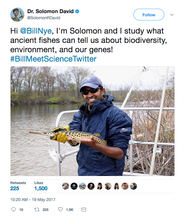
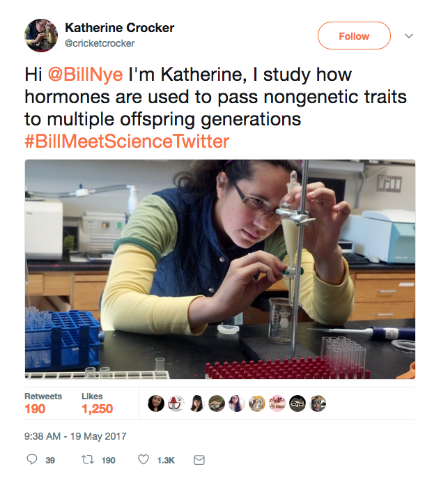
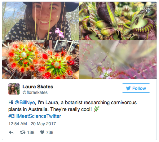
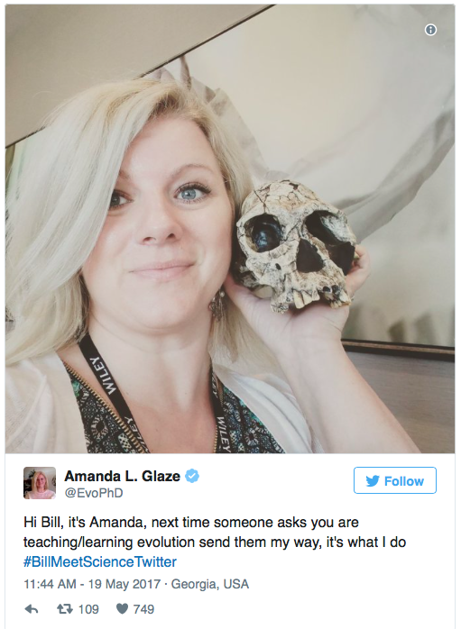
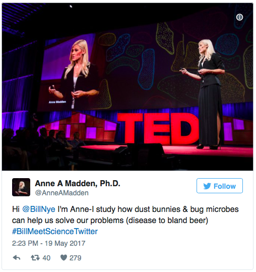
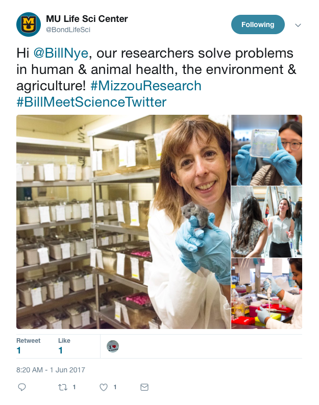
If you follow any scientists on Twitter, you may have come across the hashtag #BillMeetScienceTwitter while scrolling through your feed. Thousands of scientists on Twitter introduced themselves to the famous TV host of Bill Nye the Science Guy using the hashtag. By May 22, a mere three days after the hashtag started, more than 27,000 scientists and experts had tweeted at Nye.
#BillMeetScienceTwitter was born from a Twitter discussion between Ludwick, London-based zoologist Dani Rabaiotti and New Zealand-based marine biologist Melissa Marquez.
“Bill Nye Saves the World” is a television show currently streaming on Netflix hosted by Bill Nye. The first season explores topics such as climate change, alternative medicine and video games.
The original sentiment behind the hashtag was something scientists have long been discussing — that Nye’s television show doesn’t include a diverse array of science experts to answer questions outside Nye’s specialty. On Season One of his show, a majority of the experts Nye invited were comedians, supermodels and Hollywood stars, like Karlie Kloss and Zach Braff.
We were curious about the origins of this campaign, so we reached out to Ludwick, one of the creators of the hashtag.
Ludwick regularly uses social media to reach out and connect with other scientists. He meets other scientists on Twitter, shares ideas and often turns that conversation into a real-life, professional relationship on a global scale.
Dalton Ludwick, a Ph.D candidate in Entomology at MU and one of the creators of #BillMeetScienceTwitter.
“I talk to people from the UK, Australia and New Zealand on social media,” he said. “It’s a great way to connect with people.”
Social media is a game changer for scientists who once felt walled off from the broader world. It can be a great way to connect with people doing similar research, track grants and jobs, share exciting breakthroughs, and follow conferences.
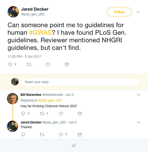
Jared Decker, an assistant professor in the College of Agriculture, Food and Natural Resources at MU, is another avid social media user on campus. He uses Facebook, Twitter and YouTube accounts to connect with other science professionals and academics, as well as his public — mainly beef and cattle producers and farmers.
Jared Decker, an assistant professor in the College of Agriculture, Food and Natural Resources at MU.
“Just the other night I was writing a grant and one of the reviewers had a specific criticism,” he said. “So, I got on Twitter and asked my question. A colleague of mine was online in Australia and was able to respond to make sure we were meeting the guidelines.”
Scientists used to have to walk down the hall to ask a colleague, or play phone tag with someone abroad.
“You can’t do that at 1 a.m.,” said Decker, “but you can go on Twitter.”
Many scientists believed that Nye’s television show wasn’t utilizing his vast array of science connections to find experts in specific fields of science.
“If you ask me about biology or oncology, I probably shouldn’t answer because that’s not my area of expertise,” said Ludwick.
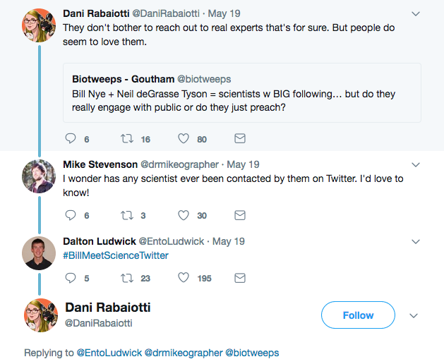
In response to a tweet by Rabaiotti, Mike Stevenson was the first to ask if anyone had reached out to Nye on Twitter. Ludwick replied to that conversation with the hashtag #BillMeetScienceTwitter, which was meant to show Nye the diversity of scientists on social media.
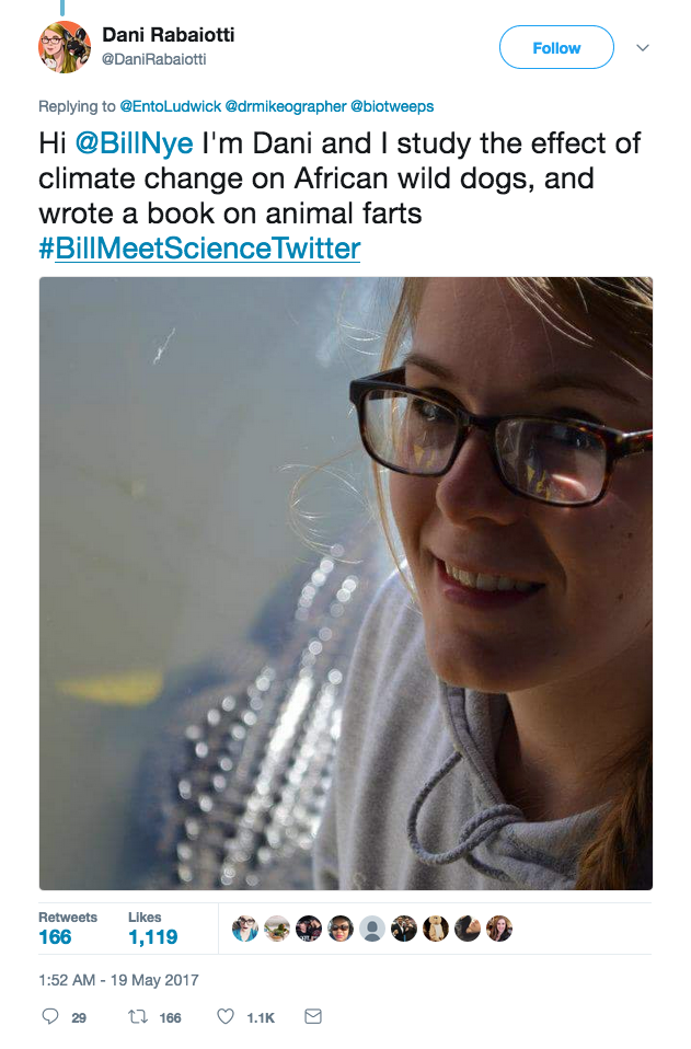
Rabaiotti – a Ph.D candidate at University College London, who studies the effects of climate change on wild dogs in Africa – was the first to introduce herself to Nye.
Overall, the tweets and engagement have been overwhelmingly positive.
We decided to tweet at Nye too!
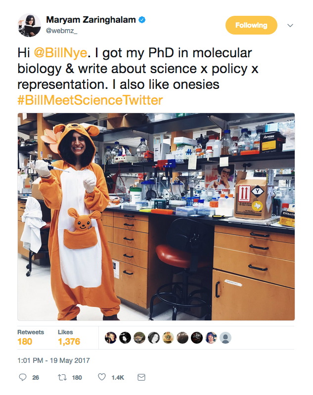
“What we were actually trying to do was reach out and offer assistance in areas outside the expertise of Bill and Neil,” said Ludwick. “We wanted to show the diversity of people doing science, as well as the diversity of the science that we do. More than 50 percent of the people tweeting on #BillMeetScienceTwitter were women — certainly not just a bunch of nerdy men in lab coats!”
Ludwick adds that the hashtag wasn’t intended as an attack on Bill Nye or Neil deGrasse Tyson, another scientist celebrity with broad reach. Instead, the point was to let them know that fellow scientists exist and can be a great source for accurate scientific information.
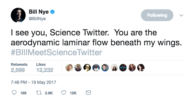
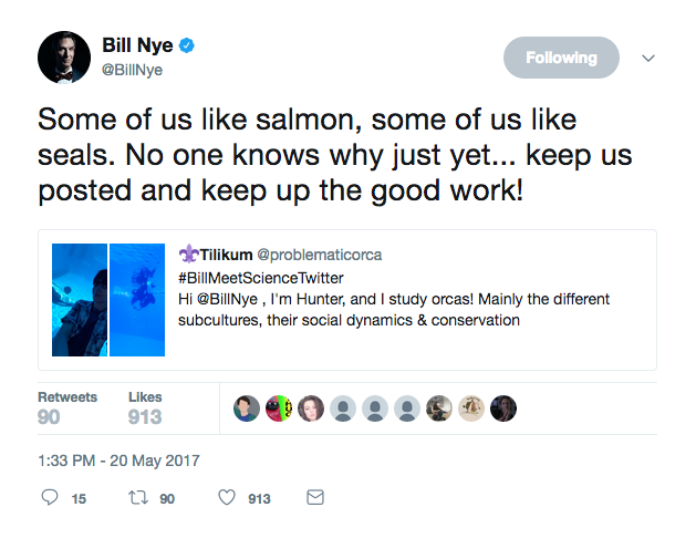
Nye responded to the hashtag, and even took the time to retweet and reply to his favorite posts.
Overall, the hashtag was a huge success, brought awareness and engaged scientific topics. But more than that, it shows how responsive and positive the scientific community can be. Some news articles noted that the campaign was “trolling in the politest way possible.”
“The scientific community on Twitter is really welcoming,” Decker said. “It doesn’t matter if you’re a first year science student or an endowed professor. People don’t treat you any differently.”
And an online presence is vital for scientists and their careers. In 2007, BioInformatics LLC conducted a survey of 1,510 scientists with regard to how they used social media. They found:
77% of life scientists participated in some type of social media
50% viewed blogs, discussion groups, online communities, and social networking as beneficial to sharing ideas with colleagues
85% saw social media affecting their decision-making
For junior scientists or researchers who are just getting started, Decker has some advice.
“Tweeting out at conferences is a good way to practice taking in an idea and getting it back out there in written form,” Decker said. “Instead of taking notes, tweet out what you would have written down.”
#BillMeetScienceTwitter also helped bridge the gap between scientists and the public. Ludwick said that this hashtag helped flip the public perception that scientists are only old men in lab coats on its head.
“People were saying, ‘Hey, I’m going to show my daughter this and inspire her,’” Ludwick said.
Ludwick also thinks that, in general, social media makes him better at communicating science to the public.
“Twitter is a great way to break things down and stop using scientific jargon,” he said. “I think it has helped me personally and it’s great practice.”
Decker agrees with Ludwick’s assessment.
“The first few months on the job it felt like I was back in Spanish class,” he joked. “I was taking the science jargon and doing mental gymnastics to translate it into the language a lay person would understand. But, now I’m fluent in both!”
So, think twice the next time you consider social media to be a waste of time. Whether it’s a hashtag that brings issues to the attention of science celebrities, platforms that connect scientists at a global level or posts that make research more accessible, social media has done a pretty cool job of advancing science.