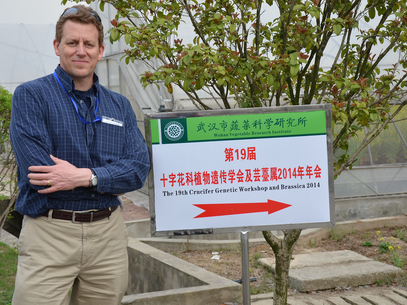
Recently, one of our investigators, J. Chris Pires traveled to Fudan University in Shanghai and the Wuhan Vegetable Research Institute for the 19th annual Crucifer Genetic Workshop and Brassica 2014 Conference in Wuhan, China.
Pires was invited to the esteemed event as the keynote speaker of the Brassica Conference. He led workshops as part of the three-day conference March 30 through April 2 and also visited Fudan University in Shanghai during the trip.
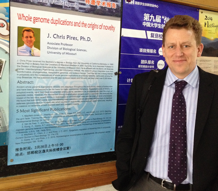
Pires is an associate professor in biological sciences and focuses much of his research on the evolution of plants. His talk at Fudan University discussed his research on whole genome duplications and the origins of novelty in plants.
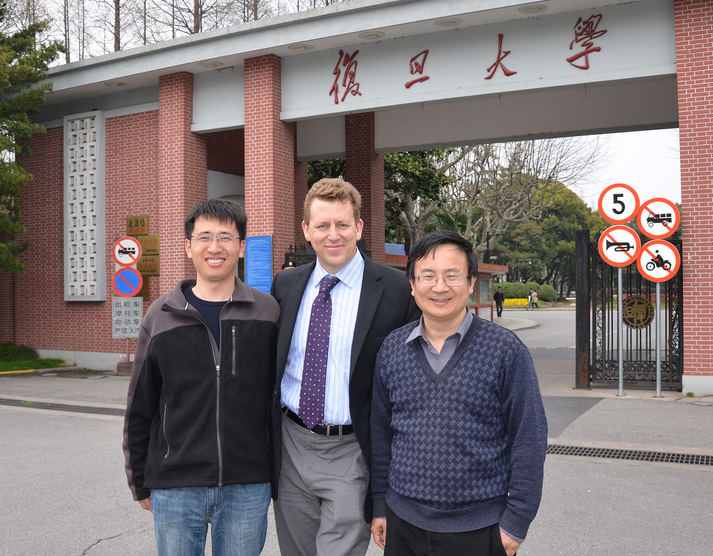
Chris Pires, investigator and associate professor in biological sciences at the Bond Life Sciences Center at Fudan University in Shanghai.Pires with Hong Ma, the dean of life sciences at Fudan University (right) and an assistant professor (left). Pires traveled to Shanghai to lead a workshop at the university over his research in whole genome duplications in plants.Pires at the 19th Crucifer Genetic Workshop and Brassica 2014 at the Wuhan Vegetable Research Institute in Wuhan, China. Pires was the keynote speaker at the conference.
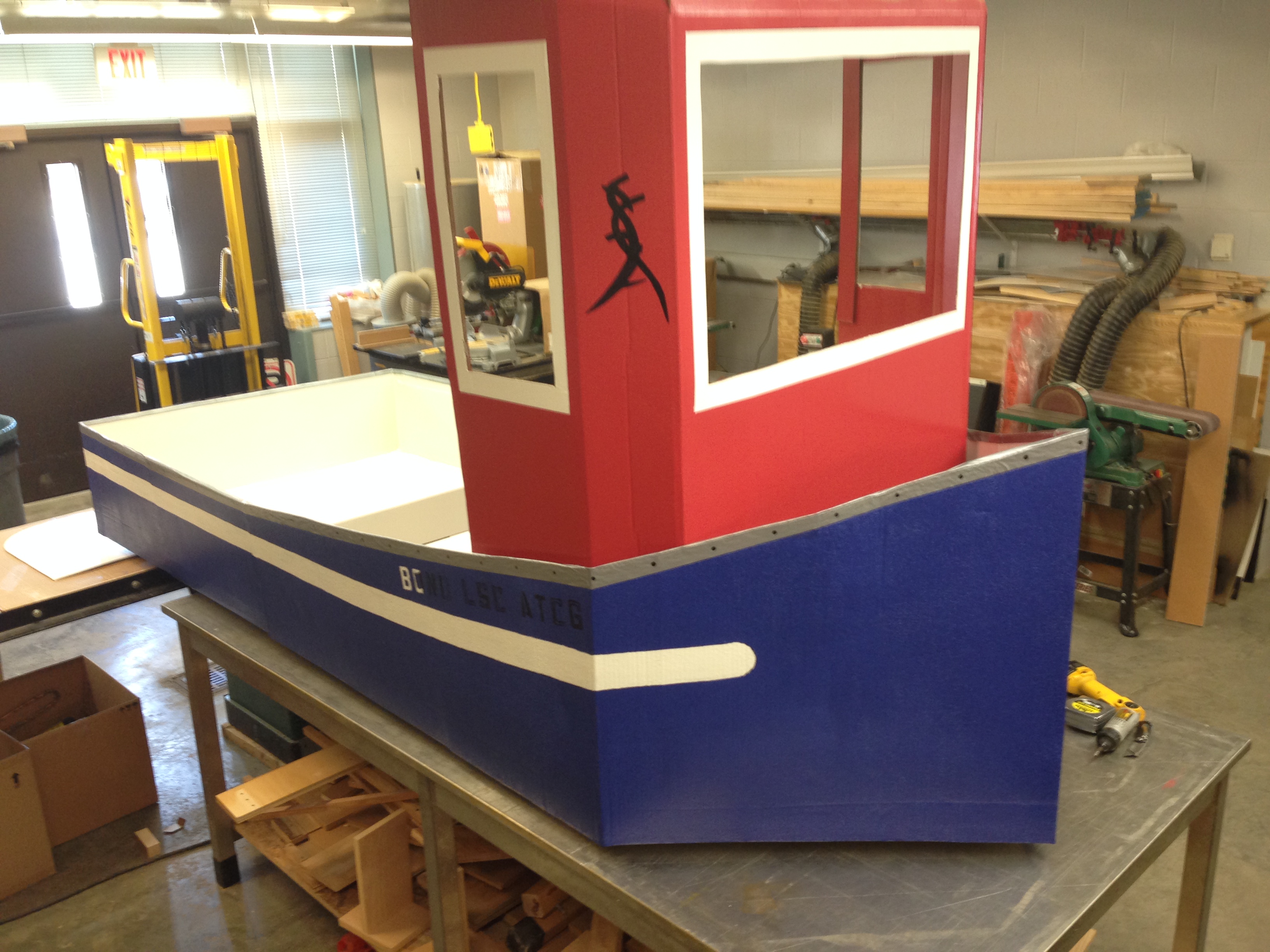
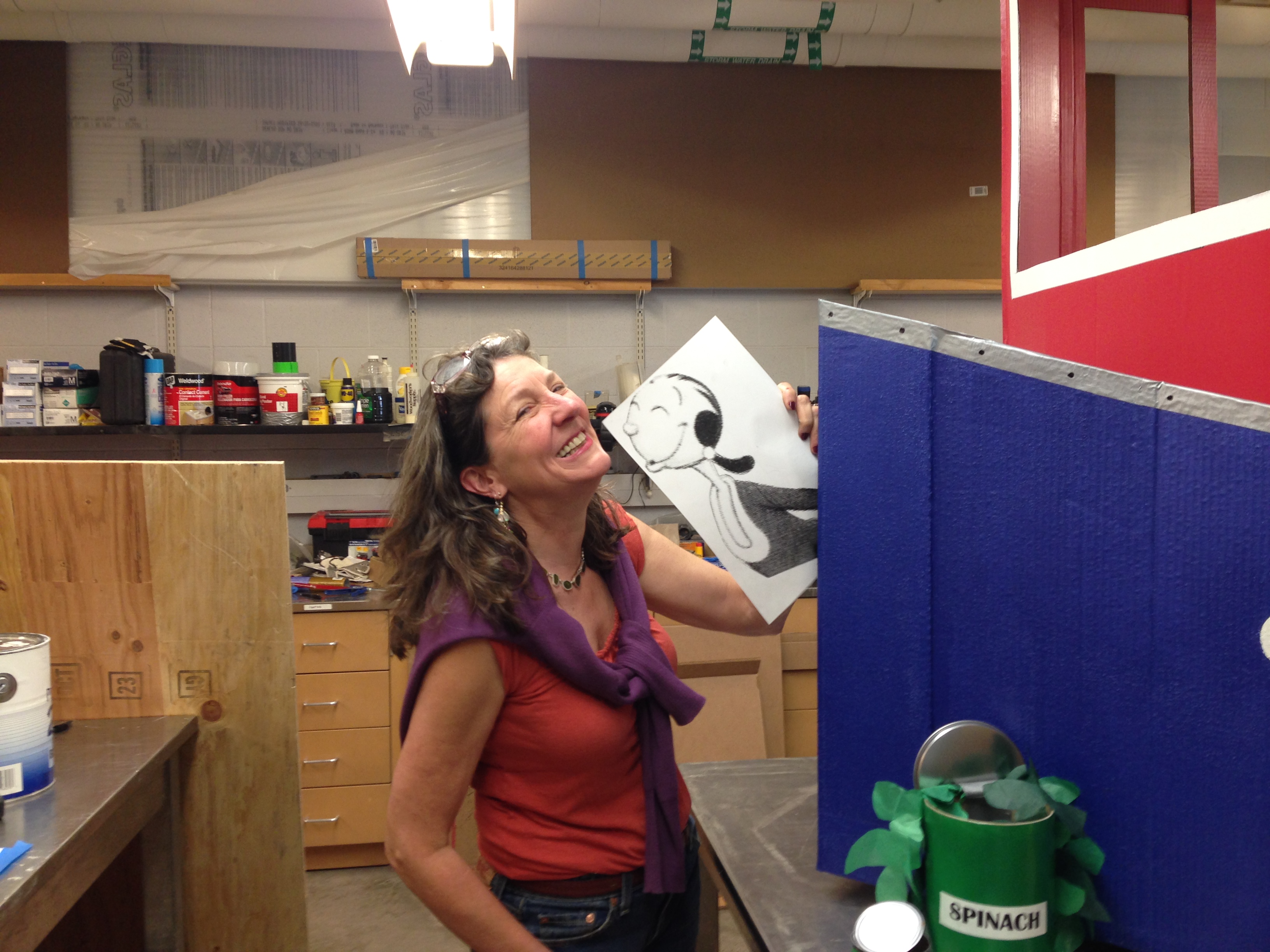
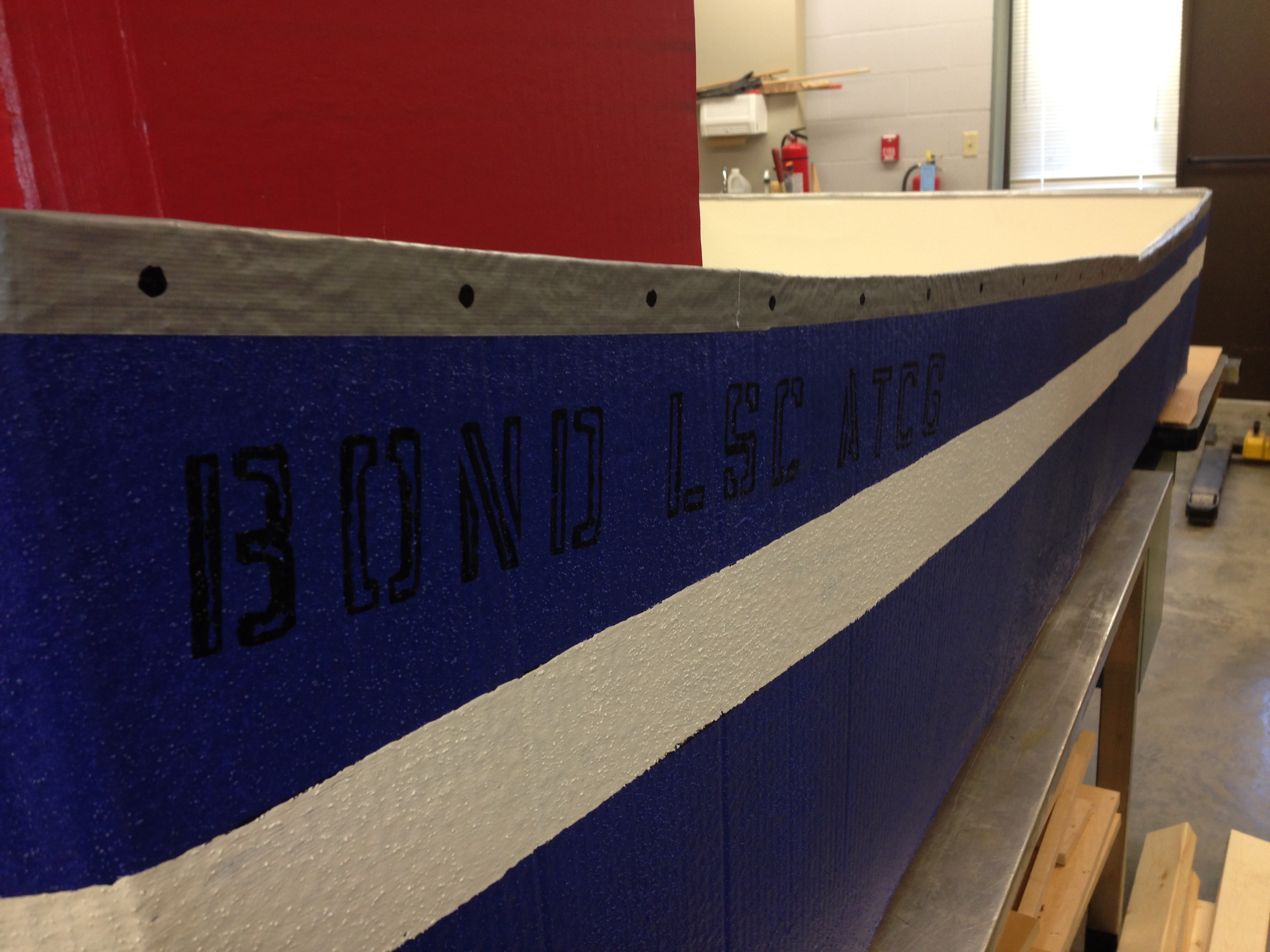
Made completely of cardboard and Popeye themed, Bond LSC facilities crew say this boat could be the winner of the 3rd Annual Flot Your Boat for the Food Bank Race on April 12 — BLANKENBUEHLER
Every year the College of Agriculture, Food and Natural Resources puts on a Float Your Boat for the Food Bank Race. All proceeds go to the Columbia Food Bank and last year, with 45 participants, more than $17,000 was donated. All participants craft their own boat and obey one golden rule: cardboard only.
The Bond LSC crew are returning to the race, this year on April 12, with a Popeye themed boat they say will win it all. Cash donations are being accepted until the race day by Maureen Kemp in 106 at the Bond Life Sciences Center. The People’s Choice Award is given to the boat with the team that raised the most money for the Food Bank.
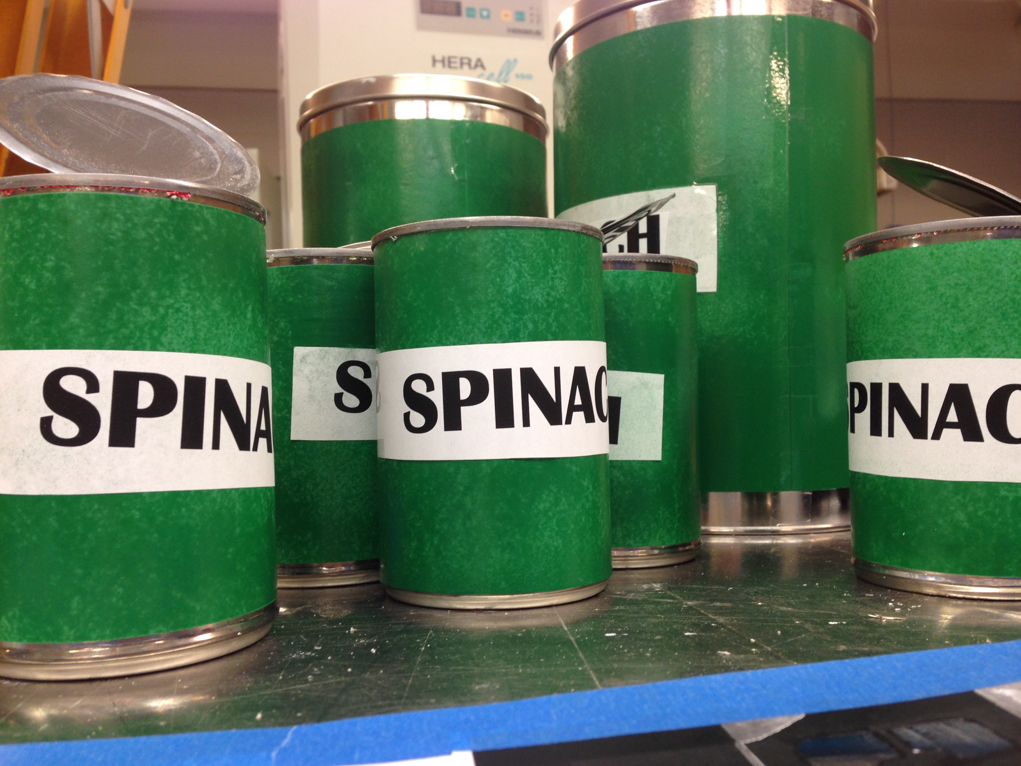
Barbie Reid, Bond LSC office support assistant, will dress as Olive Oil for this year’s race. — BLANKENBUEHLERThe Bond LSC ATCP is a streamlined, perfectly buoyant cardboard boat. ATCP are the letters of DNA sequencing. — BLANKENBUEHLERThe Bond LSC facilities department crafted mock spinach cans to play up the Popeye theme for this year’s Float Your Boat for the Food Bank Race— BLANKENBUEHLER
Jeongmin Choi (left), Gary Stacey (center) and postdoc Kiwamu Tanaka recently discovered the first plant receptor for extracellular ATP. Choi received the 2014 Distinguished Dissertation Award for her part in this work.
A former Bond LSC graduate student is being recognized for a dissertation that stands out from the crowd.
Jeongmin Choi received the 2014 Distinguished Dissertation Award this month from MU’s Graduate Faculty Senate for her work identifying the first plant receptor for extracellular ATP. The journal Science published Choi’s “Identification of a plant receptor for extracellular ATP” Jan. 17, 2014.
Choi completed her dissertation working as a member of Gary Stacey’s lab team. Stacey, a Bond LSC researcher, nominated her work for this award. This is the second year work completed in Bond LSC garnered this award after Lefteris Michailidis won in 2013 for work on the HIV drug EFdA.
Choi has since received her Ph.D. and now resides in Cambridge, England.
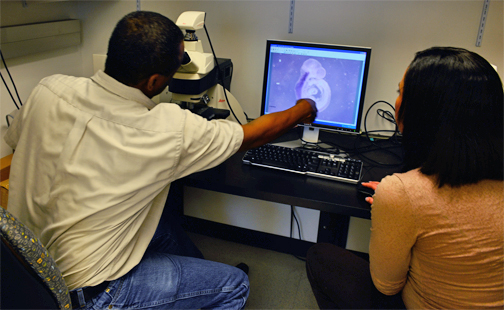
Samuel Waters and graduate researcher Desiré Buckley review stages of embryonic development. — BLANKENBUEHLER
The difference between walking and being paralyzed could be as simple as turning a light switch on and off, a culmination of years of research shows.
Recently, University of Missouri Assistant Professor of biology Samuel T. Waters isolated a coding gene that he found has profound effects on locomotion and central nervous system development.
Waters’ work with gene expression in embryonic mouse tissue could shed light on paralysis and stroke and other disorders of the central nervous system, like Alzheimer’s disease.
Waters works extensively with two coding genes called “Gbx1” and “Gbx2”. These genes — exist in the body with approximately 20,000 other protein-coding genes — are essential for development in the central nervous system.
“To understand what’s going wrong, it’s critical that we know that’s right,” Waters said.
Coding genes essentially assign functions for the body. They tell your fingernail to grow a certain way, help develop motor control responsible for chewing and, as shown in Waters’ research, help your legs work with your spinal cord to facilitate movement.
Waters and his researchers, including graduate student Desiré Buckley, investigated the function of the Gbx1 by deactivating it in mouse embryos and observing their development over a 18.5-day gestation period — the time it takes a mouse to form.
The technology could eventually contribute to developing gene therapies for paralysis that happens at birth or from a direct result of blunt trauma, like a car accident.
“Understanding what allows us to walk normally and have motor control, allows us to have better insight for developing strategies for repairing neural circuits and therapies,” Waters said.
Technology for isolating genes and their functions
Waters studies embryonic mouse development. To understand certain gene functions, he inactivates different genes using a technology called “Cre-loxP.”
Genes can be isolated, then inactivated throughout embryonic tissue. Many of Waters’s studies inactivate genes to harness a better understanding of which genes are responsible for what.
“The relevance of it to the well-being of humans, is apparently relevant to development and more importantly to the development of the central nervous system,” Waters said. “Now it’s taking me to the point where we’re getting a bird’s eye view of what’s actually regulating our ability to have locomotive control.”
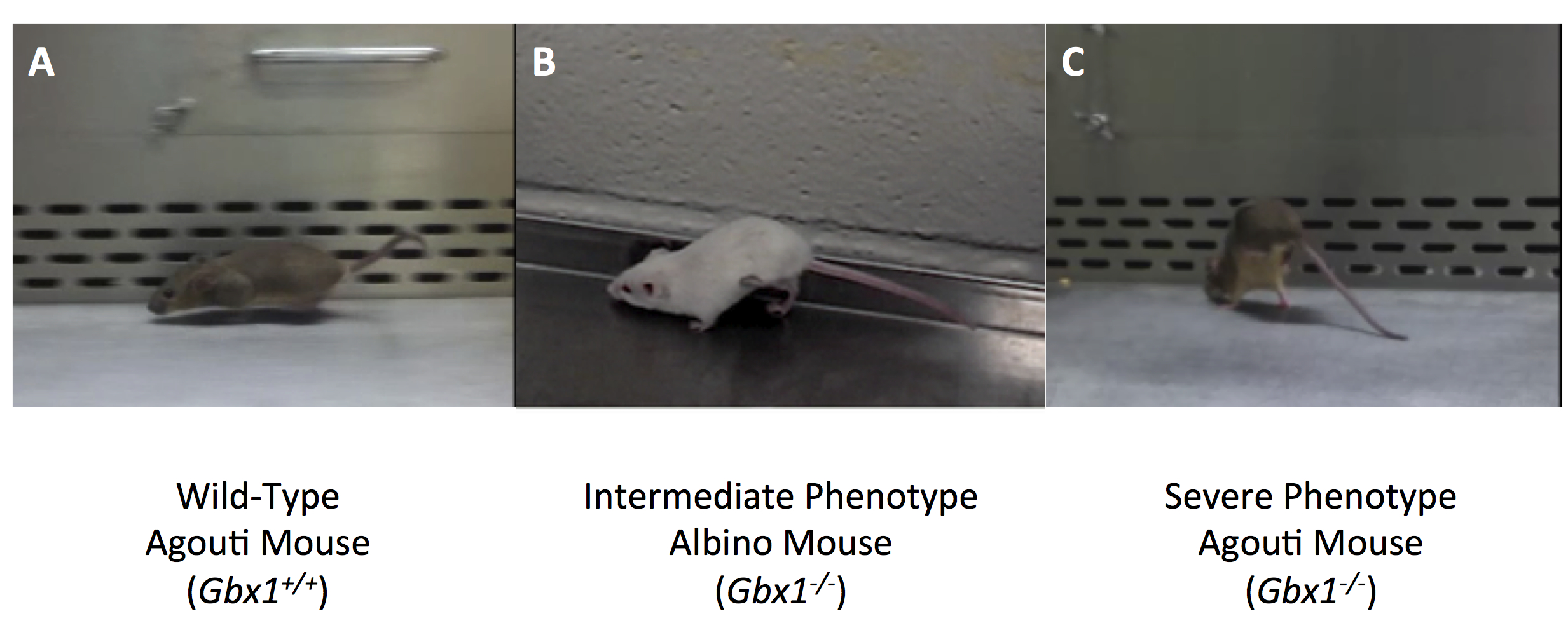
No Gbx1, no regular locomotion
Mice that Waters uses in his lab, “display a gross locomotive defect that specifically affects hind-limb gait,” according to their article published in Plos One, February, 2013.
In contrast to its family member Gbx2, when Gbx1 is inactivated, Waters concluded, the anterior hindbrain and cerebellum appear to develop normally. But neural circuit development in the spinal cord —- what allows us to walk normally —- is compromised, he said. According to an article published by
Waters, November 2013, in Methods in Molecular Biology, this occurs despite an increase in the expression level of its family member, Gbx2, in the spinal cord.
A video recording from the research, which was funded by the National Science Foundation and start-up funds from MU, show the mouse with the Gbx1 held back, with an abnormal hind-limb-gait.
Mice with this inactivated gene were otherwise normal, Waters said.
“If they were sitting there without moving, you wouldn’t know anything was wrong with them,” Waters said.” They’re able to mate, eat and appear to function normally.”
Photographs taken during the research that show the hind limb gait defect in specimen with Gbx1 held back.
No Gbx2, no jaw mobility
When Gbx2 function is impaired in the mouse, Waters observed that development of the anterior hindbrain, including the cerebellum, a region of the brain that plays an important role in motor control, didn’t form correctly.
The mice, as a result, cannot suckle, so they die at birth, Waters said.
“We’re getting a better insight into the requirements for suckling — another motor function required for our survival,” Waters said.
The research has paved the way for investigating other coding genes and their responsibilities and roles in development, Waters said.
“We have a lot to do still,” Waters said. “So, why am I so excited about it? That’s part of the reason.”
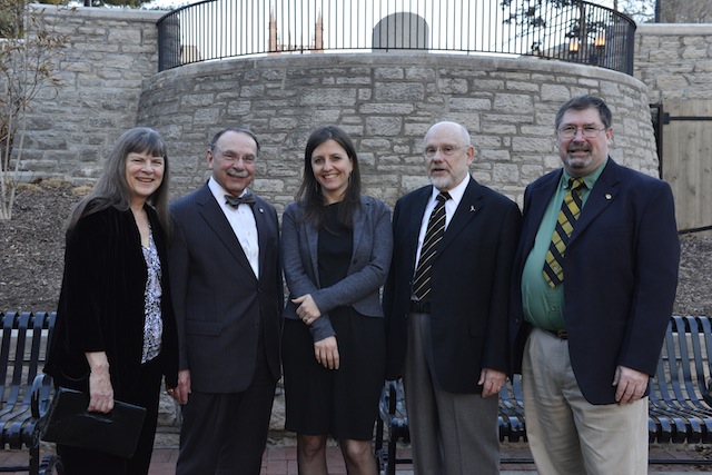
Karin Loftin, MU Chancellor R. Bowen Loftin, Bond Life Sciences Director Jack Shultz and Tim Evans pose with Rebecca Skloot at the University of Missouri Monday evening — BLANKENBUEHLER
The bridge between public knowledge and the inner-workings of the science community is one that many are reluctant to cross. Sometimes riddled with confusing terms, the most exciting discoveries aren’t always approachable.
The 10th annual MU Life Sciences & Society Symposium began Monday evening with Rebecca Skloot as she spoke to a nearly full house at Jesse Auditorium Monday. Every year the symposium erases the line between community understanding and the discoveries of the scientific community.
Skloot, the New York Times bestselling author of The Immortal Life of Henrietta Lacks, spoke about the power of science writing in making science more approachable, gave advice to scientists on spreading the word about their discoveries and gave an insight into to the decade of reporting she did for her book. Skloot autographed copies of the book following the talk.
This year’s theme, Decoding Science, speaks to the issue of communicating scientific issues and discoveries with the general public. Skloot said scientists need to keep terms and technicalities basic and exciting.
Rebecca Skloot signs copies of her book, The Immortal Life of Henrietta Lacks, after the talk Monday at Jesse Hall — BLANKENBUEHLER
Jesse Hall was filled with an eclectic mix of community, faculty and students from MU many of which lined up following the talk for nearly 30 minutes of questions.
Throughout the week, the gap between the science community and the public will be bridged with an impressive list of speakers.
The symposium, organized by the Bond Life Sciences Center which houses researchers that represent various schools at the University of Missouri, is a week-long event that features many speakers prevalent in scientific communications.
Other events to catch this week
Tuesday The “Thoughts of Plants” will be uncovered 6 p.m. at Broadway Brewery. The talk, as part of the Science Café speaker series, will be lead by Dr. Jack Shultz, director of the Bond Life Sciences Center.
Wednesday Superhero Science 11 a.m. until noon at the Colonnade in Ellis Library. Superhero submissions will be judged by the spring symposium’s own superhero: “The Antidote.” Dressed in a mask and cape, MU professor Tim Evans’ alter ego, has spicing up the field of toxicology at MU for 12 years.
Thursday James Surowiecki, a contributor to The New Yorker, will speak at 7 p.m. Thursday at Bush Auditorium, Cornell Hall. Free admission and no ticket or registration required.
Saturday All Saturday talks will be held at Jesse Hall.
10:00 a.m. Bill Nye at Jesse Auditorium, doors open at 9:00 a.m. with overflow seating available at the Monsanto Auditorium at the Bond Life Sciences Center. Tickets are sold out. Nye, most well-known for his 1990’s show Bill Nye The Science Guy, has immersed youth in “fun science” by educating in easy-to-understand terms. Nye is one of the pioneers of science communication, trying to make science more approachable by the general public.
12:30 – 1:15 p.m. Chris Mooney is a science journalist and author of Unscientific America, The Republican Brain: The Science of Why They Deny Science and Reality, and New York Times bestselling The Republican War on Science.
1:20 – 2:10 p.m. Dominique Brossard, professor and chair of the Department of Life Sciences Communication at the University of Wisconsin, Brossard studies strategic communication and public opinion in science and risk communication.
2:30 – 3:15 p.m. Liz Neeley, assistant director of Science Outreach for COMPASS, leads communications training for scientists, specializing in social media and multimedia outreach. She previously studied tropical fish evolution.
3:20 – 4:05 p.m. Barbara Kline Pope, the executive director for communications for the National Academy of Sciences, leads the Science & Entertainment Exchange, which connects top scientists with the entertainment industry for accurate science in film and TV programming.
4:10 – 5:00 p.m. A recovering marine biologist, Randy Olson is an independent filmmaker and author of Don’t Be Such a Scientist and Connection: Hollywood Storytelling Meets Critical Thinking. Olsen is a leading proponent of storytelling in science communication. His films include “Flock of Dodos” and “Sizzle,” about evolution and climate change, respectively.
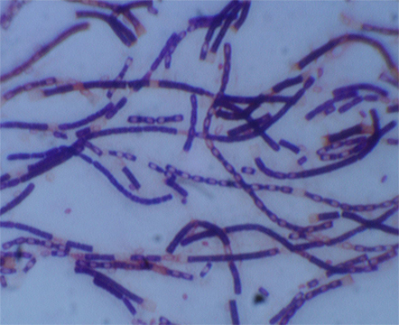
Anthrax bacteria is a rod-shaped culture. Most common forms of transmission are through abrasions in the skin and inhalation.
Imagine researchers in hazmat suits moving slowly and deliberately through a lab. One of them holds up a beaker. It’s glowing.
This light — or the absence of it — could save millions of dollars for governments and save the lives of anthrax victims.
Scientists at the University of Missouri Laboratory of Infectious Disease Research proved a new method for anthrax detection can identify anthrax quicker than any existing approach.
When the “bioluminescent reporter phage” — an engineered virus — infects anthrax bacteria, it takes on a sci-fi-movie-type glow.
George Stewart, a medical bacteriologist at MU’s Bond Life Sciences Center, and graduate student Krista Spreng, observed the virus against a variety of virulent strains of bacillus anthracis, the bacteria causing anthrax disease.
“For this technique, within a few hours, you’ll have a yes or no answer,” Stewart said.
The research, funded by the USDA, was published in the Journal of Microbiological Methods in Aug. 2013. David Schofield at Guild BioSciences, a biotech company in Charleston, S.C, created the reporter phage.
This new method could save a significant amount of money associated with the decontamination of anthrax from suspected infected areas.
Expensive clean-up from the 2001 “Letter attacks”
With the country on high-alert following Sept. 11, 2001, a slew of bioterrorists mailed anthrax letters, filled with a powder that if inhaled could cause death.
Numerous Post Offices and processing facilities were closed and quarantined.
The clean-up bill for the 2001 Anthrax Letter attacks was $3.2 million, according to a 2012 report in Biosecurity and Bioterrorism: Biodefense Strategy, Practice and Science.
Theoretically, the new detection method would alert of a negative result potentially five hours into clean-up efforts instead of two or three days into expensive decontaminating.
Current methods take anywhere from 24 hours or longer to produce a definitive answer for anthrax contamination.
A five-hour benchmark
Stewart said from contamination levels expected from a bioterrorism threat, a positive answer could be found in five hours. If contamination levels were higher, results would come back much more quickly.
Prior to this bioluminescent reporting phage, experts used techniques that were culture based or PCR (polymerase chain reaction) based. Both methods, require additional time for a definitive answer, a minimum of 24 to 48 hours, Stewart said.
“Normally to identify whether an organisms is present, you have to take the material culture, the organism and all the bacteria that might be present in the sample,” Stewart said. “You have to pick colonies that might be bacillus anthracis and do chemical testing which takes some time.”
From a bio-threat standpoint, breathing in anthrax, is the highest concern for public health and homeland security officials and has the highest fatality rate among forms of anthrax.
“If you have a situation and need a quick yes or no answer, this is a tool that will help that,” Stewart said.
Terrorists have used a powder form of anthrax, which has been slipped into letters of political persons and media. A person is infected when an anthrax spore gets into the blood system, most commonly through inhalation or an abrasion on the body, according to Centers of Disease Control and Prevention.
For low levels of contamination, the bioluminescent reporter phage would still detect the presence of the bacteria, but it would take longer.
“This method will be as quick as any of the others and quicker than most,” Stewart said.
The bioluminescent-detection method can detect low levels of anthrax bacteria and rule out false positives. The added benefit to this reporting system is its ability to show that anthrax bacteria are present and it’s alive, Stewart said.
What’s next?
The next step in the bioluminescent reporter phage is getting it approved so a product can be produced and branded. The agency that would warrant the stamp of approval would depend on the eventual use of the phage — food-related testing would likely go through the Food and Drug Administration, Stewart said.
When that happens, a product would not necessarily require a formal lab — it would need a place where cultures could grow at 37 degrees.
“Samples could be collected, brought back to the state public health lab for example and then the testing could be done within a few hours of the collection of the samples and you would have a result,” Stewart said.
The last anthrax attack was in 2001, but the possibility of one happening again, Stewart said, remains a driver for proactive research.
“In the years since the postal attacks, we haven’t had any bona fide anthrax attacks,” Stewart said. “That doesn’t mean it’s not going to happen — we have to be prepared for when it does occur again.”
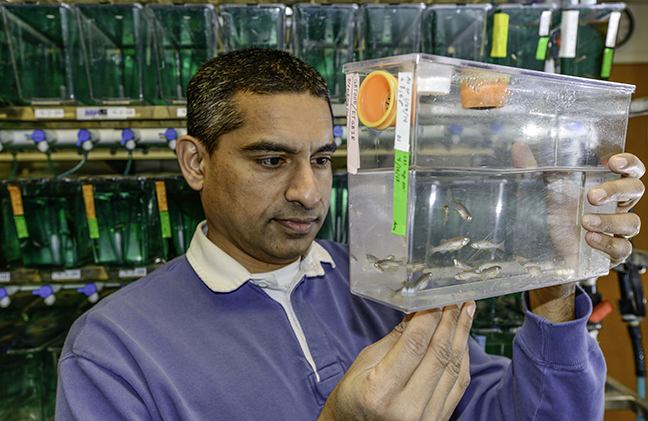
Bond LSC scientist Anand Chandrasekhar studies the zebrafish model to learn how motor neurons develop. These adult zebrafish lay eggs used to gain insight into how motor neurons arrange themselves as embryos grow into adults. Roger Meissen/ Bond LSC
Three thousand zebrafish swim circles in tanks located on the ground floor of the Bond Life Sciences Center, content to mindlessly while away their existence by eating their fill and laying eggs.
Despite their very basic higher functions, Bond LSC researcher Anand Chandrasekhar wants to understand how their brains work. More importantly, he wants to know how individual neurons end up ordered, all in the right place to support the animal’s automatic functions like breathing, swallowing and jaw movement.
This could one day lead to better understanding of specific neuronal disorders in humans.
“We are studying how cells end up where they are, and in the nervous system that’s especially critical because these neurons are assembling circuits just like in computers,” Chandrasekhar said. “If those circuits don’t form properly, and if different types of neurons don’t end up where they are supposed to, the behavior of the animal is going to be compromised.”
Zebrafish are a perfect model to study for many scientists.
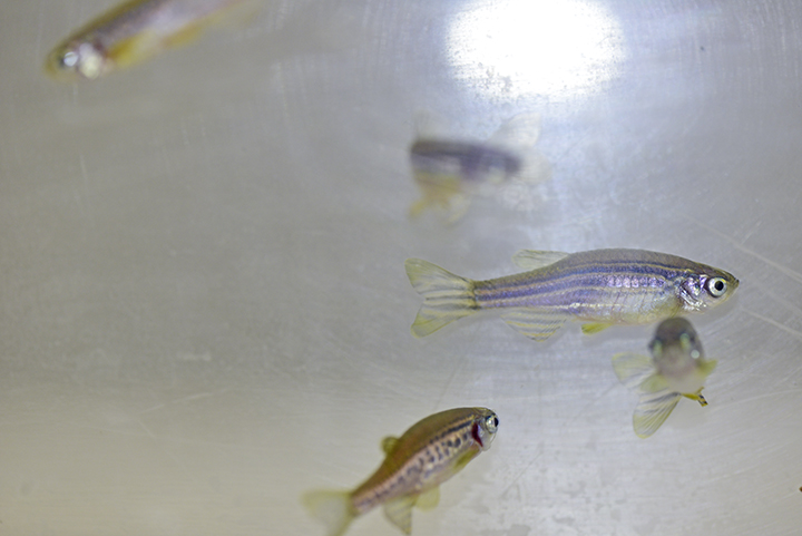
Given plentiful food, adult zebrafish lay thousands of eggs that fall through a screen in the bottom of fish tanks to be collected. These eggs turn into new embryos that are nearly transparent, allowing for easy observation of the 3-4 mm fish under a microscope.
Zebrafish embryos are nearly transparent when born, making internal processes easy to observe. Only as they mature into adults like these do their scales develop pigment. Roger Meissen/ Bond LSC
Scientists have not only sequenced the zebrafish genome, but they’ve also inserted fluorescent jellyfish protein genes into the genome. This allows easy tracking of neurons.
“It’s called a reporter, and it’s used all the time to visualize a favorite group of cells. In our case, all of these green clusters of lighted up cells are motor neurons,” Chandrasekhar said. “Some groupings are shaped like sausages, some are more round, but each cluster of 50-150 cells sends out signals to different groups of muscles.”
These motor neurons are concentrated in the hindbrain. Akin to the brain stem, it controls the gills, breathing and jaw movement in these tiny fish. Genes controlling these functions are similar in zebrafish, higher vertebrates and humans despite their evolutionary paths diverging millions of years ago.
“The brain stem is the so-called reptilian brain that has changed very little, looking very similar in a lamprey or an eel all the way up to humans. All the elaborate brain structures you see in the cerebellum and the cortex are built up on top of that,” Chandrasekhar said. “Motor neurons from the brain stem send connections to muscles of the head including the gills, jaw and, in humans, the tongue, neck, voice box and muscles for breathing. We study those motor neurons and they undergo migration in many of these vertebrates systems.”
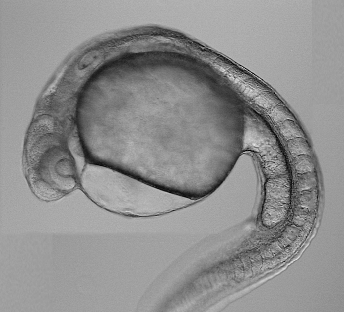
Zebrafish embryos measure only 3-4 mm seven days after they hatch. Chandrasekhar observes their motor neurons in this stage to discern how they migrate as the embryo develops. Courtesy of Anand Chandrasekhar.
This migration and signals that control it are what Chandrasekhar’s research is all about.
For example, his recent studies suggest that a protein called Vangl2 plays an important role in regulating movement of neurons through the zebrafish embryo’s matrix of tissue. Proteins like this are present in many organisms, from flies to fish to mammals.
“When I say a neuron is migrating in its environment, it’s actually pushing its way in between all these other cells,” Chandrasekhar said. “Cells in the environment of this migrating neuron secrete proteins that may diffuse away from the cell and bind to a receptor on a migrating neuron and then kind of beckon this neuron to keep moving.”
Chandrasekhar’s work contributes to a better understanding of how basic neuronal networks are created in development. That sort of knowledge could one day help with understanding the mechanistic bases of diseases like spina bifida, a nervous system disorder that results in muscle control problems due to the spinal cord not completely closing. Versions of this defect affect 1 in every 2,000 births, according to the National Institutes of Health.
“The significance of the work that we are doing is quite high for development,” Chandrasekhar said. “It is clear that even for the process of closing of the neural tube in the spinal cord and the brain, those cells closing the neural tube actually know left side from right side. The same kinds of mechanisms are going to be important and required whether you are talking about zipping together the neural tube or about allowing cells to squeeze between other cells to migrate and end up in a target position.”
Jeongmin Choi (left), Gary Stacey (center) and Kiwamu Tanaka recently discovered the first plant receptor for extracellular ATP using Arabidopsis plants. Roger Meissen/Bond LSC
It’s the genetic equivalent to discovering a new sensory organ in plants.
A team at the University of Missouri Bond Life Sciences Center found a key gene that sniffs out extracellular ATP.
Scientists believe this is a vital way plants respond to dangers, such as insects chewing on its leaves. The journal Science published their research Jan. 17.
“Plants don’t have ears to hear, fingers to feel or eyes to see. They recognize these chemical signals as a way to tell themselves they are being preyed upon or there’s an environmental change that could be possibly detrimental, and they have ways to defend themselves,” said Gary Stacey, a Bond LSC biologist. “We have evidence that extracellular ATP is probably a central signal that controls the ability of plants to respond to a whole variety of stresses.”
ATP (adenosine triphosphate) is the main energy source inside any cell. All food converts to it before being used in a cell, and ATP is necessary to power many of the cell processes that create more energy. Its value as an energy reserve is squandered outside the cell.
Scientists spent years trying to figure out what this compound did while floating outside cell walls. Animal researchers found that answer in the 1990s. They identified the first ATP receptors, now seen to play roles in muscle control, neurotransmission, inflammation and development.
Plant scientists observe similar extracellular ATP responses in plant biochemistry, but until now could not identify the exact receptor for it or what it did.
“We call this new receptor P2K, and it’s unique to plants,” Stacey said. “Even though animals and plants hold some responses in common, they have evolved totally different mechanisms to recognize extracellular ATP.”
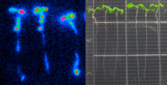
Led by Stacey, MU graduate student Jeongmin Choi and postdoc Kiwamu Tanaka screened 50,000 mutant Arabidopsis plants to find ones that didn’t respond to extracellular ATP. Using a protein called aequorin – which causes jellyfish to glow – the two-year process boiled down to whether a plant would produce light when ATP was added. Since aequorin only luminesces when it binds to calcium, those plants without extracellular ATP receptors stayed dark.
“If you add ATP to wild-type plants, calcium concentrations go up and the plants produce more blue light,” Choi said. “We found nine mutant plants with no increase in calcium and, therefore, no increase in light emission.”
These Arabidopsis plants contain aequorin. Aequorin produces blue light as a result of oxidation of the substrate, in a calcium-dependent manner. When wild-type seedlings were treated with 500 uM of ATP, intracellular calcium concentration is rapidly elevated. Increased calcium were bound to aequorin, which leads to blue light emission. Courtesy Jeongmin Choi
By comparing the genetic sequences of these nine mutants, Stacey’s lab pinpointed the gene to chromosome 5 and labeled it DORN 1, since it doesn’t respond to the nucleotide ATP.
This discovery casts a different light on previous research.
“What we think is happening is that when you wound a plant, ATP comes out in the wound and that ATP triggers gene expression, not the wounding in and of itself,” Stacey said. “We think ATP is central to this kind of wound response and probably plays a role in development, in a lot of other kinds of things.”
Future research will focus on exactly how this receptor works with ATP. Tanaka plans to study its protein structure, how it reacts to pests in lab situations and possible co-receptors that could also play a role in recognizing ATP.
Grants from the U.S. Department of Energy Office of Basic Energy Sciences and the Republic of Korea supported this research.
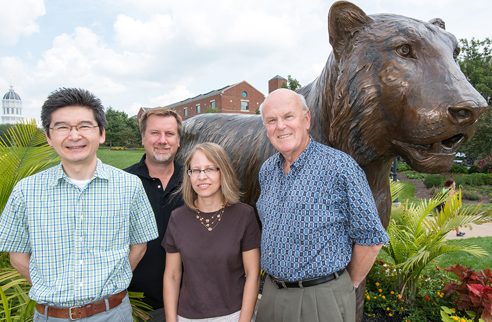
Toshihiko Ezashi (left), Danny Schust (middle), Laura Schulz (middle) and Michael Roberts (right) collaborate on new research to discover the causes of preeclampsia. Roger Meissen/ Bond LSC
You can’t see the resemblance, but cells in Michael Roberts’ lab share a family tree with some newborns.
Their common genetics may help explain severe, early-onset preeclampsia, an inherited disorder that leads to a placenta that is often small and inefficient and possibly due to the mother’s body not fully welcoming her pregnancy.
University of Missouri Health Center scientists such as Danny Schust and Laura Schulz, work with Roberts and Toshihiko Ezashi, both Bond Life Sciences Center reproductive biologists, to search for its complex cause.
That starts in the delivery room. OBGYN’s Danny Schust and his residents save small pieces of umbilical cord from preeclampsia pregnancies, allowing Roberts and Ezashi to grow cells with the disease.
“We’re essentially recreating the previous pregnancy, going back in time as far as that baby is concerned,” Roberts said. “That allows us to look at the disease in a Petri dish, look at the properties of these cells to try and figure out what’s wrong with them.”
Preeclampsia affects 3-7 percent of births worldwide, and leads to around 50,000 deaths annually. Symptoms like high blood pressure and protein in the urine tip off doctors to the disorder, but it can go unnoticed until late in the pregnancy unless the symptoms are severe. Left unchecked the disease can lead to seizures and death. The only cure for the serious early-onset form of the disease is to deliver the baby prematurely, usually between 28-33 weeks. This leaves the newborn underweight and with complications like underdeveloped lungs.
Creating useful cells from the collected umbilical cords takes a little bit of work. Cells grown from the umbilical cords are converted into induced pluripotent stem cells, with potential to become any type of cell in the body. Researchers then use a series of hormones, growth factors and other conditions to create placental cells mirroring the previous pregnancy.
Normally the placenta – containing genes from both mother and father – grows into the wall of the uterus to establish a supply of blood, nutrients and oxygen to support the embryo. But in preeclampsia, those cells encounter problems.
Roberts and Schulz focus on extravillous trophoblasts, placental cells that invade the wall of the womb, while collaborators Toshihiko Ezashi and Danny Schust study synctiotrophoblasts, placental cells responsible for uptake of oxygen and nutrients from the mother’s blood.
“The question becomes do these placental cells grow too much or too little, and it appears that in preeclampsia they don’t grow enough,” Roberts said. “We’re just beginning to look at their ability to move and grow through a jelly-like substance that impedes a cell’s mobility and ability to pass through small pores on a membrane. We’re also comparing the gene expression of the cells from preeclamptic patients with those from normal births..”
The Roberts/Ezashi/Schulz/Schust team still has several years left on two five-year National Institutes of Health grants and hopes to narrow the search for genes linked to preeclampsia. If they pinpoint the culprit genes, scientists could one day potentially correct the problem by developing drugs that restore normality to the placental cells or even by using an induced pluripotent stem cell approach.
“It might mean you could go back to correct the defect in those cells or take that patient’s cells, make some normal cells and perhaps substitute them back in to effect a cure,” Roberts said. “My own feelings are that we’re a very long way from doing that, but that is the thought.”
Michael Roberts is a Curators’ Professor of Animal Science, Biochemistry and Veterinary Pathobiology in the College of Agriculture, Food and Natural Resources (CAFNR) and the College of Veterinary Medicine, respectively. Toshihiko Ezashi is a research associate professor in CAFNR. Danny Schust is an associate professor and Laura Schulz an assistant professorof Obstetrics, Gynecology, & Women’s Health, at MU’s School of Medicine.