Tommy Langdon waits for a bee to land on a flower. // photo by CALEB O’BRIEN/BondLSC
Emily Fulcher came face-to-face with science while dissecting a hackberry gall: “Ewww,” she exclaimed, “it’s peeking out a little bit!”
Fulcher and 12 other high school students were observing plant galls as part of a Summers @ Mizzou camp exploring “The Arts as a Portal to Science Communication.”
The camp, which ran Sunday through Thursday afternoon, was facilitated by three interdisciplinary scholars and artists adept at bringing together science and the arts: Milbre Burch, an award-winning storyteller, poet and artist; Lee Ann Woolery, leader of the MU Extension Community Arts Program; and Suzanne Burgoyne, director of MU’s Center for Applied Theatre and Drama Research.
The goal of the camp was “to strengthen interdisciplinary research,” Woolery said, and to encourage “imaginative, creative minds from different disciplines to work together.”
Heidi Appel and Jack Schultz explain the complicated, contentious relationship between plants and insects during the Summers @ Mizzou camp “The Arts as a Portal to Science Communication.” // photo by CALEB O’BRIEN/BondLSC
On Monday, Heidi Appel, co-director of the Schultz-Appel Chemical Ecology Lab, and Jack Schultz, director of the Bond Life Sciences Center, were invited to give the students a crash-course on the complex realm of plant-insect interactions and the equally nuanced world of scientific research. They delved into the nature and process of science as a knowledge-generating enterprise: How do we know what we know? What is peer review? Why do grants matter?
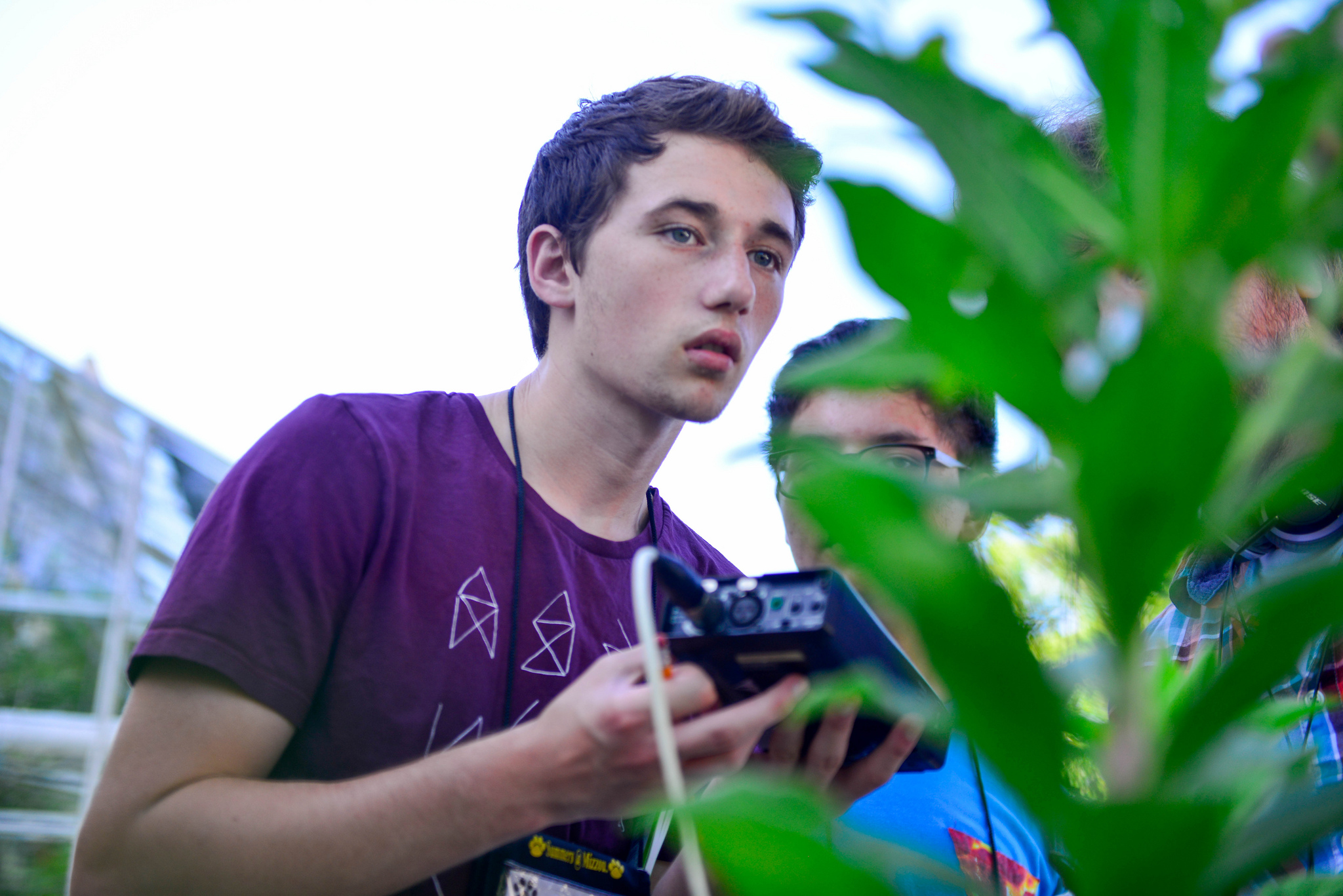
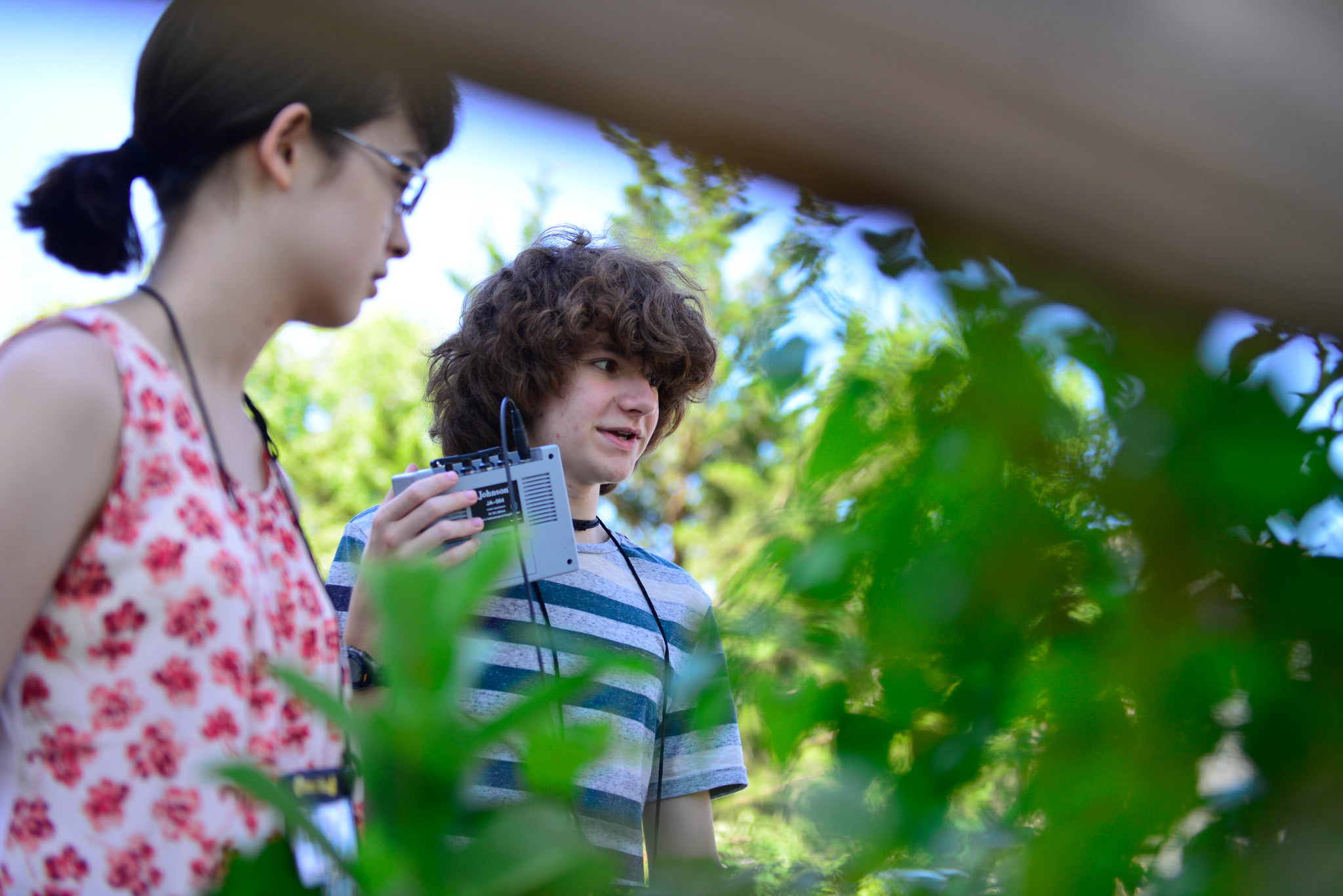
After the talk, the students split into groups — half dissected galls and scrutinized their contents under the microscope, while the other group ventured out into the Tucker greenhouse and to the tiny prairie patch next door, where they recorded the sounds of insects pollinating and eating plants. And they visited the lab of Rex Cocroft, professor of biological sciences at MU, where they used lasers to record the vibrations produced when caterpillars munch on Arabidopsis leaves.
Drawings and collages, such as this illustration of a gall by Emily Fulcher, lined the entrance to Ellis Auditorium, where students gave presentations on the the final day of the Summers @ Mizzou camp “The Arts as a Portal to Science Communication.” //photo by CALEB O’BRIEN/BondLSC
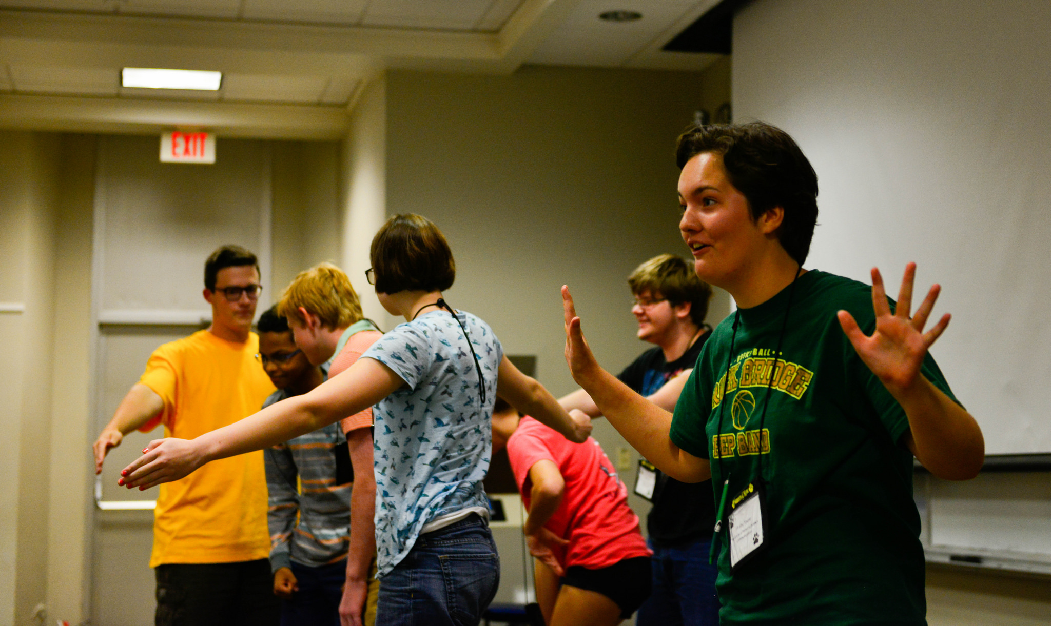
The rest of the week, the students explored different artistic media and disciplines to communicate science and deepen their own understanding of research. They painted, wrote, created videos and performed.
On the final afternoon of the camp, they gathered in Ellis Auditorium with the other Summers @ Mizzou camp participants, facilitators and family members for a final presentation. The arts and sciences group read brief, poetic explanations of the research they’d encountered earlier in the week, accompanied by amusing theatrical interpretations.
For Appel, the benefits of the camp go both ways: “Explaining my work to non-scientists makes me a better scientist because I have to clarify and distill my work down to a few key points,” Appel said. Illuminating her work for others affords an opportunity to step back and take a fresh look at the broader context of her research.
Cameron Christensen, a fifteen-year-old student from Jefferson City, said the camp offered a unique opportunity to learn how to convey information in different formats. The challenge of their final presentation, for example, was to “take cold data and make it lifelike,” Christensen said.
Appel, who has worked with all three of the camp’s facilitators in the past, said the camp “helps students understand that science is more than just a collection of facts.”
Cameron Christensen listens to the dulcet vibrations produced by a bug on a plant during the Summers @ Mizzou camp “The Arts as a portal to Science Communication.” // photo by CALEB O’BRIEN/BondLSC
Communicating science is increasingly recognized as a key step in the process of conducting and disseminating research, and invoking the arts can be a valuable tool in that process.
But in a way, putting the arts back in the sciences is just a long-overdue return to a rich heritage of creative polymathy, Woolery said. In the days before ubiquitous photography, keen powers of observation and adroit draftsmanship were de rigueur for anyone doing research — witness Audubon’s exquisite watercolors of America’s birds or Santiago Ramón y Cajal’s Miró-esque drawings of neurons.
For her part, Appel sees deep parallels between the arts and sciences.
“The processes themselves are not that different,” Appel said. “We all make observations and construct mental models. Then we either test a model (if we’re scientists), or we figure out a way to depict that model if we’re artists. Hypothesis testing is peculiar to science, but the other components are not that different.”
On the final day of the Summers @ Mizzou camp “The Arts as a Portal to Science Communication,” the students performed theatrical and literary interpretations of the science they studied. //photo by CALEB O’BRIEN/BondLSC
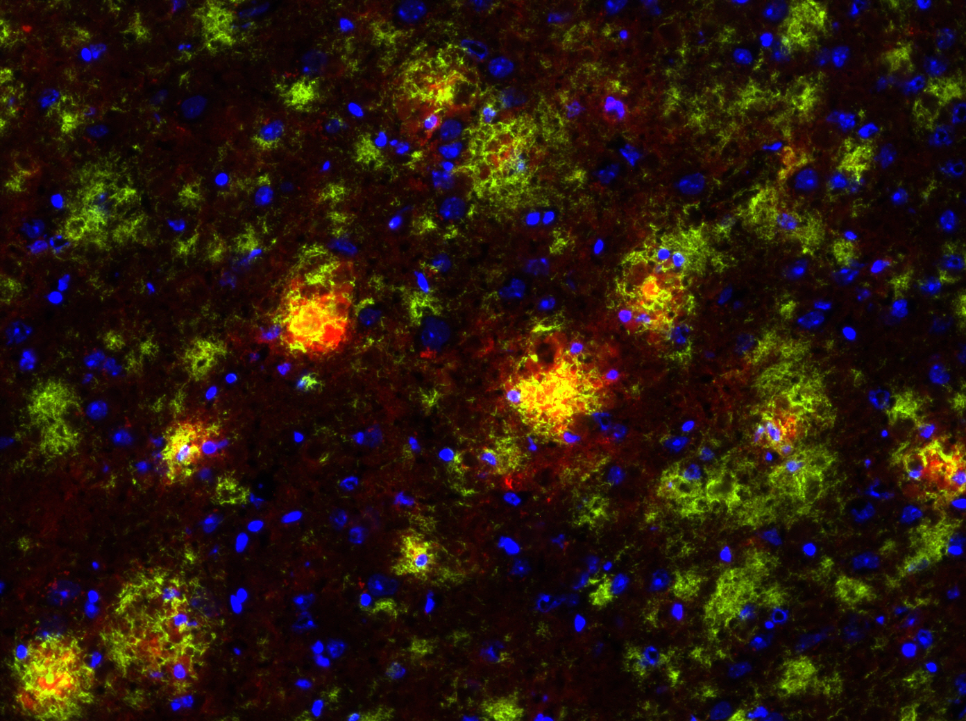
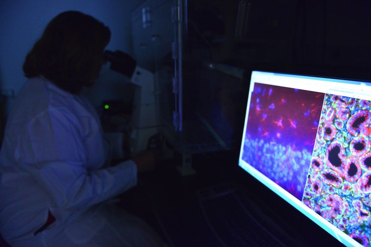
This immunofluorescence picture shows the brain of an Alzheimer’s disease mouse model, also known as the TgCRND8 mouse. In the picture, the amyloid beta plaques are stained green and the microglia, or immune cells of the brain, are stained red. Image courtesy of Luke Woods.
By Caleb O’Brien | MU Bond Life Sciences Center
Jean Camden and Luke Woods have an ant’s-eye view of Alzheimer’s disease.
Both are bench scientists in the laboratory of Gary Weisman, a professor of biochemistry at the Bond Life Sciences Center. Jean has spent the past 12 of her 35 years at the University of Missouri in the Weisman lab, running experiments, managing the lab and working with students. Luke joined the Weisman lab six years ago, doing what he call’s the dirty work of science: “Gary does the writing and the NIH stuff, but down in the trenches — that’s me and Jean.”
Weisman’s lab studies Alzheimer’s and other diseases, so I sat down recently with Jean and Luke to talk about their research for Alzheimer’s & Brain Awareness Month.
Q: What does your lab do, and how does it involve Alzheimer’s?
Luke: We primarily have two projects. One, which has been a very longstanding project, is focused on salivary glands and salivary gland inflammation. The other is the Alzheimer’s project. The link between them is a particular type of cell surface receptor called a nucleotide receptor — more specifically, a P2 nucleotide receptor called P2Y2. These P2 receptors function in a lot of different ways, but the link is with inflammation: We look at P2 receptors in salivary gland inflammation and in Alzheimer’s disease, which has a very large inflammation component that often gets glossed over. In a lot of Alzheimer’s articles that the public reads, you hear about amyloid beta plaques and tau tangles and neurodegeneration, but a large component of that is inflammation, where some of the resident non-neurons in the brain start responding like there’s inflammation in the brain, and it actually kills neurons. That’s been the focus in Gary’s lab for the past 30-plus years.
JEAN: The P2 receptors — especially the P2X7 and P2Y2 which we focus on — Gary during his postdoctoral work started studying these receptors without really knowing that they existed. At the time, he just knew that there was a pore formed in cells caused by the addition of the nucleotide ATP which eventually leads to apoptosis (cell death). Eventually, we cloned the human P2Y2 receptor gene with another group in North Carolina, so we call it “our receptor.” It only appears in cells under inflammatory conditions, such as Alzheimer’s disease, salivary gland autoimmune disease and cardiovascular disease. Any time you have tissue damage, it looks like the P2Y2 receptor is up-regulated. And then once the damage is healed, the receptor goes away.
Inflammation is good — we want inflammation, that’s how we heal — it’s the chronic inflammation that’s bad. But we really don’t know how these receptors work and what their role is during chronic inflammation. Do we want to activate them, or do we want to inhibit them?
LUKE: Scientists have investigated P2X7 receptor antagonists in the treatment of Crohn’s disease and rheumatoid arthritis — there are several clinical trials that have been focused on these receptors, evaluating whether you want to block or activate them. If you block them, you prevent the acute inflammatory responses that are good for wound healing; if you activate them, you may extend those responses past the healing phase into a chronic inflammatory phase that can be quite damaging. So unraveling that fine line of what you want to be doing to these receptors in disease settings is sort of what we do here.
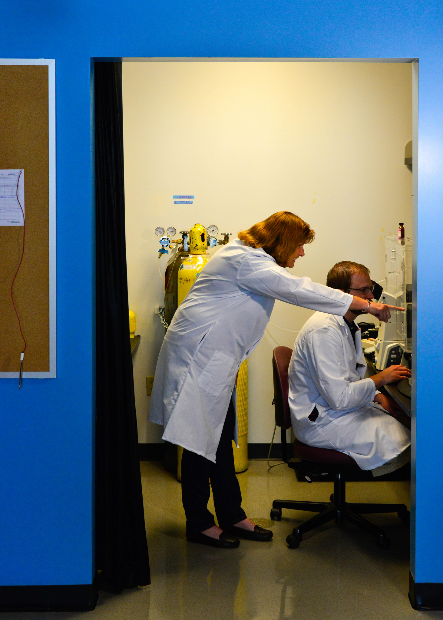
Jean Camden and Luke Woods look at images of a mouse brain with Alzheimer’s disease. // Photo by CALEB O’BRIEN/Bond LSC
Q: When I think of Alzheimer’s, I think of a shriveled, shrunken brain, but I associate inflammation with swelling. Why the difference?
LUKE: I think the distinction is acute versus chronic inflammation. With acute inflammation, you get swelling. The body has different types of immune responses: acute responders like neutrophils and macrophages are immune cells that act quickly. They come in, for example, if you have a scratch, and there can be swelling. Along with macrophages neutrophils can protect cells from bacteria. The macrophages can also clean up damaged tissue and then the repair cells go to work. Cells come in that lay down a new matrix, whereas undamaged cells then migrate onto the matrix and regenerate. Well, what happens after you’re done repairing is that there are signals that tell the inflammation to stop. In chronic inflammation, that’s where you have continued cell death, and the tissue would then shrivel up. The shriveled brain that you’re referring to is during chronic inflammation, and that’s an end-of-life case, after a very long bout with Alzheimer’s.
JEAN: What we think of as inflammation is often a cut or a wound. It’s only been in recent years that Alzheimer’s disease has been considered an inflammatory disease. We have a phenomenal immune system, but when it goes awry, you have problems. In the other disease we look at — an autoimmune disease — your immune system starts to attack your own body. It’s hard to treat and understand the underlying mechanism.
Q: So how are you trying to unravel the role of inflammation in Alzheimer’s?
JEAN: To study Alzheimer’s, we have an Alzheimer’s mouse model. It overexpresses a gene for the amyloid precursor protein that enables the brain to accumulate high amounts of beta-amyloid plaques that you always hear about. So we’re using this mouse model that we’ve crossed with a mouse that does not express any P2Y2 receptor, so it’s called a knockout mouse. The P2Y2 receptor knockout mouse by itself is fine, and the Alzheimer’s mouse does develop Ab plaques, but it lives to approximately 6 months old before it will develop behavioral defects. The interesting thing is that when we cross the P2Y2 receptor knockout mouse with the Alzheimer’s mouse, the offspring that are Alzheimer’s mice without P2Y2 receptors prematurely die. So at least in this Alzheimer’s mouse model, it looks like the presence of the P2Y2 receptor is protective, because without it, the Alzheimer’s mice die much earlier. But we don’t really know which cell type is most important: Is it the P2Y2 receptor up-regulated on neurons that acts to repair them —which we’ve already shown happens — or is it the P2Y2 receptor on microglia (an immune cell of the brain), or is it the P2Y2 receptor on blood vessels in the brain that help recruit immune cells from the cardiovascular system to help with repair?
So we’re using this mouse model to investigate the role of the P2Y2 receptor, plus we also use cell lines because we can easily control the environment for these cell lines in culture. We isolate primary neurons, we can prepare primary microglial cells or we can purchase cell lines that comprise blood vessels. We can then utilize these tools to investigate cell signaling mechanisms for the P2Y2 receptor in individual cell types.
LUKE: One of the findings that we have found interesting in these primary cells is when you take them fresh out of the mouse, put them in a dish and then treat them as you wish. We’ve shown that if you activate the P2Y2 receptor in primary microglia from the mouse, they will actually engulf and chew up beta-amyloid. And so one of the things we think might be happening in this Alzheimer’s mouse model is that P2Y2 receptor activation in these microglial immune cells in the brain is working to break down those beta-amyloid plaques. And when you lose the P2Y2 receptor in that mouse model, those plaques develop quicker because the immune cells are no longer offering protection by chewing up that beta-amyloid. That’s one of the hypotheses we’re exploring right now.
Q: So you’d bet that these receptors are actually protective against Alzheimer’s?
JEAN: Yes. Going back to the human — it’s hard to get human tissues, especially brain tissues, but there is one published study that has shown that in Alzheimer’s patients who have passed away the P2Y2 receptor is down-regulated, meaning there’s not much left. Which would make sense. If it’s down-regulated, the plaques aren’t able to be chewed up, per se, by these microglia. There’s a correlation between low levels of P2Y2 receptors and Alzheimer’s disease that is apparent at the end of life.
LUKE: It’s very difficult to do some of these studies in humans because most of the available Alzheimer’s tissues are from end of life cases where you can only look at the end result of the disease without looking at the progression of the disease. Obviously you can’t take brain tissue from a living person, so the ability to study live cells from Alzheimer’s patients is limited. We rely very heavily on mouse models.
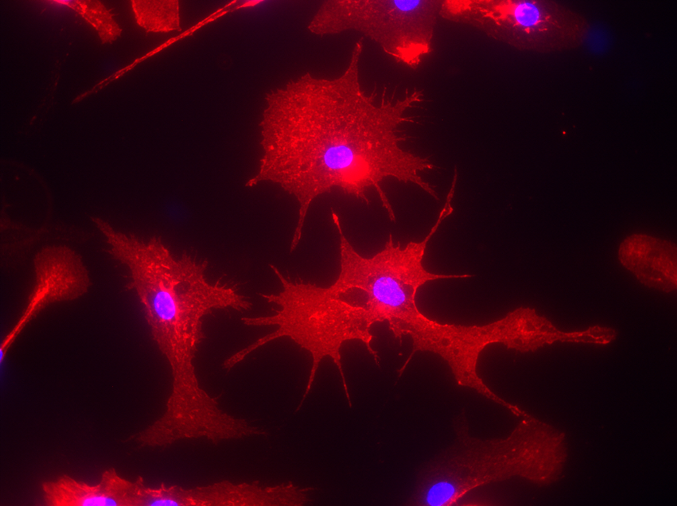
This immunofluorescence picture shows microglia cells that were isolated from the brain of an Alzheimer’s mouse model called TgCRND8 and cultured in a dish for further analysis. Image courtesy of Luke Woods.
Q: What have been the biggest shifts in our understanding of Alzheimer’s in recent years?
LUKE: Maybe one shift — I may not be the best expert to speak about it — is the idea that the beta-amyloid plaques are the cause of disease. It is now being mostly recognized that they’re really the tombstones of the disease. They’re not the initial cause, but rather the end result of the disease. For a long time investigators were focused on trying to prevent the buildup of beta-amyloid because that was one aspect of Alzheimer’s disease that you could see and measure. Now the thinking is that maybe the beta-amyloid does not contribute as much to disease progression as originally thought, and rather is the end result of a complicated mechanism that is actually causing the neurodegeneration.
JEAN: There’s still debate on what causes Alzheimer’s disease. There is a small percentage of patients where it’s actually related to a genetic alteration in the amyloid precursor protein gene.
LUKE: Another link has been with the ApoE (apolipoprotein E) gene, which makes a lipoprotein and cholesterol transporter. We inherit 1 copy of the ApoE gene from each of our parents and it has been shown that individuals who have at least 1 copy of a particular variant of the gene called ApoE4 are at increased risk of developing Alzheimer’s disease.
Q: From the perspective of a lab scientist, why do you care about Alzheimer’s?
JEAN: We care about any disease, really, and if we can show that our receptors have anything to do with any disease, we’d be proud to have a role in that.
LUKE: We don’t do much clinical science here, it’s mostly basic science. We contribute to the basic understanding of the disease so that drug companies and medicinal chemists who develop drugs for clinical use in Alzheimer’s patients can say, “Hey, this group’s research found a new mechanism related to Alzheimer’s disease, so let’s target this pathway to treat the disease.” It’s always nice to contribute to that sort of ground-level science.
JEAN: That would be the ideal, to show that whether you have to activate or inhibit the P2Y2 receptor, it does something to improve the clinical outcome in Alzheimer’s patients. A better understanding of Alzheimer’s and other diseases is what’s needed — we’re just working to provide a piece of the puzzle.
Q: How has being down in the trenches changed your perspective on research and Alzheimer’sin general?
JEAN: We’re the ones who are hoping to clarify the direction for science to go. We do the experiments and we are the first ones to see the data. We collect the data that becomes the cornerstone for deciding the direction our research goes. I think Gary would agree with that — he depends on us a lot to collect the data and we depend on him to help determine which scientific findings to chase and which ones not to chase.
I’ve been doing this for 35 years, and I really do enjoy the science. I’ve seen the science of these nucleotide receptors come a long way. These receptors have in common their use of extracellular nucleotides, particularly ATP (or adenosine triphosphate, more commonly known as the intracellular high energy molecule of all cells). And this ATP, is at a high concentration inside cells, so when it is released by cell damage, it can easily activate nucleotide receptors on nearby cells. It was Dr. Geoffrey Burnstock, now considered to be the grandfather of nucleotide receptors, who claimed a long time ago that there are receptors on the outside of cells that respond to ATP. Everybody kind of laughed at him, “Yeah, sure, right. There’s no way: ATP belongs inside the cell.” So for me personally, to come in on the ground level for these receptors and find a role for them in a variety of diseases has been exciting for me.
LUKE: ATP is the energy currency inside of all cells, so it’s use outside cells would be like tossing money out the window. Why would they want ATP outside the cell? It didn’t make any sense at the time, but looking back I think it does. What happens if you damage or rupture a bunch of cells during an injury? You get the release of a high concentration of ATP that neighboring cells recognize as a danger signal telling them that an injury has occurred. In that sense, ATP makes the perfect signaling molecule to tell other cells that an injury has occurred and they need to start the repair work by recruiting immune cells to the damaged tissue.
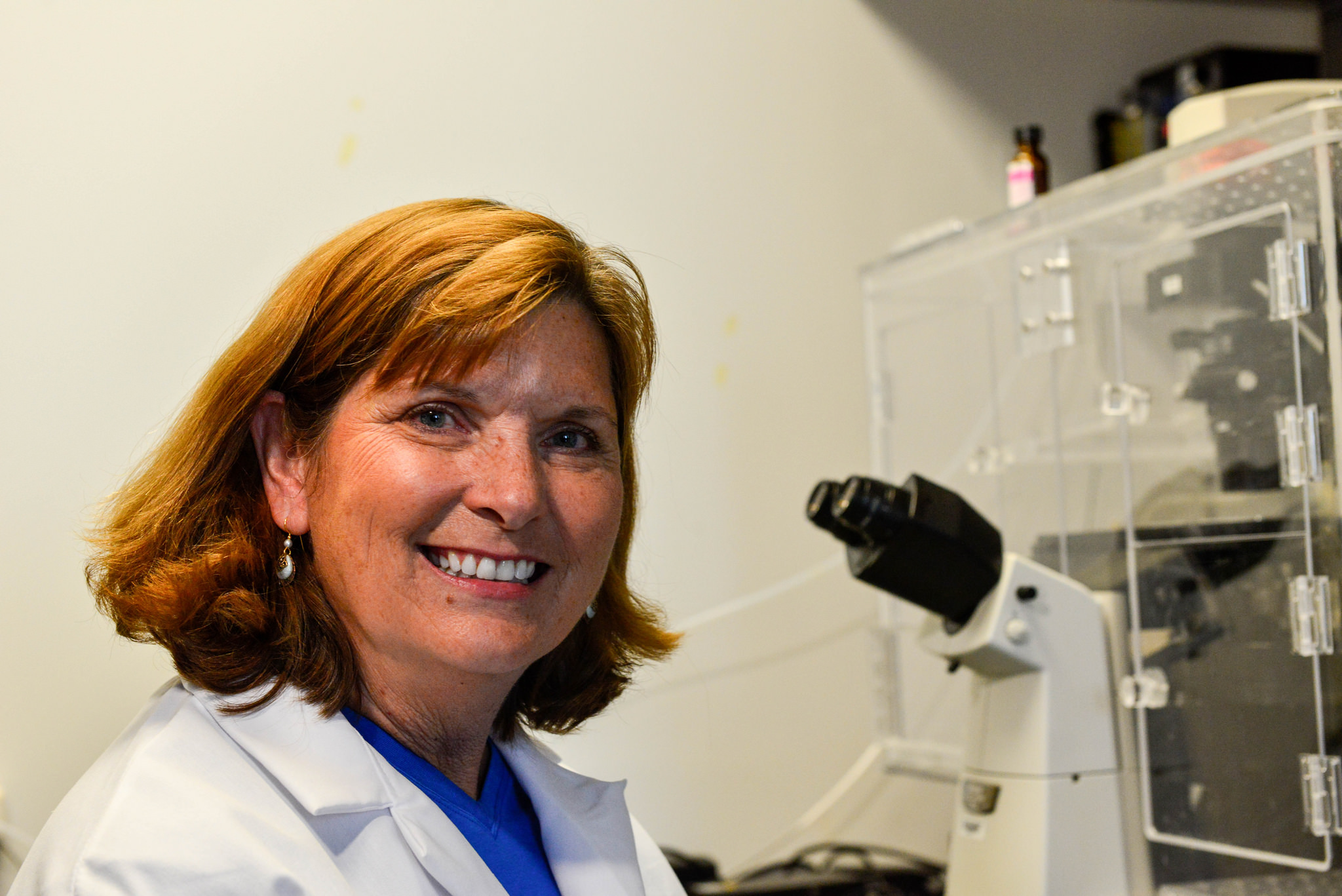
Jean Camden has spent 35 years working at the University of Missouri and more than a decade in Gary Weisman’s lab. // Photo by CALEB O’BRIEN/Bond LSC
Q: Where would you like to be in five years with this research?
JEAN: I talked about determining how the P2Y2 receptor in this mouse model was protective. It would be nice to find out which cell type on which the P2Y2 receptor is expressed in contributes most to neuroprotection. Our hypothesis would be that the microglial cells are very important, since they gobble up beta-amyloid, but other cell types including neurons and endothelial cells are likely involved. We’re also anxious to look at other inflammatory diseases to see if the P2Y2 receptor plays a similar role there.
LUKE: From somebody who does a lot of bench work, something I would like to see is a really good tool, a specific agonist or antagonist of the P2Y2 receptor that could be used in the clinic. There are a few suitable compounds available that we use to investigate the P2X7 receptor— I’ve told you that some have been tested in clinical trials — but the P2Y2 receptor has been sort of an enigma, due to the lack of selective inhibitors and agonists that are specific enough for clinical use. I’d like to see the development of a specific agonist or antagonist that could eventually be used to treat inflammatory diseases. There’s no reliable drug that is currently suitable to investigate the P2Y2 receptor in animals or humans, so clearly more work is needed there.
This interview has been edited for length and clarity.
The next time you slather mustard on your hotdog or horseradish on your bun, thank caterpillars and brassica for that extra flavor.
While these condiments might be tasty to you, the mustard oils that create their flavors are the result of millions of years of plants playing defense against pests. But at the same time, clever insects like cabbage butterflies worked to counter these defenses, which then started an arms race between the plants and insects.
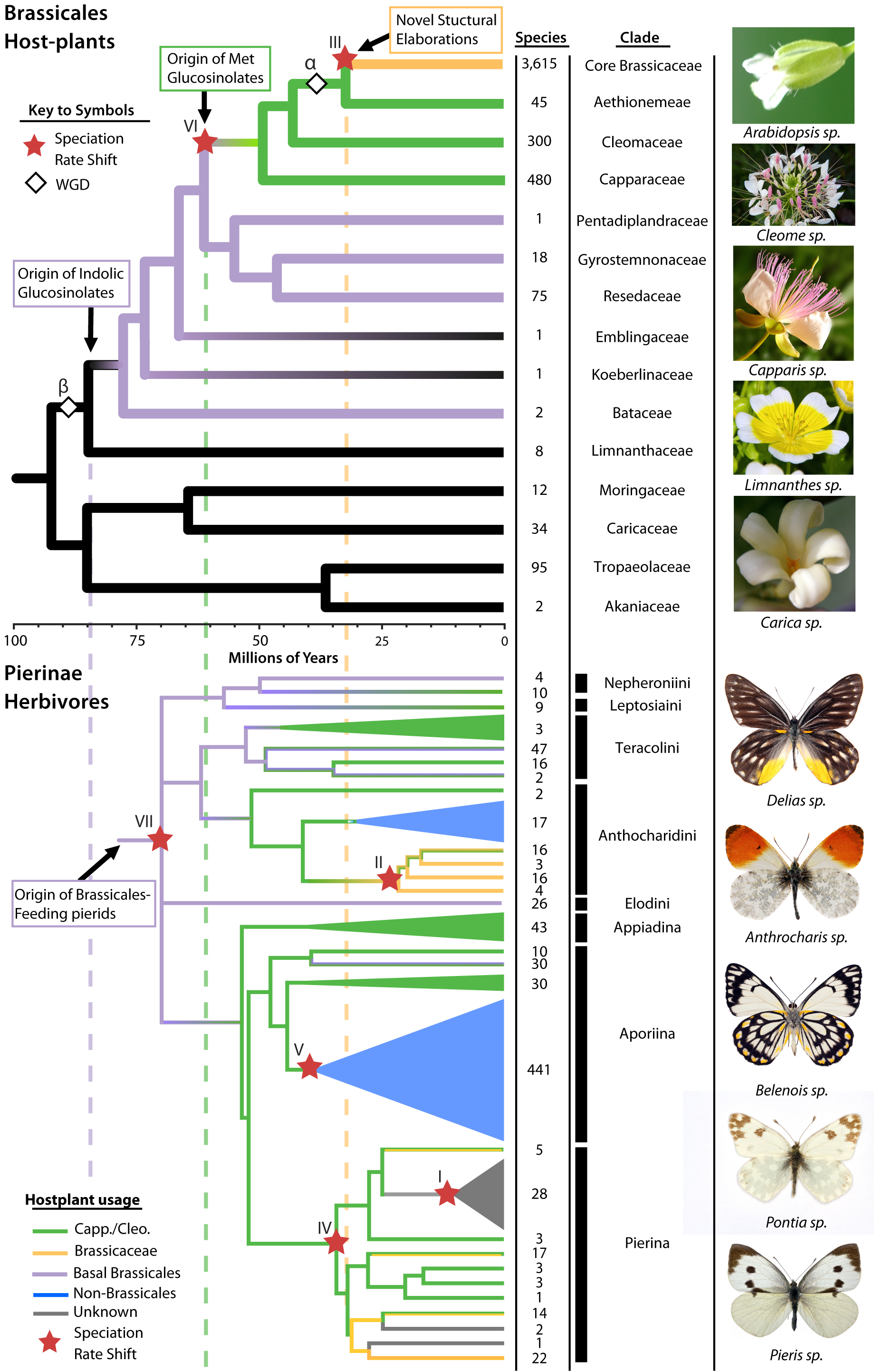
An international research team led by University of Missouri Bond Life Sciences Center researchers recently gained insight into a genetic basis for this co-evolution between butterflies and plants in Brassicales, an order of plants in the mustard family that includes cabbage, broccoli and kale.
“We found the genetic evidence for an arms race between plants like mustards, cabbage and broccoli and insects like cabbage butterflies,” said Chris Pires, an MU Bond Life Sciences Center researcher and associate professor of biological sciences in the College of Arts and Sciences. “These plants duplicated their genome and those multiple copies of genes evolved new traits like these chemical defenses and then cabbage butterflies responded by evolving new ways to fight against them.”
A biting taste
While you might like the zing in mustard, insects don’t.
Compounds, called glucosinolates, create these sharp flavors in plants to defend against caterpillars, butterflies and other pests. Brassicales species first evolved glucosinolate defenses around the KT Boundary — when dinosaurs went extinct — and eventually diversified to synthesize more than 120 different types of this compound.
For most insects, these glucosinolates prove toxic, but certain ones like the cabbage butterfly evolved ways to detoxify the compounds.
“Seeing the variation in the detoxification mechanisms among species and their gene copies gave us important evolutionary insights,” said Hanna Heidel-Fischer, a lead author on the study based at the Max Plank Institute for Chemical Ecology in Germany.
To look at these genetic differences, the team used 9 existing Brassicales genomes and also generated transcriptomes — the set of all RNA in a cell — across 14 Brassicales families. This allowed the team to map an evolutionary family tree of sorts over the millennia, seeing where major defense changes occurred. This family tree was compared with the family tree of 9 key species of Pieridae butterflies, which includes the cabbage butterfly.
Pires and his colleagues identified three significant evolutionary waves over 80 million years, where plants developed defenses and insects evolved counter tactics.
Pat Edger | Image by Roger Meissen, Bond LSC
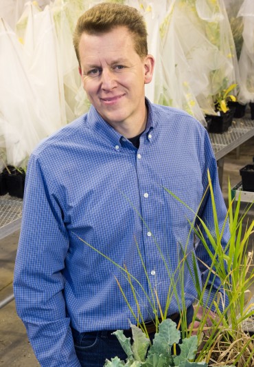
“We found that the origin of brand-new chemicals in the plant arose through gene duplications that encode novel functions rather than single mutations,” said Pat Edger, a former MU post doc and lead author on the study. “Given sufficient amounts of time the insects repeatedly developed counter defenses and adaptations to these new plant defenses.”
This back-and-forth pressure resulted in the evolution of many more species of plants and butterflies than in other groups without glucosinolate pressures.
Proving an old concept
Co-evolution is not a new idea.
About 50 years ago two now-renowned biologists, Peter Raven and Paul Erhlich, introduced the idea of co-evolution to science. Using cabbage butterflies and Brassica plants as a prime example, the two published a landmark study in 1964 advancing the idea that two species can mutually influence the development and evolution of each other.
“Using Ehrlich and Raven’s principles and models, we looked at the evolutionary histories of these plants and butterflies side-by-side and discovered that major advances in the chemical defenses of the plants were followed by butterflies evolving counter-tactics that allowed them to keep eating these plants,” Wheat said.
Chris Pires and colleagues mapped the evolution of Brassicales and butterflies to find how each evolved to combat the defenses of the other. | Courtesy Chris Pires
This research provides striking support for the ideas of Ehrlich and Raven published 50 years ago.
“We looked at the patterns 50 years ago, and found conclusions that proved correct,” said Peter Raven, professor emeritus of the Missouri Botanical Garden and a former University of Missouri Curator. “The wonderful array of molecular and other analytical tools applied now under leadership of people like Chris Pires, provide verification and new insights that couldn’t even have been imagined then.”
Understanding more about how plants and insects co-evolve could one day lead to advances in crops.
“If we can harness the power of genetics and determine what causes these copies of genes, we could produce plants that are more pest-resistant to insects that are co-evolving with them—it could open different avenues for creating plants and food that are more efficiently grown,” said Pires.
Proceedings of the National Academy of Sciences (PNAS) published the study, “The butterfly plant arms-race escalated by gene and genome duplications,” in June. The National Science Foundation (PGRP 1202793), the Knut and Alice Wallenberg Foundation and the Academy of Finland provided the funding for this research.
The HIV capsid protein (shown above in an array of hexagons) plays a critical role in the virus life cycle. Bond LSC researchers recently developed the most complete model yet of this vital protein. Image by Karen Kirby and Anna Gres
Seeing the whole picture can mean a lot when it comes to figuring out HIV.
Researchers at the University of Missouri Bond Life Sciences Center are gaining a clearer idea of what a key protein in HIV looks like, which will help explain the flexible protein’s vital role in the virus life cycle.
“The capsid acts as an invisibility cloak that hides the virus’ genetic information, the genome, while it is being copied in a hostile environment for the virus,” said Stefan Sarafianos, a virologist at Bond LSC and lead author of the study. “Fine-tuned capsid stability is critical for successful infection: too stable a capsid shell and the cargo is never delivered properly; not stable enough and the contents are detected by our immune defenses, triggering an antiviral response. Capsid stability is a key to the puzzle, and to solve it you have to understand its structure.”
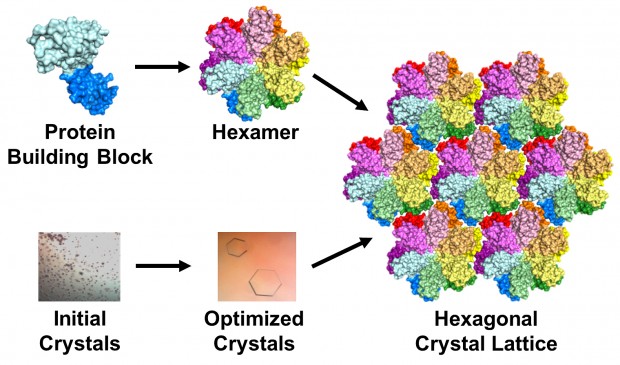
This is the most complete model yet of an HIV-1 capsid protein. In a virus, the protein combines in groups of five or six — called pentamers and hexamers, respectively — that assemble into a mosaic that forms the capsid shell. Roughly 1,500 copies of the protein, grouped into about 250 hexamers and 12 pentamers, comprise the capsid.
The protein building block of HIV capsid (top left) can assemble to form a hexamer (top middle). Crystals grown using this building block (bottom left and middle) contain an array or lattice of hexamers (right). | Image by Karen Kirby and Anna Gres
HIV, or human immunodeficiency virus, is the retrovirus that leads to AIDS — acquired immunodeficiency syndrome. Roughly 1.2 million people live with HIV in the United States, according to the Centers for Disease Control and Prevention. Globally, about 35 million people were living with HIV in 2013.
A lucky break
Over the years, scientists have employed various techniques and tricks to figure out the structure of the capsid protein. But until now, the clearest image had been made of a mutated version of the protein. It was a compromise: the mutation made the protein stable enough that the scientists could get a good snapshot, but they couldn’t see the detailed interactions between hexamers.
Sarafianos’ lab figured out how to get the full picture: a detailed image of the unmodified proteins that filled all the gaps between hexamers.
The team used a technique called X-ray crystallography to unravel the protein’s secrets. Basically, they took many copies of the protein and coaxed them into forming a patterned, crystalline lattice. Next they shot high-powered X-ray beams at the crystal. By interpreting how the X-rays scattered when they ricocheted off the proteins, the researchers made a 3-D map of the protein.
Karen Kirby
“But it doesn’t make sense until we make an atomic model of the protein to fit in that map,” said Karen Kirby, a research scientist at Bond LSC. “The map is just a grid that you can’t really interpret unless you put a model into it to see ‘Ok, it looks like this part is here, and that part is there, and this is how the protein is put together.’”
The researchers altered, tested and honed their 3-D model until it exactly matched the map produced by the X-ray diffraction pattern. This can be difficult and painstaking, but the researchers’ greatest challenge was creating the protein crystals in the first place: Scientists had been trying to crystallize the unmodified version of the HIV protein for decades without success.
To make a crystal, proteins are suspended in a liquid then slowly precipitated out, just like a “grow your own crystals” kit. But there are a lot of variables that control the process, from salts and additives in the liquid to the amount of protein in the mixture.
“It’s a very delicate balance to grow crystals,” Kirby said. “Many people call it more of an art than a science. It’s frustrating because you can never predict which solution will grow crystals. There are a large number of variables.”
Initially, most arrangements the researchers tried resulted in useless brown junk, Kirby said, caused by the proteins forming solids too quickly. Anna Gres, an MU chemistry grad student who led the project, used a crystallization robot to screen roughly 2,500 conditions.
That was the easy part, Gres said: “The real challenge begins afterwards, as one needs to manually optimize the initial crystallization conditions to find the one that will produce protein crystals of desired quality. This process can take years. In our case, I think we were lucky: It took approximately 500 manual screenings and about 6 month.” But the hard work paid off when she was finally able to produce lovely, hexagonal crystals. Surprisingly, the crystals formed in groups of six proteins, which matched their formation in the viral capsid.
The transition from tiny, useless particulate to invaluable crystals was tremendously exciting, Kirby said. But even to Kirby and Sarafianos, why their attempts succeeded when many others failed remains a little mysterious.
“I still don’t know what are the fine details that made the difference,” Sarafianos said.
“That’s the million dollar question,” said Kirby. “We really don’t have a good answer for that.”
Although solving the enigmatic crystal structure of the native full-length capsid protein was really rewarding, Gres said, she will continue to tinker with her technique: “I am still trying to optimize crystallization conditions, hoping to improve the quality of the crystals and diffraction.”
Water, water everywhere
Once the researchers got a good look at the interactions between hexamers, they were surprised by what they found.
Based on the genetic sequence of the protein, scientists speculated that they would be hydrophobic, or repel water. Instead, they found that “ordered” water molecules at specific sites played a crucial structural role by bridging interactions at the interface between hexamers.
“We thought, ‘How could these lowly waters really be of consequence?’” Sarafianos said. “But if you think about it, there’s 256 of these hexamers in the whole capsid and all kinds of interfaces among them: There’s thousands of water molecules that stabilize the whole structure. We hypothesize that this is an essential part of the stability of the whole capsid molecule.”
To test that hypothesis, they took the crystals, dehydrated them and checked to see if their shape changed. Although the protein lattices may look like sturdy crystals, they’re more like jello, Sarafianos said.
“The protein molecules are precariously touching each other and forming a lattice that is very, very sensitive. It’s held together in this case by water molecules in addition to other interactions.”
The change in shape suggested that water molecules are important in that they allow the capsid to assume different shapes. Moreover, Sarafianos said, the capsid’s malleability and plasticity could be critical to the life cycle of the virus and allow it to act as a multi-functional molecular Swiss army knife.
Onward with research
A clearer image of the capsid protein, could help Sarafianos’ lab gain a better understanding of how the body combats the virus and to discover new ways to disrupt the viral capsid.
“Now we have a system to study effects of capsid-targeting compounds with novel mechanism of action,” Gres said.
Working with a medicinal chemist, Sarafianos’ lab will undertake an iterative process of making compounds, solving their structures, testing them against HIV and then refining the molecules, with the ultimate aim of producing new and effective antiviral drugs.
Endocrine disruptors alter parent behavior in California mice
California mice exposed to bisphenol A (BPA) or ethinyl estradiol changed their parenting behavior, according to an MU Bond LSC study. | Photo by Roger Meissen, Bond LSC
By Roger Meissen | MU Bond Life Sciences Center
What if a chemical changes the way an animal parents?
That could happen due to endocrine disruptors like bisphenol A (BPA).
A recent study of California mice exposed to BPA showed parents spend less time feeding, grooming and interacting with their babies, according to University of Missouri research. Even mother mice not exposed to the chemical parented differently if their male partner was exposed during development.
Most studies only use laboratory mice and rats — where the mother is the sole parental provider — so how early contact to BPA may affect the father and his partner remained a critical gap in existing research.
Bond LSC researcher Cheryl Rosenfeld | Photo by Roger Meissen, Bond LSC
“The nature and extent of care received by an infant is important because it can affect social, emotional and cognitive development,” said Cheryl Rosenfeld, a researcher in MU’s Bond Life Sciences Center and associate professor of biomedical sciences in the College of Veterinary Medicine. “We found that females who were exposed early on to BPA spent less time nursing, so the pups likely did not receive the normal health benefits ascribed to nursing. Likewise, we found that developmental exposure of males and females resulted in them spending more time out of the nest and away from their pups, further suggesting that biparental care was reduced.”
BPA and other endocrine disrupting chemicals like ethinyl estradiol (EE) — found in birth control — concern scientists because they build up in the environment and mimic natural hormones produced by animals, including humans. Everyday exposure to these chemicals can impact offspring development and now have been found to alter adult behavior in test animals.
California mice have special significance for studying parental behavior. Unlike most lab mice, Californian mice pair up to mate and care for offspring. This monogamous behavior could give researchers insight into child rearing behavior found in most human societies and other biparental animals that would be impossible to measure in lab mice and rats.
MU graduate student and primary author Sarah Johnson worked with Rosenfeld to design the study to look at both sexes. Female and male mice were fed one of three diets — food supplemented with BPA or ethinyl estradiol or endocrine-free (control) food — two weeks before breeding. The mice were then randomly paired with the same mate for the entire study. The behavior of both sexes was then tracked for activities like time spent grooming pups, time spent in the nest and time mothers spent nursing.
But how do you measure the behavior of parents?
Rosenfeld’s team depended on hundreds of hours of video footage, taken at particular times of day and night for seven days, starting two days prior to birth. By using infrared cameras they tracked all 56 litters of mice, logging the number of and duration of activities mothers and fathers completed. During this time, the body weight and temperature of the F2 pups, who were not directly or fetally exposed to any chemicals, was logged to monitor their development.
While results showed reduced pup attention from BPA/EE exposed mother mice, the most intriguing result showed that unexposed moms mated with exposed fathers reduced the time they groomed and cared for offspring.
“These female mice have not been exposed here, but if you can see they are still reducing parental care when paired with the BPA/EE-exposed males,” Rosenfeld said. “And what’s even more interesting is that if a mother and father are both exposed, that parental care diminishes further, and becomes even more statistically significant.”
Researchers hope these results will spur others to look at long-term effects of endocrine disruptors on parenting behavior from generation to generation in animal models and, more importantly, in humans, to see if these chemicals can disrupt parental behavior of mothers and fathers, and if so, whether these effects can be transmitted to subsequent generations.
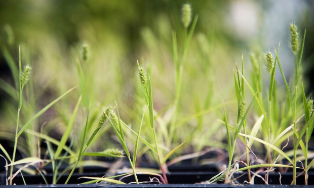
Bond LSC researchers showed for the first time ever that a grass, Setaria viridis, can receive 100 percent of its nitrogen needs from bacteria when associated with plant root surfaces. This grass will now serve as model for research into biological nitrogen fixation that could benefit crop development. | Photo by Roger Meissen, Bond LSC
By Roger Meissen | MU Bond Life Sciences Center
As farmers spend billions of dollars spreading nitrogen on their fields this spring, researchers at the University of Missouri are working toward less reliance on the fertilizer.
Less dependence on nitrogen could start with a simple type of grass, Setaria viridis, and its relationship with bacteria. The plant promises to lay groundwork for scientists exploring the relationship between crops and the fixing nitrogen bacteria that provide them the nitrogen amount plants need daily.
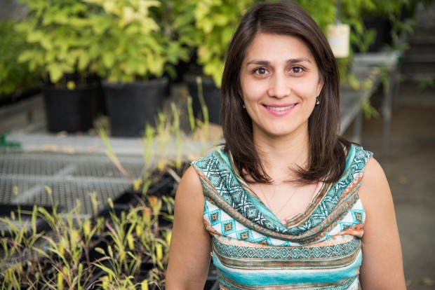
“In science sometimes you have to believe because we often work with such small microorganisms and DNA that you cannot see,” said Fernanda Amaral, coauthor and MU postdoctoral fellow at Bond Life Sciences Center. “Before this research no one had actually proved such evidence that nitrogen excreted by bacteria could be incorporated into plants like this.”
Fernanda Amaral, coauthor and MU postdoctoral fellow at Bond Life Sciences Center. | Photo by Roger Meissen, Bond LSC
Biological Nitrogen fixation — where diazotrophic bacteria fix atmospheric nitrogen and convert it to ammonium — provides a free way for plants to alter and absorb the nutrient. Farmers have long known that legumes like soybean fix nitrogen due to the symbiosis with bacteria in the soil through development of nodules on their roots, but since grasses like corn and rice don’t form this specialized structures that relationship has been trickier to explore.
Yet in fact, this team’s experiments showed the grass Setaria viridis received 100 percent of its nitrogen needs from the bacteria Azospirillum brasilense when associated with plant root surfaces.
“I believed in these bacteria’s ability, but I was really surprised that the amount of nitrogen fixed by the bacteria was 100 percent,” Amaral said. “That’s really cool, and that nitrogen can make so much of a difference in the plant.”
Worldwide farmers used more than 100 million tons of nitrogen on fields in 2011, according to the United Nations Food and Agriculture Organization. In the same year, the U.S. alone produced and imported more than $37 billion in nitrogen.
This grass can serve as a simple model for research, standing in for grass relatives such as corn, rice and sugarcane to explore a similar relationship in those crops. This research, “Robust biological nitrogen fixation in a model grass–bacterial association,” was published in the March 2015 issue of The Plant Journal.
A nutrient, a nuclear reactor and a model plant
Proving that this grass actually uses nitrogen excreted from the bacteria took some clever experiments, a global collaboration and a nuclear reactor.
MU researchers in the lab of Gary Stacey, a Bond LSC investigator, partnered with scientists in Brazil and at Brookhaven National Laboratory in New York to find a robust plant model system.
They screened more than 30 genotypes of Setaria viridis grass, looking for a strong nitrogen fixing response when colonized with three different bacteria strains. They germinated the seeds in Petri dishes and inoculated those three days after germination with a bacterial solution. Then plants were transplanted into soil containing no nutrients. By eliminating nitrogen in the soil, the scientists were able to make sure that the bacteria was the only source of nitrogen for plant.
The team settled on Azospirillum brasilense bacteria, which has been used commercially in South America to improve crop plant growth. It colonizes the surface of the roots and showed the greatest amount of plant growth when associated with plant roots.
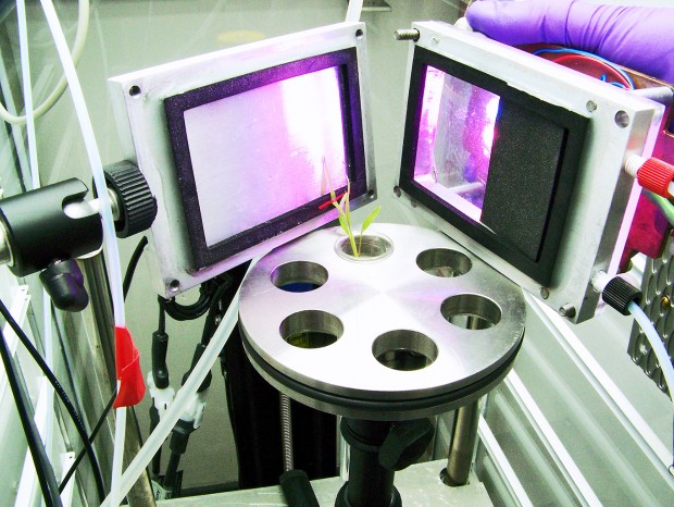
Proving that the bacteria truly fixed the nitrogen used by the plant, required exposing plants to radioactive isotopes at Brookhaven National Laboratory. That began with Nitrogen 13, an unstable radio isotope that showed exactly where and how quickly this nutrient was taken up from the bacteria.
A radio tracer chamber at Brookhaven National Laboratory was needed to test if Setaria viridis actually used nitrogen produced by the bacteria. The scientists allowed only one leaf to contact the radioactive nitrogen, so they could truly tell if it was being used. | Photo provided by Fernanda Amaral
“Nitrogen 13 is really sensitive matter with a half-life of less than 10 minutes, and we first thought there wouldn’t be that much nitrogen fixed by the plant,” Amaral said. “We administered Nitrogen 13 only on the roots, quickly scanned the samples and calculated how much of the nitrogen the plants assimilated based on the decay analysis of the tracer.”
This experiment, paired with several others, showed that this model grass truly incorporated the nitrogen released by the bacteria and metabolizes it in several components.
Model (plant) citizen
But why does a type of grass that doesn’t produce food matter so much?
The answer is time and simplicity.
“Corn is really good at responding to bacterial inoculation, but it’s very big and takes a long time to produce seeds and also the genome is complex,” said Beverly Agtuca, an MU Ph.D. student who worked on the study. “Setaria viridis is a small plant that can produce a lot of seeds faster, has a pretty simple genome and can serve as a model for research.”
That makes it perfect to explore how the plant actually uses its bacterial partners, and labs around the world are already using this plant model for research.
For the Stacey lab, the next step is to pinpoint the gene in the model grass that makes this possible.
“We want to identify the genes responsible for the interaction between plant and bacteria and meanly the ones involved with the nitrogen uptake,” Fernanda said. “We hope that will allow us to improve plant growth based on the gene to further study.” We believe that our findings can stimulate others studies at this area, which seems to be a promise plant friendly way to apply for promoting a sustainable agriculture, especially to crop systems including bioenergy grass.
Amaral and Agtuca work in the lab of Gary Stacey at Bond LSC. Stacey is a Bond LSC investigator and a Curators Professor of Plant Sciences in the College of Agriculture, Food and Natural Resources at the University of Missouri. Collaborators included researchers at Brookhaven National Laboratory, State University of New York, Federal University of Paraná in Brazil and Federal University of Santa Catarina in Brazil.
Funding for this project came from the National Institute of Science and Technology- Biological Nitrogen Fixation, INCT-FBN, the Brazilian Research Council, Ciência Sem Fronteiras Program, The Department of Energy and SUNY School of Environmental Science and Forestry Honors Internship Program.
An MU student uses his cell phone while in Costa Rica. | Photo by Jack Schultz, Bond LSC
By Jack Schultz | Director of MU Bond Life Sciences Center
“Fieldwork” means many things to researchers, but in the past it often meant working without easy access to communication.
Now cell phones allow my students visiting the La Selva Biological Station in the lowland rainforest of Costa Rica to remain connected.
While our science and journalism majors learn to report on biological research, I find that I can be replaced. As an experienced biologist who has taught and worked in the Costa Rican tropics for some time, I normally serve as a biology resource. After all, our journalism students have little or no science background.
Yet, as students interview scientists working in a rainforest, learn about the forest’s biology and write about it daily, they now can go online to find the answer. Everything from ecological theory to species lists for our forest site are accessible to any student with a WiFi connection. Fortunately, the biological station has good WiFi service.
While I need to prompt searches to help students know what to look for, the answer to “what was that animal?” is just a hyperlink away. I’m carrying a bulky field guide to the birds, but most often find myself online, checking my own recollection of animals, plants, and facts and figures.
Students return from the forest with evidence of what they’ve seen, which is much better than a hand-waving verbal description. Group meals are eaten with one hand on the phone and the other on a fork. The day’s plans can be refined at breakfast by checking the weather forecast for our rainforest site.
Any good journalist acquires as much background as possible before an interview. Our students can do that in short order by visiting websites of the people we meet in the field. Over several days, they can refine their knowledge and questions to get the most from conversations with researchers. When a term or concept arises in interviews, clarification is right there on the phone.
Cell phone use goes well beyond fact checking.
Paper maps melt in the rain, but the students took photos of the maps we were given and use their cell phones to find their way on the forest trails. Many actually take notes on their phones, and some compose essays there. The improving quality of cell phone cameras produces excellent pictures to post with blogs and articles. Some of the students are producing photos that rival the quality of photos I take with my bulky DSLR. And the videos they produce are high quality and easy to edit.
While computers and tablets are the instruments of choice for uploading larger essays, cool observations can go direct from a cell phone to Twitter, Instagram or even Facebook. And posting to personal Facebook pages keep family and friends updated on each day’s adventures. Everyone in our group is in close contact with home, even if home is in Saudi Arabia (in one case).
While I will admit to feeling, at first, that cell phones could ruin the fieldwork experience, my perspective has changed to value it as a professional tool and not just a personal toy.
Now I’ll be in line for a cell phone upgrade when I return home.
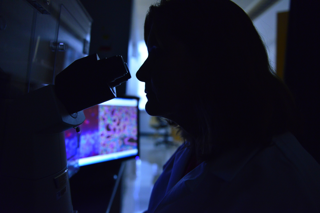
White coat, dark room. Jean Camden, a senior technician in the Weisman lab, reviews salivary gland and brain tissue samples for research on inflammation. | Photo by Paige Blankenbuehler, Bond LSC
By Paige Blankenbuehler | MU Bond Life Sciences Center
There’s a criminal on the loose, striking every day. Millions fall victim, but there’s still no way to stop it. And, in all likelihood, you have been hurt by it.
If inflammation is an unsolved criminal case of the last three decades, then Gary Weisman has been the detective. He’s certain there’s an accomplice — perhaps many — that may be triggering the discomfort.
The Bond Life Sciences Center investigator is slowly revealing what makes inflammation tick and what makes it strike. Each epiphany brings another question. He’s certain there’s a way to prevent negative effects of unsolved inflammation.
Bond LSC investigator and MU professor of biochemistry, has been studying the ins-and-outs of inflammation for the last 30 years. | Photo by Paige Blankenbuehler, Bond LSC
Weisman has dedicated his career to understanding the micro-processes behind inflammation. He’s become so specialized that his techniques can be as hard to crack as the case itself.
“I would not ask anyone to explain what I do,” Weisman says. Nonetheless, he’s been able to divide the process of inflammation into two categories: components that repair the body and components that lead to its destruction. This will help find inflammation’s many accomplices to figure out why humans work, and what their bodies do when they don’t work so well.
“I am interested in the meaning of life,” Weisman says. “Life has become simpler for me because the scientific method carries everywhere. I’ve become aware of how simple we are as a machine.”
Criminal or just misunderstood?
Most criminals adopt patterns, but inflammation stands as a signpost for mysterious, underlying problems.
Its effects are usually localized: an arm, a joint, the brain or a gland. You feel a temperature spike then the skin reddens in a part of your body. Later still, the skin tightens and pain comes at a snail’s pace.
Not even cells are safe. Inflammation even strikes on the molecular level.
But really, inflammation can be a good thing. It’s part of the immune system’s bag of tricks to signal the body to bring in reinforcements to fight off the invasion. Normally, inflammation corrects a physical problem, but if it is not successful in repairing a problem, inflammation can become chronic and accelerate tissue destruction.
Just like in an episode of CSI, Weisman puts the pieces of the inflammation puzzle together in his office by applying the expertise of Laurie Erb, Jean Camden and Lucas Woods — all donned in white lab coats, eyes pressed to the microscope examining evidence and building molecular evidence in the case.
The MU associate professor of biochemistry and his team have become a sort of grant-wielding wizards to sustain his pursuit of inflammation triggers. National Institutes of Health grant awards have sustained his lab for decades. The funding has come from varied sources such as the MU Food for the 21st Century Program, the Bond LSC, the Bright Focus Foundation, the American Heart Association and the Cystic Fibrosis Foundation. In recent years, research funding for Alzheimer’s disease and Sjogren’s syndrome (a disease of the salivary gland that causes dryness) have contributed, too.
But the funding source doesn’t matter because inflammation is the tie that binds.
Jean Camden processes samples under the Weisman lab’s microscope. | Photo by Paige Blankenbuehler, Bond LSC
Advancements, like recent mapping of the human genome, have moved his work forward to understand inflammation’s complexity. Each experiment he completes fills in another blank slate in the “human owner’s manual.”
“As humans, we’re so intent on the fact that we’re superior to all, but really we’re not,” Weisman says. “With the Human Genome project, we’ve come to understand that all living things have similar designs … we are on the verge of finding revolutionary solutions to preventing or reversing human diseases.”
A receptor all our own
One specific player in the body’s immune system has kept Weisman’s attention for most of his career. The P2Y2 protein is a nucleotide receptor, and his lab team members affectionately refer to it as “our receptor.”
Nucleotide receptors are regulatory molecules in red blood cells. What they regulate is nuanced, mostly undetermined and of great interest to scientists. Answering that question has become Weisman’s wheelhouse.
The body manufactures 15 different types of nucleotide receptors, all similar in construction, but each are believed to have subtly different functional roles. It’s as if Weisman and his lab is on the case of a highly organized crime ring.
“Our receptor is mainly present when inflammation occurs, and we’re trying to figure out its role in a variety of diseases,” Weisman says.
The P2Y2 receptor has been observed in Alzheimer’s patients, along with a plaque build-up in the brain, and the receptor was suspected of playing a role in the disease’s progression.
Weisman and his colleagues found that the deletion of the P2Y2 receptor in a mouse model of Alzheimer’s disease accelerates progression of plaque build-up, neurological symptoms and death. This suggests that the receptor has anti-inflammatory effects rather than being “guilty by association” with the tissue-destructive aspects of inflammation.
“It’s like I have this 30,000-piece jigsaw puzzle in front of me that I have to put together,” Weisman says. “What’s the difference between you and me? As a machine, surprisingly very little.”
This simplicity drives Weisman to continue solving the mysteries of inflammation and search for its underlying chemical processes. By understanding the body’s chemical reactions, he believes treatments can be developed to focus the immune system on repairing damaged tissues.
Through studying his receptor, Weisman is breaking up inflammation’s crime ring.
“You’ve probably never really seen a fat plant before, right?” said Salie, a fourth year MU graduate student in biochemistry. “Humans, we make plenty of extra fat and store that as energy. But plants don’t really need to do that — they make just as much as they need, and that’s about it.”
Salie studies plant metabolism with Bond LSC researcher Jay Thelen, an associate professor of biochemistry. He’s one of 25 winners honored for research presented during Missouri Life Sciences Week 2015.
The Thelen lab looks for ways to increase the amount of vegetable oil that crops such as corn and soybean can produce. Salie focused on an enzyme that is the first step in the pathway to producing fatty acid in plants.
The idea was that if he could reduce metabolic limits at the beginning of the process, then the downstream production of oil would increase.
“I found these new proteins that no one has ever really studied before,” Salie said. “As I started to look into them over the last year or two, it turns out that they actually seem to incorporate themselves into the enzyme and slow down it’s activity.”
Four separate proteins normally combine to form the functional enzyme, but the new proteins Salie identified mimic those components and can take their place, like a cuckoo bird replacing another species’ eggs with its own. The more mimics that replace proteins, the fewer functional enzymes the plant produces, which means less oil.
It’s a simple, nuanced way for the plant to fine-tune the production of fatty acids.
“Instead of being an on-off switch, it’s more like a thermostat,” Salie said. And if he can adjust that thermostat in a plant, it should start packing on the pounds.
Although Salies work was only recently submitted for publication, it’s already receiving recognition. His poster, “The BADC proteins — a novel paradigm for regulation of de novo fatty acid synthesis in plants,” won first place in the Molecular and Cellular Biology category during the Life Sciences Week poster competition in April.
Salie relished the opportunity to share his findings with researchers and non-scientists alike.
“It’s a great experience, because it helps you realize what’s really important about the work that your doing,” he said. “It also really encourages you to work harder. It’s like, ‘Wow, this is actually meaningful stuff!’ which can be hard to see when you’re working 60 or 70 hour weeks at the lab, just sitting there by yourself.”
Salie was among more than 300 students who presented their research during the 31st annual Life Sciences Week poster sessions.
The winners in each of the five categories are:
Molecular and Cellular Biology
Matthew Salie, Matthew Muller, Stephanie Bowers
Organismal Biology
Miqdad Dhariwala, Ryan Sheldon, Carine Collins
Genetics, Evolution and Environment
Julianna Jenkins, Nathan Harness, and a tie for third between Sharon Kuo and Susheel Bhanu Busi
Life Science and Biomedical Engineering Technologies and Informatics
Jamie Hibbard, Hang Xu, Brittany Hagenhoff
Social and Behavioral Sciences
Vaness Cox and Ian George tied for first place
Undergraduate winners are Vincent Farinella, James Mrkvicka, Anette van Swaay, Romanus Hutchins, Dallas Pineda, Kelsey Boschert, Anthony Onuzuruike, Clare Diester, Adam Kidwell and Sean Rogers.
Honorable mention:
Social and Behavioral Sciences
Undergrad Honorable Mention – Kelsey Clark
Undergrad Honorable Mention – Louie Markovits
Genetics, Evolution, and Environment
Grad Honorable Mentions: Megan Murphy (Schul) and Amanda Smolinsky (Holliday)
Undergrad Honorable mention: Anthony Spates (Holliday)
Organismal Biology
Grad Honorable Mention: Kathleen Pennington
Grad Honorable Mention: Kasun Kodippili
Grad Honorable Mention: Christopher Tracy
Undergrad Honorable mention: Chelsie Todd
Undergrad Honorable mention: Holly Doerr
Undergrad Honorable mention: Zeina Zeida
Molecular and Cellular Biology
Grad Honorable mention, Khalid Alam [Burke lab]
Grad Honorable mention, Zhe Li [Sarafianos lab]
Undergrad Honorable mention: Vincent Markovitz [Guo lab]
Additional prizes were awarded for communication prowess and poster design chops.
For photos of some of this year’s winner, check out this Flickr album
The environmental build-up of bisphenol A (BPA) can result in a life-changing shift for aquatic animals.
For painted turtles, exposure to this chemical can disrupt sexual differentiation, according to new research in General and Comparative Endocrinology.
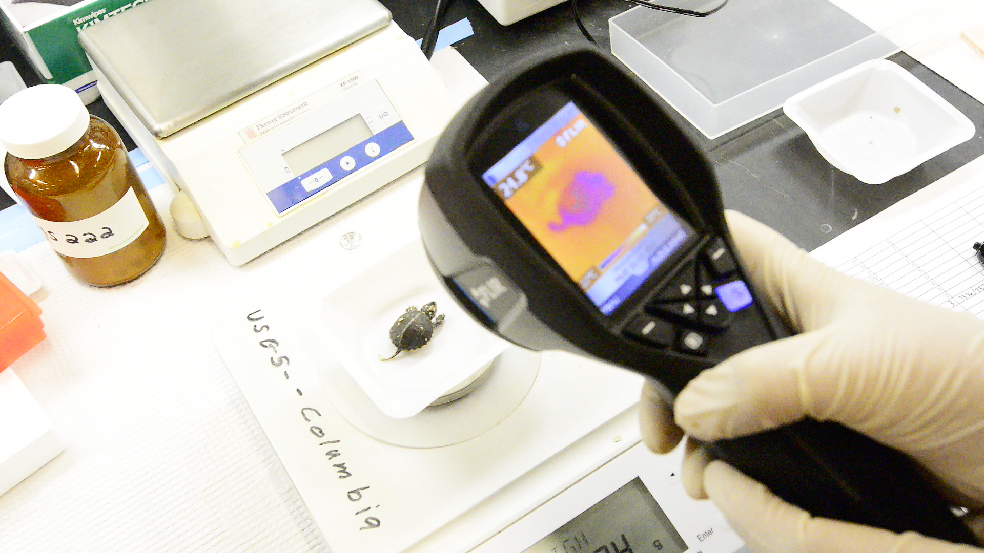
Scientists at the University of Missouri have teamed up to show how low levels of certain endocrine disruptors like BPA can cause males to possess female gonadal structures in newly-hatched turtles. This collaboration between MU, Westminster College, the U.S. Geological Survey (USGS) and the Saint Louis Zoo exposed turtle eggs to levels of BPA similar to those currently found in the environment.
“It’s important because this is one of the first times we’ve seen low doses of BPA causing disorganization or reorganization of the male gonad to resemble females,” said Dawn Holliday, adjunct assistant professor of pathology & anatomical sciences at MU’s School of Medicine and assistant professor of biology at Westminster College. “We’re not sure what this means in terms of population-level effects, but certainly it can cause some reproductive dysfunction for turtles.”
Endocrine disruptors leach into rivers and streams and concern scientists because of potential effects on animals and humans. While BPA is used as a hardening agent in plastics, it also is used to line cans and in manufacturing where more than 15 billion tons are produced each year.
In the case of painted turtles, these chemicals have potential to alter sex ratios, which are normally regulated by temperature during incubation. Eggs exposed to cooler temperatures normally produce males and those hatched at warmer temperatures produce females.
In this experiment, turtle eggs were incubated at temperatures known to rear males and dosed with low, medium and high levels of BPA. BPA-exposed turtles were compared to hatchlings not exposed to chemicals as well as a group exposed to high levels of ethinyl estradiol — an endocrine disruptor found in birth control — at the USGS Columbia Environmental Research Center.
These doses resulted in turtle sex organs that should have been male , but abnormally contained female gonadal elements. The low dose represented BPA concentrations found in fields where turtles can nest while the mid and high doses approximate BPA levels near contaminated sites like landfills.
“We exposed the eggs for a limited amount of time right when they were most vulnerable to the effects,” said Cheryl Rosenfeld, a researcher at MU’s Bond Life Sciences Center and an associate professor of biomedical sciences in the College of Veterinary Medicine. “We found that we got partial feminization in more than 30 percent of turtle eggs exposed to BPA despite being incubated at male-permissive temperatures.”
These results give the team a look into what real-world exposure levels might mean in the wild and a starting point for comparison.
“Turtles are the most endangered vertebrate taxa and they have all sorts of conservation issues coming at them from people harvesting them to disease, and endocrine disruptors are another potentially big whammy they have against their conservation status,” said Sharon Deem, director of the Saint Louis Zoo’s Institute for Conservation Medicine. “This research is a stepping stone, and we are hoping we can apply these results to populations of turtles throughout the state and use these results as a marker to look at endocrine disruptors in the wild.”
Future studies plan to look at the underlying mechanisms behind sexual disruption and will extend the study to animals including fish and mammals. Rosenfeld’s laboratory is in the process of examining how early exposure of turtles to endocrine disruptors might affect cognitive behaviors, including spatial navigation ability.
Fred vom Saal, Curators Professor of Biological Sciences in the College of Arts and Science at MU, Don Tillitt, an adjunct professor of biological sciences at MU and a research toxicologist with the USGS, Ramji Bhandari, an assistant research professor of biological sciences and a visiting scientist with the USGS at MU and Caitlin Jandegian, a senior research technician at MU, all collaborated on the study.
Funding was provided by Mizzou Advantage, an MU initiative that fosters interdisciplinary collaboration among faculty, staff, students and external partners to solve real-world problems in four areas of strength identified at the University of Missouri. These areas include Food for the Future, Sustainable Energy, Media for the Future and One Health/One Medicine.