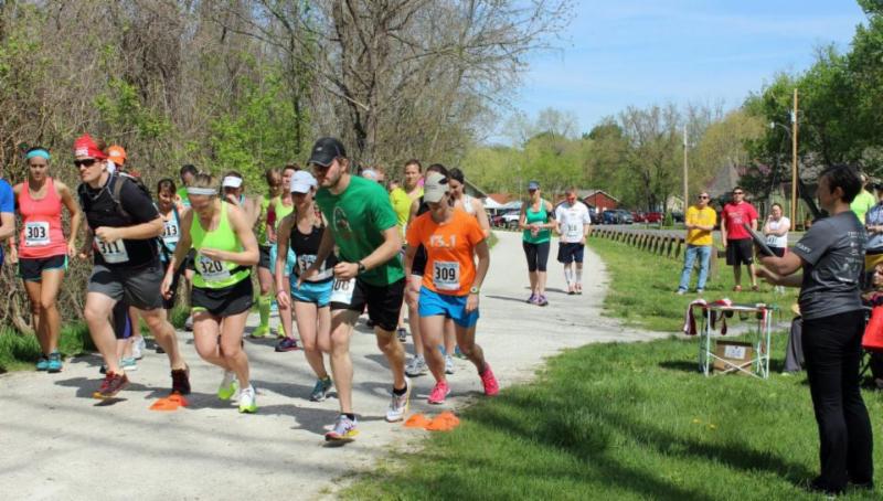
Over the weekend, Bond LSC HIV researchers Stefan Sarafianos, Marc Johnson and Donald Burke-Aguero joined Trail to a Cure, Inc., a Columbia nonprofit organization that helped fund important HIV research.
Since 2008, the organization has raised $74,000 for HIV/AIDS research, with some of that funding going directly to the Bond LSC providing additional hours of lab research. The 2014 online fundraising is still open and donations can be made to Trail to a Cure, Inc. until the end of the month.
The funding from Trail to a Cure helps Bond LSC researchers train future scientists and physicians in labs and in some cases, training them on the development of next generation therapies, Johnson said.
Bond LSC HIV researchers Stefan Sarafianos (left), Marc Johnson (center), and Donald Burke-Aguero (right) at the 7th annual Trail to a Cure along Katy Trail in Rocheport on May 3. | Credit: Trail to a Cure, Inc..Trail to a Cure, Inc., a Columbia-based nonprofit, has raised $74,000 for HIV/AIDS since 2008. Part of the raised funds go to HIV research in the LSC, providing additional hours of lab research. | Credit: Trail to a Cure, Inc.
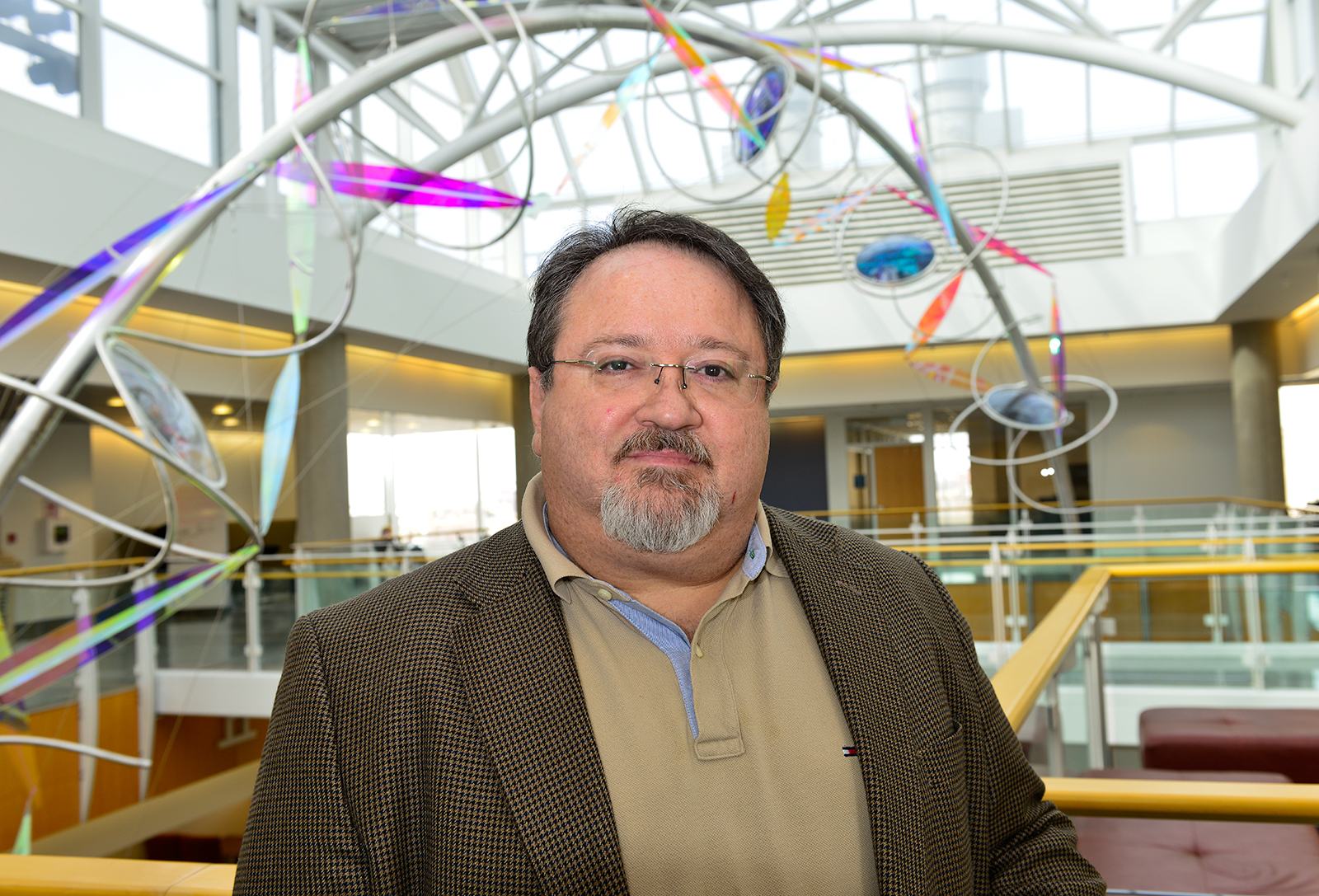
Bond LSC researcher Stefan Sarafianos stands in the LSC atrium. The virologist is an associate professor of molecular microbiology and immunology and Chancellor’s Chair of Excellence in Molecular Virology with appointments in MU’s School of Medicine and the Department of Biochemistry.
Resistance is the price of success when it comes to treating HIV.
Virologists at the Bond Life Sciences Center are helping to test the next generation of anti-AIDS medication to quell that resistance.
Stefan Sarafianos’ lab recently proved that EFdA, a compound that stops HIV from spreading, is 70 times more potent against some HIV that resists Tenofovir – one of the most used HIV drugs.
“HIV in patients treated with Tenofovir eventually develop a K65R RT mutation that causes a failure of this first line of defense,” said Sarafianos, virologist at Bond LSC. “Not only does EFdA work on resistant HIV, but it works 10 times better than on wild-type HIV that hasn’t become Tenofovir resistant.”
Sarafianos and a team of researchers found that EFdA (4′-ethynyl-2-fluoro-2′-deoxyadenosine) is activated by cells more readily and isn’t broken down by the liver and kidneys as quickly as similar existing drugs.
“These two reasons make it more potent than other drugs, and so our task is to look at the structural features that make it such a fantastic drug,” he said.
From soy sauce to virus killer
The path from EFdA’s discovery to current research is a bit unorthodox.
A Japanese soy sauce company named Yamasa patented this molecule, which falls into a family of compounds called nucleoside analogues that are very similar to existing drugs for HIV and other viruses. EFdA was designed and synthesized by Hiroshi Ohrui (Chem Rec. 2006; 6 (3), 133-143; Org. Lett. 2011; 13, 5264) and shown by Hiroaki Mitsuya, Eiichi Kodama, and Yamasa to have potential usefulness against HIV. Samples sent for further testing confirmed EFdA’s potential usefulness against HIV. This started more than a decade of research to pinpoint what makes the compound special.
EFdA joins a class of compounds called nucleoside reverse transcriptase inhibitors (NRTIs) that includes eight existing HIV drugs. Like all NRTIs, EFdA hijacks the process HIV uses to spread by tricking an enzyme called reverse transcriptase (RT). RT helps build new DNA from the RNA in HIV, assembling nucleoside building blocks into a chain. Since EFdA looks like those building blocks, RT is tricked into using the imposter. When this happens the virus’ code cannot be added to the DNA of white blood cells it attacks.
“NRTIs are called chain terminators because they stop the copying of the DNA chain, and once incorporated it’s like a dead end,” Sarafianos said.
A little help from some friends
Sarafianos isn’t alone in studying EFdA.
The virologist’s lab works closely with University of Pittsburgh biochemist Michael Parniak and the National Institutes of Health’s Hiroaki Mitsuya to explore the molecule’s potential. Mitsuya had a hand in discovering the first three drugs to treat HIV and Parniak has spent years evaluating HIV treatments using cultured white blood cells.
Sarafianos’ focus requires him to take a very close look at EFdA to define how it works on a molecular level. He uses virology, crystallography and nuclear magnetic resonance to piece together the exact structure, bonding angles and configuration of the compound.
By looking at subtle differences in EFdA’s sugar-like ring, his lab identified the best structure that looks the most like actual nucleosides, doesn’t break down easily and is activated readily by CD4+ T lymphocyte white blood cells.
“The structure of this compound is very important because it’s a lock and key kind of mechanism that can be recognized by the target,” Sarafianos said. “We’re looking at small changes and the ideal scenario is a compound bound very efficiently by the target and activating enzyme but not efficiently by the degrading enzymes.”
Treatment for the future
The research of Sarafianos, Parniak and Mitsuya continue to uncover the magic of EFdA. In 2012, they showed that the drug worked incredibly well to treat the HIV equivalent in monkeys.
“These animals were so lethargic, so ill, that they were scheduled to be euthanized when EFdA was administered,” said Parniak. “Within a month they were bouncing around in their cages, looking very happy and their virus load dropped to undetectable levels. That shows you the activity of the molecule; it’s so active that resistance doesn’t come in as much of a factor with it.”
HIV prevention is the newest focus in their collaboration.
By recruiting formulation expert Lisa Rohan at the University of Pittsburgh, they are now putting EFdA in a vaginal film with a consistency similar to Listerine breath strips.
“The only way we are going to make a difference with HIV is prevention,” Parniak said. “If we can prevent transmission, this approach could make a huge difference in minimizing the continued spread of the disease when combined with existing therapies for people already infected.”
While AIDS in the U.S. occurs mostly in men, the opposite is true in sub-Saharan Africa where more than 70 percent of HIV cases occur. Since a film has a better shelf life than creams or gels, it could benefit those at risk in extreme climates and third-world countries.
“We have nearly 30 drugs approved for treating HIV infected individuals, but only one approved for prevention,” Sarafianos said. “Women in Africa would benefit from a formulation like this as a means to protect themselves.”
Despite this success, Sarafianos and Parniak aren’t slowing down in figuring out how EFdA works so well.
“We want to understand how long EFdA stays in the bloodstream and cells,” Parniak said. “If we understand structurally why this drug is so potent it allows us to maybe develop additional molecules equally potent, and a combination of those molecules could be a blockbuster.”
Grants from the National Institutes of Health fund this research.
In 2013 and 2014, the journals Retrovirology, Antimicrobial Agents and Chemotherapy and The International Journal of Pharmaceutics published this group’s work on EFdA. Sarafianos is an associate professor of molecular microbiology and immunology and Chancellor’s Chair of Excellence in molecular virology with MU’s School of Medicine and a joint associate professor of biochemistry in the MU College of Agriculture, Food and Natural Resources.
Powdery mildew on a cabernet sauvignon grapevine leaf. | USDA Grape genetics publications and research
A princess kisses a frog and it turns into a prince, but when a scientist uses a frog to find out more information about a grapevine disease, it turns into the perfect tool narrowing in on the cause of crop loss of Vitis vinifera, the world’s favorite connoisseur wine-producing varietal.
MU researchers recently published a study that uncovered a specific gene in the Vitis vinifera varietal Cabernet Sauvingon, that contributes to its susceptibility to a widespread plant disease, powdery mildew. They studied the biological role of the gene by “incubating” it in unfertilized frog eggs.
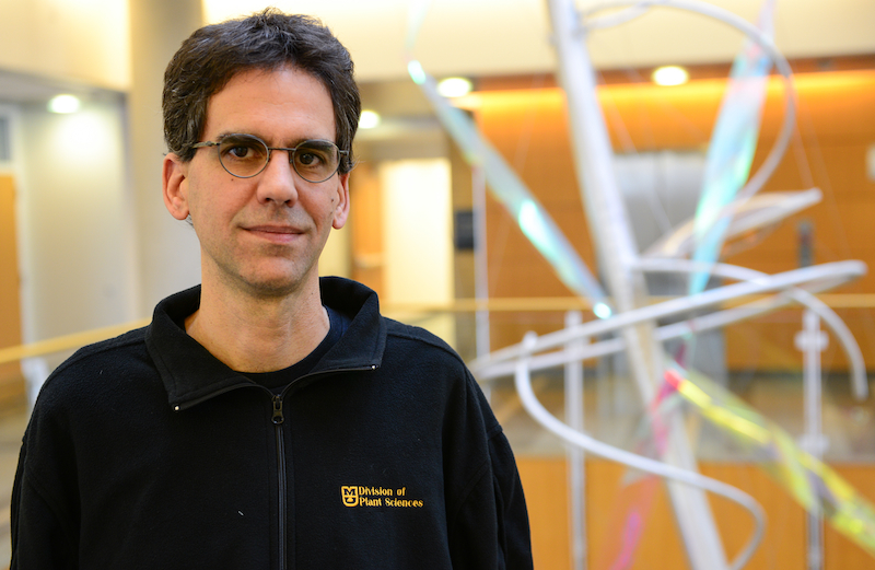
The study, funded by USDA National Institute of Food and Agriculture grants, was lead by Walter Gassmann, an investigator at the Bond Life Sciences Center and University of Missouri professor in the division of plant sciences.
The findings show one way that Vitis vinifera is genetically unable to combat the pathogen that causes powdery mildew.
Gassmann said isolating the genes that determine susceptibility could lead to developing immunities for different varietals and other crop plants and contribute to general scientific knowledge of grapevine, which has not been studied on the molecular level to the extent of many other plants.
The grapevine genome is largely unknown.
“Not much is known about the way grapevine supports the growth of the powdery mildew disease, but what we’ve provided is a reasonable hypothesis for what’s going on here and why Cabernet Sauvingon could be susceptible to this pathogen,” Gassmann said.
The research opens the door for discussion on genetically modifying grapevine varietals.
Theoretically, Gassmann said, the grapevine could be modified to prevent susceptibility and would keep the character of the wine intact — a benefit of genetic modification over crossbreeding, which increases immunity over a lengthy process but can diminish character and affect taste of the wine.
Grapevine under attack
Gassmann’s recent research found a link between nitrate transporters and susceptibility through a genetic process going on in grapevine infected with the powdery mildew disease.
Infected grapevine expressed an upregulation of a gene that encodes a nitrate transporter, a protein that regulates the makes it possible for the protein to enter the plant cell.
Once the pathogen is attracted to this varietal of grapevine, it tricks grapevine into providing nutrients, allowing the mildew to grow and devastate the plant.
As leaves mature, they go through a transition where they’re no longer taking a lot of nutrients for themselves. Instead, they become “sources” and send nutrients to new “sink” leaves and tissues. The exchange enables plants to grow.
The powdery mildew pathogen, which requires a living host, tricks the grapevine into using its nutrient transfer against itself. Leaves turn into a “sink” for the pathogens, and nutrients that would have gone to new leaves, go instead, to the pathogen, Gassmann said.
“We think that what this fungus has to do is make this leaf a sink for nitrate so that nitrate goes to the pathogen instead of going to the rest of the plant,” Gassmann said.
Walter Gassmann, of the Bond Life Sciences Center at the University of Missouri was the lead investogator on the research. Much of his work has been on grapevine susceptibility to pathogens. | Roger Meissen, Bond Life Sciences Center
According to a report by the USDA, powdery mildew can cause “major yield losses if infection occurs early in the crop cycle and conditions remain favorable for development.”
Powdery mildew appears as white to pale gray “fuzzy” blotches on the upper surfaces of leaves and thrives in “cool, humid and semiarid areas,” according to the report.
Gassmann said powdery mildew affects grapevine leaves, stems and berries and contributes to significant crop loss of the Vitas vinifera, which is cultivated for most commercial wine varietals.
“The leaves that are attacked lose their chlorophyll and they can’t produce much sugar,” Gassmann said. “Plus the grape berries get infected directly, so quality and yield are reduced in multiple ways.”
Pinpointing a cause
Solutions to problems start with finding the reason why something is happening, so Gassmann and his team looked at a list of genes activated by the pathogen to find transporters that allowed compounds like peptides, amino acids, and nitrate to pass.
Genes for nitrate transporters, Gassmann said, pointed to a cause for vulnerability to the mildew pathogen.
Over-fertilization of nitrate increases the severity of mildew in many crop plants, according to previous studies sited in Gassmann’s article in the journal of Plant Cell Physiology.
The testing system for isolating and analyzing the genes began with female frogs.
Gassmann used frog oocytes (unfertilized eggs), to verify the similar functions of nitrate transporters in Arabidopsis thaliana, a plant used as a baseline for comparison.
A nitrate transporter, he hypothesized, would increase the grapevine’s susceptibility to mildew.
“The genes that were upregulated in grapevine showed similarity to genes in Arabidopsis that are known to transport nitrate,” Gassmann said. “We felt the first thing we had to do was verify that what we have in grapevine actually does that.”
The eggs are very large relative to other testing systems and act as “an incubating system” for developing a protein. Gassmann and his team of researchers injected the oocyte with RNA, a messenger molecule that contains the information from a gene to produce a protein. The egg thinks it’s being fertilized and protein reproduces and is studied.
“The oocyte is like a machine to crank out protein,” Gassmann said. “We use that technique to establish what we have is actually a nitrate transporter.”
The system confirmed that the gene isolated from grapevine encodes a nitrate transporter.
“We contributed to the general knowledge of the nitrate transporter family,” Gassmann said. “It turned out to be the first member of one branch of nitrate transporters that, even in Arabidopsis haven’t been characterized before.”
The mounting knowledge of Vitis vinifera genes could make genetically modifying the strain to prevent the susceptibility easier.
“Resistance is determined sometimes by a single gene,” Gassmann said. “Until people are willing to have the conversation of genetic modification, the only way to save your grapevines is to be spraying a lot.”
Sharon Pike, Gassmann, other investigators from the MU Christopher S. Bond Life Sciences Center and post-doctoral student, Min Jung Kim from Daniel Schachtman’s lab at the Donald Danforth Plant Science Center in Saint Louis, Mo. contributed to the report.
The article was accepted November 2013 into the Plant Cell Physiology journal.
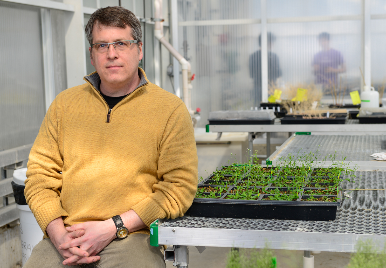
Scott Peck, Bond LSC scientist and associate professor of biochemistry, studies Arabidopsis and how bacteria perceive it before initiating an infection. Roger Meissen/ Bond LSC
Sometimes plants inadvertently roll out the red carpet for bacteria.
Researchers at the University of Missouri Bond Life Sciences Center recently discovered how a plant’s own chemicals act as a beacon to bacteria, triggering an infection. Proceedings of the National Academy of Sciencespublished their study April 21.
“When bacteria recognize these plant chemicals it builds a needle-like syringe that injects 20-30 proteins into its host, shutting down the plant’s immune system,” said Scott Peck, Bond LSC plant scientist and lead investigator on the study. “Without a proper defense response, bacteria can grow and continue to infect the plant. It looks like these chemical signals play a very large role in mediating these initial steps of infection.”
The question of how bacteria actually know they are in the presence of a plant has puzzled scientists for years. Being able to identify the difference between a plant cell and, say, a rock or a piece of dirt, means the bacteria saves energy by only turning on its infection machinery when near a plant cell.
“Our results show the bacteria needs to see both a sugar – which plants produce quite a bit of from photosynthesis – and five particular acids at the same time,” Peck said. “It’s sort of a fail-safe mechanism to be sure it’s around a host before it turns on this infection apparatus.”
Peck’s work started with one mutant plant called Arabidopsis mkp1.
Discovered several years ago by Peck’s lab, this little mustard plant acts differently than others by rebuffing the advances of bacteria. Lab tests confirmed that this mutant didn’t get infected by Pseudomonas syringae pv. tomato DC3000, a bacterial pathogen that causes brown spots on tomatoes and hurts the model plant Arabidopsis. Along with MU biochemistry research scientist Jeffrey Anderson and post doc Ying Wan, they showed that this mutant didn’t trigger the bacteria’s Type III Secretion System, the needle-like syringe and associated proteins that lead to infection.
Pacific Northwest National Laboratory (PNNL) worked with Peck’s team to compare levels of metabolites between the mutant Arabidopsis and normal plants. This comparison helped Peck identify a few of these chemicals – created from regular plant processes – that existed in much lower levels in their special little mutant.
Using the PNNL work as a guide, the team found five acids collectively had the biggest effect in turning on a bacteria’s infection: aspartic, citric, pyroglutamic, 4-hydrobenzoic and shikimic acid.
“The key experiment involved us simply adding these acids back into the mutant,” Peck said. “Suddenly we saw the mutant plant wasn’t resistant anymore and the bacteria were once again capable of injecting proteins to turn off the plant’s immune system.”
First contact and recognition means all the difference, whether bacteria or plant. Just a slight jump out of the starting blocks by one or the other could change who will win a battle of health or infection.
While low concentrations of these five acids trigger the bacteria’s attack, high levels blind it to the plant’s presence, leading Peck to believe it could be used to hinder bacterial growth. If this actually thwarts the bacteria’s head start, it could mean stopping disease in crops and could lead to a different approach in the field.
“A lot of the winning and losing occurs within the first 2-6 hours and it seems to be that if the microbe is too slow to turn off the immune system, the plant can actually fight off the infection,” Peck said. “In the future we could possibly make a new generation of anti-microbial compounds that don’t try to kill the bacteria, but rather just make them no longer virulent by blocking these chemical signals so the natural plant immune system can basically take over.”
Peck’s team believes at least some other bacteria will respond to these chemical signals, and he plans to test other bacterial pathogens to make certain. They also want to test bacteria to see if they are more virulent in humans once primed for attack by these plant chemical signals.
“In the long run the question is how far this extends. A lot of people get salmonella or listeria infections through a food source,” Peck said. “The question is do other bacteria that come in through plant food sources have similar perception systems and end up being more infectious in humans because they are already primed for infection.”
A $500,000 grant from the National Science Foundation supported this research.
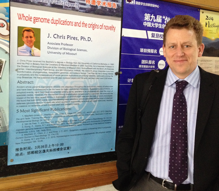
Recently, one of our investigators, J. Chris Pires traveled to Fudan University in Shanghai and the Wuhan Vegetable Research Institute for the 19th annual Crucifer Genetic Workshop and Brassica 2014 Conference in Wuhan, China.
Pires was invited to the esteemed event as the keynote speaker of the Brassica Conference. He led workshops as part of the three-day conference March 30 through April 2 and also visited Fudan University in Shanghai during the trip.
Pires is an associate professor in biological sciences and focuses much of his research on the evolution of plants. His talk at Fudan University discussed his research on whole genome duplications and the origins of novelty in plants.
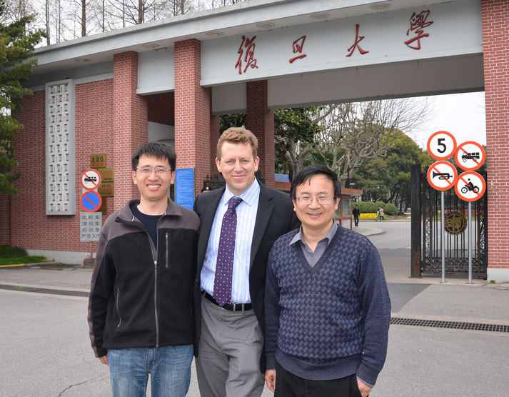
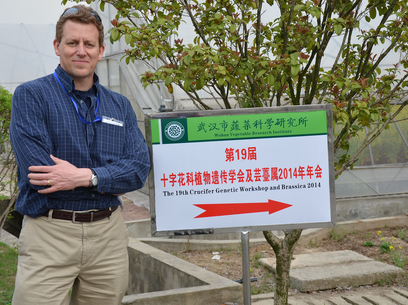
Chris Pires, investigator and associate professor in biological sciences at the Bond Life Sciences Center at Fudan University in Shanghai.Pires with Hong Ma, the dean of life sciences at Fudan University (right) and an assistant professor (left). Pires traveled to Shanghai to lead a workshop at the university over his research in whole genome duplications in plants.Pires at the 19th Crucifer Genetic Workshop and Brassica 2014 at the Wuhan Vegetable Research Institute in Wuhan, China. Pires was the keynote speaker at the conference.
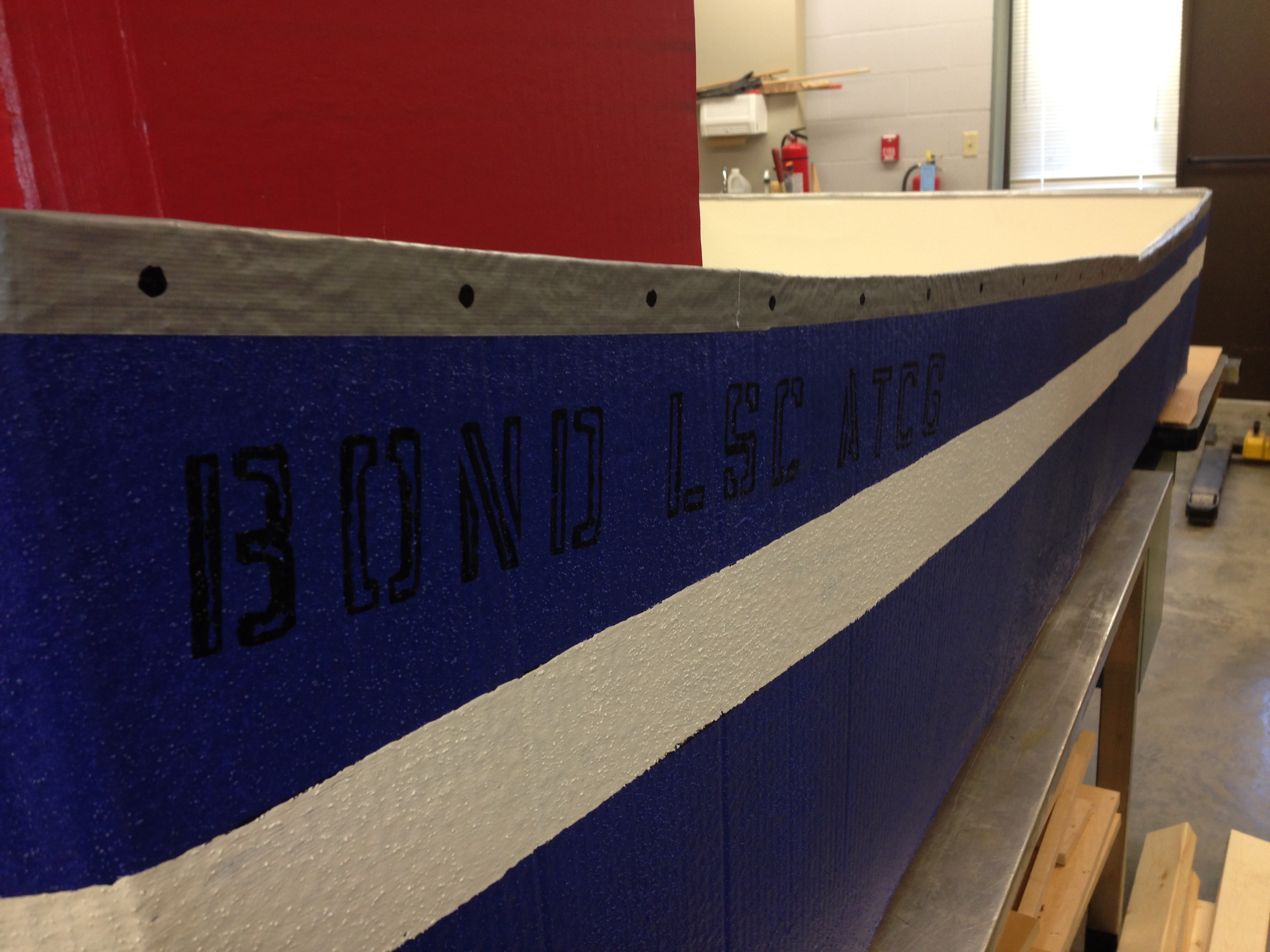
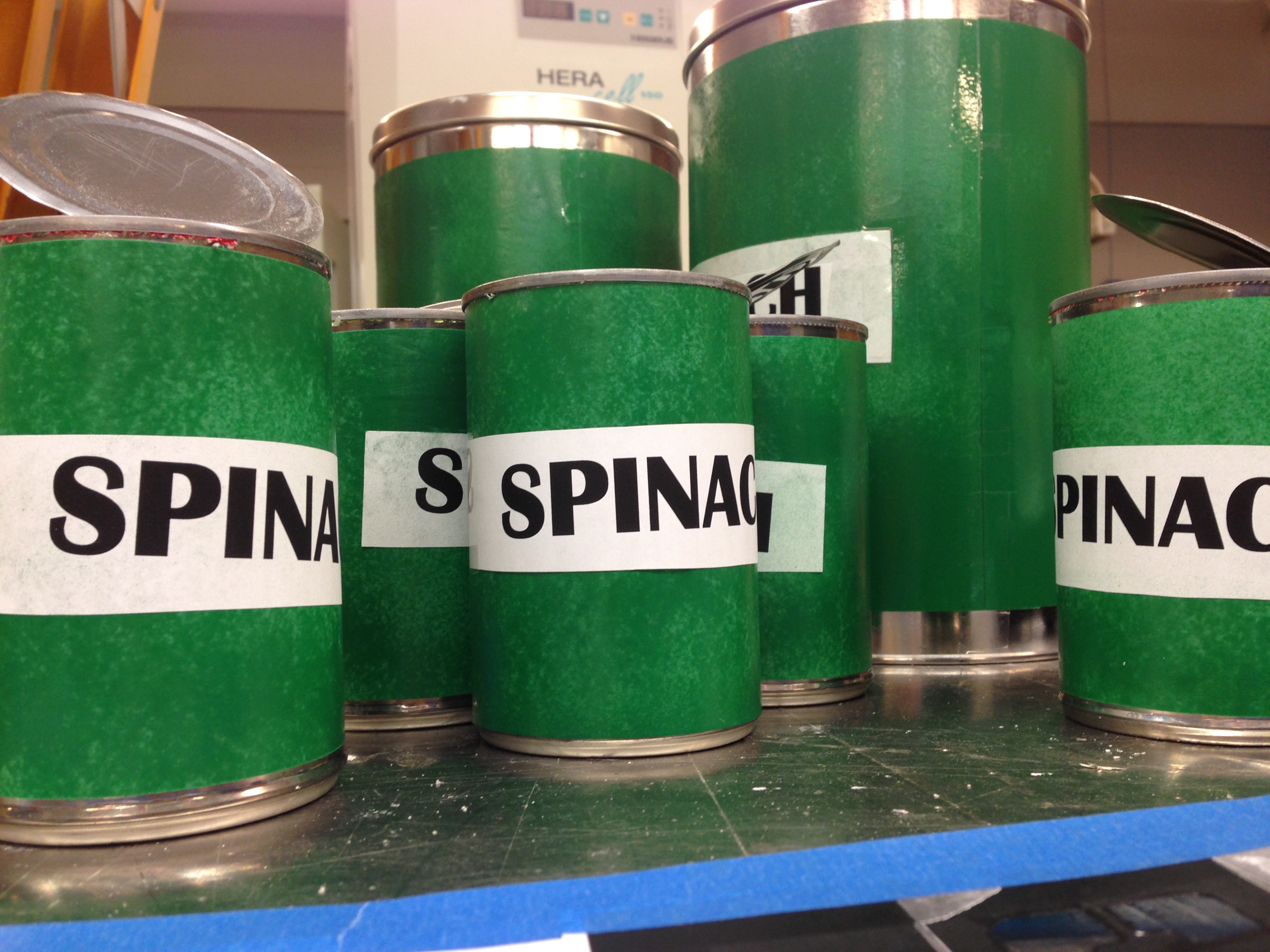
Made completely of cardboard and Popeye themed, Bond LSC facilities crew say this boat could be the winner of the 3rd Annual Flot Your Boat for the Food Bank Race on April 12 — BLANKENBUEHLER
Every year the College of Agriculture, Food and Natural Resources puts on a Float Your Boat for the Food Bank Race. All proceeds go to the Columbia Food Bank and last year, with 45 participants, more than $17,000 was donated. All participants craft their own boat and obey one golden rule: cardboard only.
The Bond LSC crew are returning to the race, this year on April 12, with a Popeye themed boat they say will win it all. Cash donations are being accepted until the race day by Maureen Kemp in 106 at the Bond Life Sciences Center. The People’s Choice Award is given to the boat with the team that raised the most money for the Food Bank.
Barbie Reid, Bond LSC office support assistant, will dress as Olive Oil for this year’s race. — BLANKENBUEHLERThe Bond LSC ATCP is a streamlined, perfectly buoyant cardboard boat. ATCP are the letters of DNA sequencing. — BLANKENBUEHLERThe Bond LSC facilities department crafted mock spinach cans to play up the Popeye theme for this year’s Float Your Boat for the Food Bank Race— BLANKENBUEHLER
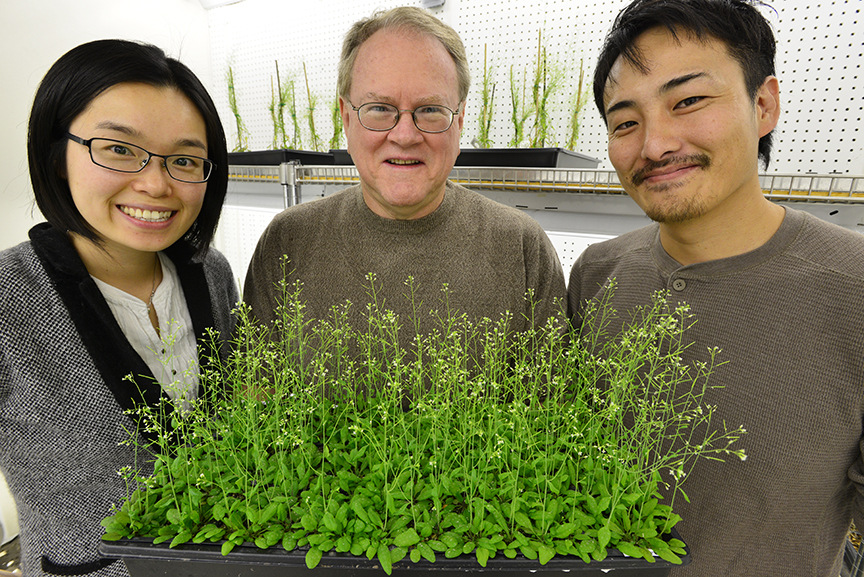
Jeongmin Choi (left), Gary Stacey (center) and postdoc Kiwamu Tanaka recently discovered the first plant receptor for extracellular ATP. Choi received the 2014 Distinguished Dissertation Award for her part in this work.
A former Bond LSC graduate student is being recognized for a dissertation that stands out from the crowd.
Jeongmin Choi received the 2014 Distinguished Dissertation Award this month from MU’s Graduate Faculty Senate for her work identifying the first plant receptor for extracellular ATP. The journal Science published Choi’s “Identification of a plant receptor for extracellular ATP” Jan. 17, 2014.
Choi completed her dissertation working as a member of Gary Stacey’s lab team. Stacey, a Bond LSC researcher, nominated her work for this award. This is the second year work completed in Bond LSC garnered this award after Lefteris Michailidis won in 2013 for work on the HIV drug EFdA.
Choi has since received her Ph.D. and now resides in Cambridge, England.
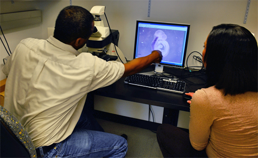
Samuel Waters and graduate researcher Desiré Buckley review stages of embryonic development. — BLANKENBUEHLER
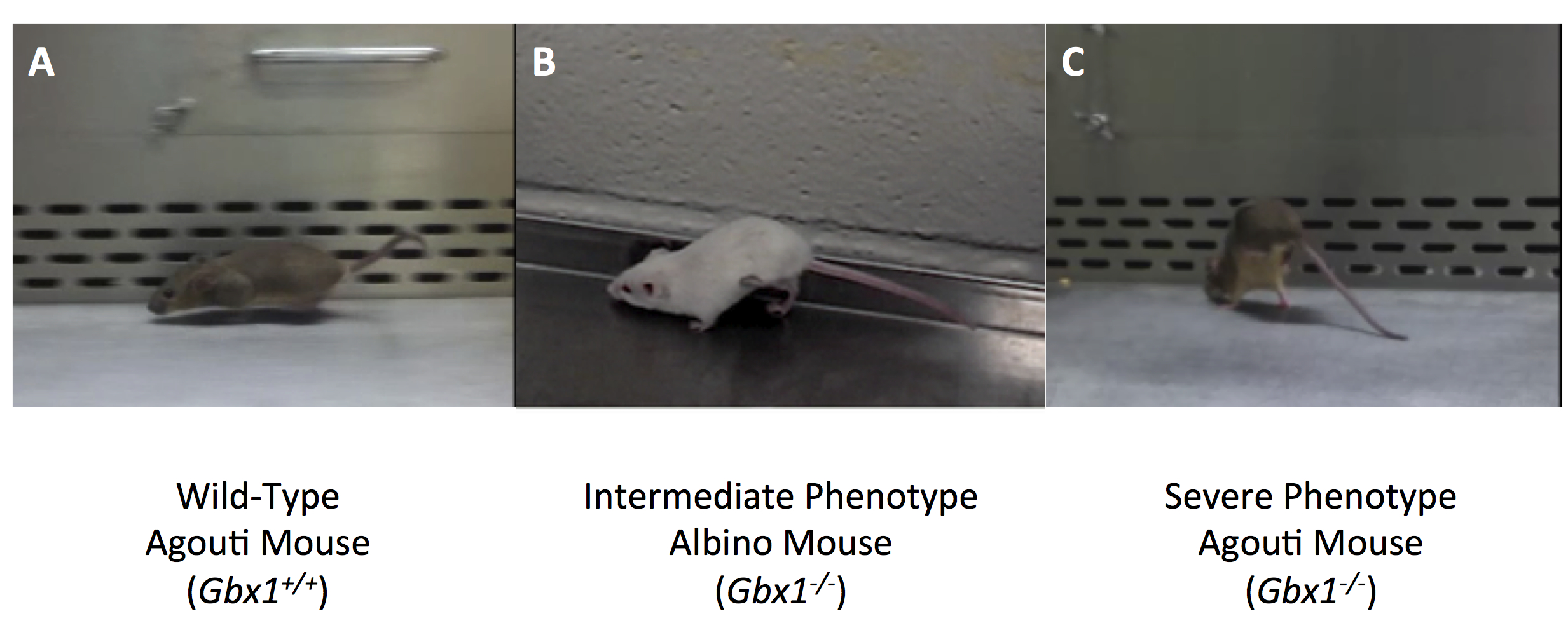
The difference between walking and being paralyzed could be as simple as turning a light switch on and off, a culmination of years of research shows.
Recently, University of Missouri Assistant Professor of biology Samuel T. Waters isolated a coding gene that he found has profound effects on locomotion and central nervous system development.
Waters’ work with gene expression in embryonic mouse tissue could shed light on paralysis and stroke and other disorders of the central nervous system, like Alzheimer’s disease.
Waters works extensively with two coding genes called “Gbx1” and “Gbx2”. These genes — exist in the body with approximately 20,000 other protein-coding genes — are essential for development in the central nervous system.
“To understand what’s going wrong, it’s critical that we know that’s right,” Waters said.
Coding genes essentially assign functions for the body. They tell your fingernail to grow a certain way, help develop motor control responsible for chewing and, as shown in Waters’ research, help your legs work with your spinal cord to facilitate movement.
Waters and his researchers, including graduate student Desiré Buckley, investigated the function of the Gbx1 by deactivating it in mouse embryos and observing their development over a 18.5-day gestation period — the time it takes a mouse to form.
The technology could eventually contribute to developing gene therapies for paralysis that happens at birth or from a direct result of blunt trauma, like a car accident.
“Understanding what allows us to walk normally and have motor control, allows us to have better insight for developing strategies for repairing neural circuits and therapies,” Waters said.
Technology for isolating genes and their functions
Waters studies embryonic mouse development. To understand certain gene functions, he inactivates different genes using a technology called “Cre-loxP.”
Genes can be isolated, then inactivated throughout embryonic tissue. Many of Waters’s studies inactivate genes to harness a better understanding of which genes are responsible for what.
“The relevance of it to the well-being of humans, is apparently relevant to development and more importantly to the development of the central nervous system,” Waters said. “Now it’s taking me to the point where we’re getting a bird’s eye view of what’s actually regulating our ability to have locomotive control.”
No Gbx1, no regular locomotion
Mice that Waters uses in his lab, “display a gross locomotive defect that specifically affects hind-limb gait,” according to their article published in Plos One, February, 2013.
In contrast to its family member Gbx2, when Gbx1 is inactivated, Waters concluded, the anterior hindbrain and cerebellum appear to develop normally. But neural circuit development in the spinal cord —- what allows us to walk normally —- is compromised, he said. According to an article published by
Waters, November 2013, in Methods in Molecular Biology, this occurs despite an increase in the expression level of its family member, Gbx2, in the spinal cord.
A video recording from the research, which was funded by the National Science Foundation and start-up funds from MU, show the mouse with the Gbx1 held back, with an abnormal hind-limb-gait.
Mice with this inactivated gene were otherwise normal, Waters said.
“If they were sitting there without moving, you wouldn’t know anything was wrong with them,” Waters said.” They’re able to mate, eat and appear to function normally.”
Photographs taken during the research that show the hind limb gait defect in specimen with Gbx1 held back.
No Gbx2, no jaw mobility
When Gbx2 function is impaired in the mouse, Waters observed that development of the anterior hindbrain, including the cerebellum, a region of the brain that plays an important role in motor control, didn’t form correctly.
The mice, as a result, cannot suckle, so they die at birth, Waters said.
“We’re getting a better insight into the requirements for suckling — another motor function required for our survival,” Waters said.
The research has paved the way for investigating other coding genes and their responsibilities and roles in development, Waters said.
“We have a lot to do still,” Waters said. “So, why am I so excited about it? That’s part of the reason.”
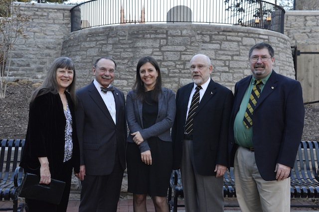
Karin Loftin, MU Chancellor R. Bowen Loftin, Bond Life Sciences Director Jack Shultz and Tim Evans pose with Rebecca Skloot at the University of Missouri Monday evening — BLANKENBUEHLER
The bridge between public knowledge and the inner-workings of the science community is one that many are reluctant to cross. Sometimes riddled with confusing terms, the most exciting discoveries aren’t always approachable.
The 10th annual MU Life Sciences & Society Symposium began Monday evening with Rebecca Skloot as she spoke to a nearly full house at Jesse Auditorium Monday. Every year the symposium erases the line between community understanding and the discoveries of the scientific community.
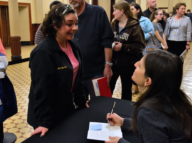
Skloot, the New York Times bestselling author of The Immortal Life of Henrietta Lacks, spoke about the power of science writing in making science more approachable, gave advice to scientists on spreading the word about their discoveries and gave an insight into to the decade of reporting she did for her book. Skloot autographed copies of the book following the talk.
This year’s theme, Decoding Science, speaks to the issue of communicating scientific issues and discoveries with the general public. Skloot said scientists need to keep terms and technicalities basic and exciting.
Rebecca Skloot signs copies of her book, The Immortal Life of Henrietta Lacks, after the talk Monday at Jesse Hall — BLANKENBUEHLER
Jesse Hall was filled with an eclectic mix of community, faculty and students from MU many of which lined up following the talk for nearly 30 minutes of questions.
Throughout the week, the gap between the science community and the public will be bridged with an impressive list of speakers.
The symposium, organized by the Bond Life Sciences Center which houses researchers that represent various schools at the University of Missouri, is a week-long event that features many speakers prevalent in scientific communications.
Other events to catch this week
Tuesday The “Thoughts of Plants” will be uncovered 6 p.m. at Broadway Brewery. The talk, as part of the Science Café speaker series, will be lead by Dr. Jack Shultz, director of the Bond Life Sciences Center.
Wednesday Superhero Science 11 a.m. until noon at the Colonnade in Ellis Library. Superhero submissions will be judged by the spring symposium’s own superhero: “The Antidote.” Dressed in a mask and cape, MU professor Tim Evans’ alter ego, has spicing up the field of toxicology at MU for 12 years.
Thursday James Surowiecki, a contributor to The New Yorker, will speak at 7 p.m. Thursday at Bush Auditorium, Cornell Hall. Free admission and no ticket or registration required.
Saturday All Saturday talks will be held at Jesse Hall.
10:00 a.m. Bill Nye at Jesse Auditorium, doors open at 9:00 a.m. with overflow seating available at the Monsanto Auditorium at the Bond Life Sciences Center. Tickets are sold out. Nye, most well-known for his 1990’s show Bill Nye The Science Guy, has immersed youth in “fun science” by educating in easy-to-understand terms. Nye is one of the pioneers of science communication, trying to make science more approachable by the general public.
12:30 – 1:15 p.m. Chris Mooney is a science journalist and author of Unscientific America, The Republican Brain: The Science of Why They Deny Science and Reality, and New York Times bestselling The Republican War on Science.
1:20 – 2:10 p.m. Dominique Brossard, professor and chair of the Department of Life Sciences Communication at the University of Wisconsin, Brossard studies strategic communication and public opinion in science and risk communication.
2:30 – 3:15 p.m. Liz Neeley, assistant director of Science Outreach for COMPASS, leads communications training for scientists, specializing in social media and multimedia outreach. She previously studied tropical fish evolution.
3:20 – 4:05 p.m. Barbara Kline Pope, the executive director for communications for the National Academy of Sciences, leads the Science & Entertainment Exchange, which connects top scientists with the entertainment industry for accurate science in film and TV programming.
4:10 – 5:00 p.m. A recovering marine biologist, Randy Olson is an independent filmmaker and author of Don’t Be Such a Scientist and Connection: Hollywood Storytelling Meets Critical Thinking. Olsen is a leading proponent of storytelling in science communication. His films include “Flock of Dodos” and “Sizzle,” about evolution and climate change, respectively.
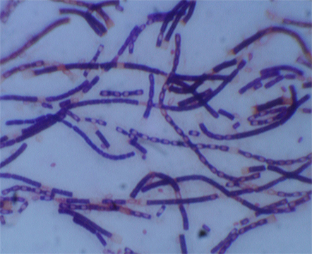
Anthrax bacteria is a rod-shaped culture. Most common forms of transmission are through abrasions in the skin and inhalation.
Imagine researchers in hazmat suits moving slowly and deliberately through a lab. One of them holds up a beaker. It’s glowing.
This light — or the absence of it — could save millions of dollars for governments and save the lives of anthrax victims.
Scientists at the University of Missouri Laboratory of Infectious Disease Research proved a new method for anthrax detection can identify anthrax quicker than any existing approach.
When the “bioluminescent reporter phage” — an engineered virus — infects anthrax bacteria, it takes on a sci-fi-movie-type glow.
George Stewart, a medical bacteriologist at MU’s Bond Life Sciences Center, and graduate student Krista Spreng, observed the virus against a variety of virulent strains of bacillus anthracis, the bacteria causing anthrax disease.
“For this technique, within a few hours, you’ll have a yes or no answer,” Stewart said.
The research, funded by the USDA, was published in the Journal of Microbiological Methods in Aug. 2013. David Schofield at Guild BioSciences, a biotech company in Charleston, S.C, created the reporter phage.
This new method could save a significant amount of money associated with the decontamination of anthrax from suspected infected areas.
Expensive clean-up from the 2001 “Letter attacks”
With the country on high-alert following Sept. 11, 2001, a slew of bioterrorists mailed anthrax letters, filled with a powder that if inhaled could cause death.
Numerous Post Offices and processing facilities were closed and quarantined.
The clean-up bill for the 2001 Anthrax Letter attacks was $3.2 million, according to a 2012 report in Biosecurity and Bioterrorism: Biodefense Strategy, Practice and Science.
Theoretically, the new detection method would alert of a negative result potentially five hours into clean-up efforts instead of two or three days into expensive decontaminating.
Current methods take anywhere from 24 hours or longer to produce a definitive answer for anthrax contamination.
A five-hour benchmark
Stewart said from contamination levels expected from a bioterrorism threat, a positive answer could be found in five hours. If contamination levels were higher, results would come back much more quickly.
Prior to this bioluminescent reporting phage, experts used techniques that were culture based or PCR (polymerase chain reaction) based. Both methods, require additional time for a definitive answer, a minimum of 24 to 48 hours, Stewart said.
“Normally to identify whether an organisms is present, you have to take the material culture, the organism and all the bacteria that might be present in the sample,” Stewart said. “You have to pick colonies that might be bacillus anthracis and do chemical testing which takes some time.”
From a bio-threat standpoint, breathing in anthrax, is the highest concern for public health and homeland security officials and has the highest fatality rate among forms of anthrax.
“If you have a situation and need a quick yes or no answer, this is a tool that will help that,” Stewart said.
Terrorists have used a powder form of anthrax, which has been slipped into letters of political persons and media. A person is infected when an anthrax spore gets into the blood system, most commonly through inhalation or an abrasion on the body, according to Centers of Disease Control and Prevention.
For low levels of contamination, the bioluminescent reporter phage would still detect the presence of the bacteria, but it would take longer.
“This method will be as quick as any of the others and quicker than most,” Stewart said.
The bioluminescent-detection method can detect low levels of anthrax bacteria and rule out false positives. The added benefit to this reporting system is its ability to show that anthrax bacteria are present and it’s alive, Stewart said.
What’s next?
The next step in the bioluminescent reporter phage is getting it approved so a product can be produced and branded. The agency that would warrant the stamp of approval would depend on the eventual use of the phage — food-related testing would likely go through the Food and Drug Administration, Stewart said.
When that happens, a product would not necessarily require a formal lab — it would need a place where cultures could grow at 37 degrees.
“Samples could be collected, brought back to the state public health lab for example and then the testing could be done within a few hours of the collection of the samples and you would have a result,” Stewart said.
The last anthrax attack was in 2001, but the possibility of one happening again, Stewart said, remains a driver for proactive research.
“In the years since the postal attacks, we haven’t had any bona fide anthrax attacks,” Stewart said. “That doesn’t mean it’s not going to happen — we have to be prepared for when it does occur again.”